



Phospho-PLK1 (Thr210) Antibody (YA161)
(Synonyms: PLK, PLK1, Serine/threonine-protein kinase PLK1, Polo-like kinase 1, Serine/threonine-protein kinase 13, PLK-1, STPK13)Based on 2 publication(s) in Google Scholar
Phospho-PLK1 (Thr210) Antibody (YA161) is a Rabbit-derived and non-conjugated IgG monoclonal antibody, targeting to Phospho-PLK1 (Thr210).
-
Host:
Rabbit
-
Isotype:
IgG
-
Application:
WB, ICC/IF, FC
-
Reactivity :
Human
-
Formulation:
Supplied in 1*TBS (pH7.4), 0.05% BSA and 40% Glycerol. Preservative: 0.05% Sodium Azide.
-
Conjugation:
Non-conjugated


To place orders, for customer services and technical support, please contact: MedChemExpress USA
Tel: 609-228-6898 E-mail: [email protected] [email protected]



Publications Citing Use of MedChemExpress (MCE) Phospho-PLK1 (Thr210) Antibody (YA161)
More

Applications
| Application |
WB

WB: Western Blot
|
ICC/IF
ICC/IF: Immunocytochemistry/
Immunofluorescence |
FC
FC: Flow Cytometry
|
|---|---|---|---|
| Dilution Ratio | 1:500-1:1000 | 1:10-1:50 | 1:10-1:50 |


Product Details
Phospho-PLK1 (Thr210) Antibody (YA161) is a Rabbit-derived and non-conjugated IgG monoclonal antibody, targeting to Phospho-PLK1 (Thr210).
-
Host Rabbit

-
Clonality Recombinant,Monoclonal

-
Species ReactivityHuman
-
Observed Molecular WeightObserved band size: 68 kDa
Note: Due to possible protein modifications or aggregation, the molecular weight should be confirmed by actual measurement, and the predicted value is for reference only.

-
Calculated Molecular Weight Predicted band size: 68 kDa

Synthetic phosphopeptide corresponding to residues surrounding Thr210 of Human PLK1.AA range:181-230.
Endogenous
Protein A affinity purified.
Non-conjugated
Phosphorylated
IgG


Product Properties
-
Appearance
Solution
-
Formulation
Supplied in 1*TBS (pH7.4), 0.05% BSA and 40% Glycerol. Preservative: 0.05% Sodium Azide.
-
Concentration
Batch-dependent, Please check the COA for the concentration of each lot. Check Lot Concentration
-
Storage & Stability
Stored at -20°C for 1 year. Avoid repeated freeze / thaw cycles.
-
Shipping
Shipping with blue ice.


Publications (2)
-
Journal Impact Factor
-
Most Recent
-
J Neurooncol
2026 Jun 8;178(2):50. PMID: 42257837


Verification Images
-
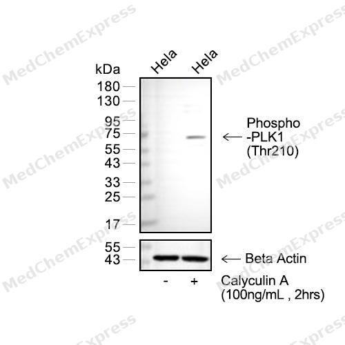 Western blot analysis of extracts from Hela (lane 2(20μg) and Hela (lane 3(20μg), Calyculin A treated (HY-18983, 100ng/mL, 2hrs) as indicated using Phospho-PLK1 (Thr210)(HY-P80474) Rabbit mAb. Proteins were transferred to a PVDF membrane and blocked with 5% non-fat milk in TBST for 2 hour at room temperature. The primary antibody (1/1000) and Loading control antibody (Beta Actin, HY-P80438, 1/10000) was used in 5% non-fat milk in TBST at 4°C overnight. Goat Anti-Mouse/Rabbit IgG-HRP Secondary Antibody (1/10000) was used for 1 hour at room temperature.
 -
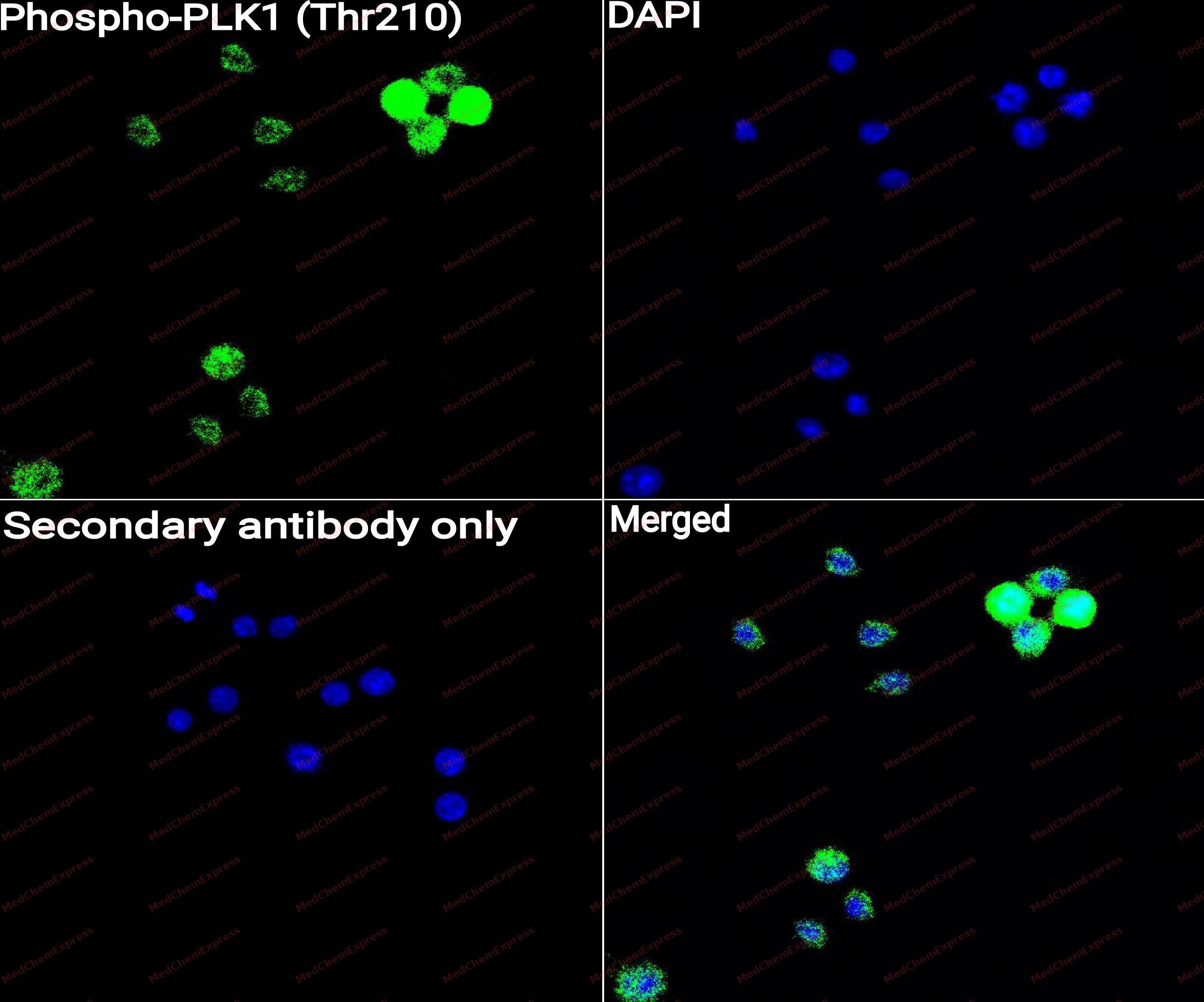 Immunocytochemistry analysis of RAW264.7 cells labeling Phospho-PLK1 (Thr210) with Phospho-PLK1 (Thr210) antibody (HY-P80474) at 1/50 dilution. Cells were fixed in 4% paraformaldehyde for 15 minutes at room temperature, permeabilized with 0.1% Triton X-100 for 10 minutes at room temperature, then blocked with QuickBlock™ Blocking Buffer for Immunol Staining for 10 min at room temperature. Cells were then incubated with Phospho-PLK1 (Thr210) antibody (HY-P80474) at 1/50 dilution in QuickBlock™ Blocking Buffer for Immunol Staining at 4 ℃. Alexa Fluor® 488-conjugated AffiniPure Goat Anti-Rabbit IgG H&L(HY-P8002, Green) was used as the secondary antibody at 1/1,000 dilution. PBS instead of the primary antibody was used as the secondary antibody only control. The Nuclear counterstain was DAPI (Blue).
 -
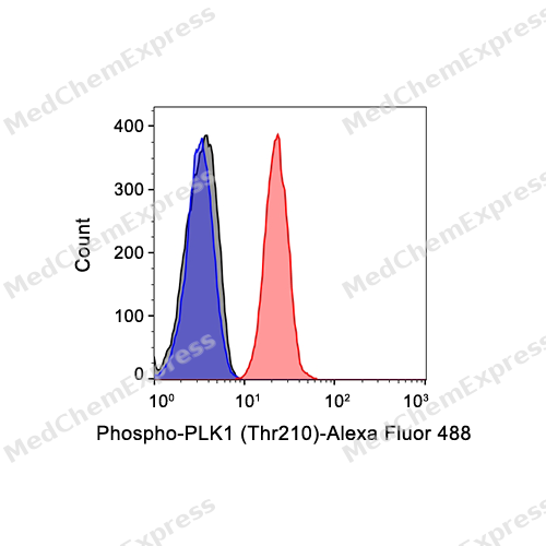 Flow cytometric analysis of 1X10^6 MCF-7 cells labeling Phospho-PLK1 (Thr210) Antibody(HY-P80474, red). Cells were fixed with 4% paraformaldehyde and permeabilised with 90% methanol. Then stained with the primary antibody at 1/50 dilution for an hour at 4℃. Alexa Fluor® 488-conjugated AffiniPure Goat Anti-Rabbit IgG H&L (HY-P8002) was used as the secondary antibody at 1/1,000 dilution for 30 minutes at 4℃. Rabbit IgG Isotype Control (HY-P80879, blue) was used as the isotype control, cells without incubation with primary antibody were used as the unlabeled control (black).


Background
-
Function
PLK1 is a serine/threonine Polo-like kinase that orchestrates cell division through mitotic entry, spindle-pole functions, and cytokinesis[1]. Mechanistically, mammalian PLK1 controls centrosome maturation, spindle assembly, and microtubule attachment to kinetochores in early mitosis[2]. This mitotic role makes PLK1 relevant to cancer biology, because dysfunction may promote cancerous transformation and drive progression, and overexpression occurs in diverse human cancers with poor prognosis[3]. Compared with related isoforms, PLK1-PLK4 participate in cell-cycle processes, but reported functional emphasis differs: PLK2 and PLK4 regulate centriole duplication, PLK3 regulates DNA replication, and PLK1 regulates centrosome separation and maturation[4]. For experimental applications, PLK1 inhibition provides a direct way to perturb mitosis, because BI 2536 inhibits mammalian PLK1 at low nanomolar concentrations, induces mitotic arrest and apoptosis in human cancer cell lines, and inhibits human tumor xenograft growth in nude mice[5].
-
Subcellular Localization
Nucleus; Chromosome, centromere, kinetochore; Cytoplasm, cytoskeleton, microtubule organizing center, centrosome; Cytoplasm, cytoskeleton, spindle; Midbody
-
Expression
Tissue_specificity:placenta and colon
Induction:By growth-stimulating agents -
Subunit
Interacts with CEP170 (PubMed:15616186). Interacts with EVI5 (PubMed:16439210). Interacts with FAM29A (PubMed:19029337). Interacts with SLX4/BTBD12 (PubMed:19596235). Interacts with TTDN1 (PubMed:17310276). Interacts (via POLO-box domain) with the phosphorylated form of BUB1, CDC25C and CENPU (PubMed:14532005, PubMed:16760428, PubMed:17307877, PubMed:19597481). Interacts with KIF2A (PubMed:19351716). Interacts with CYLD (PubMed:17495026). Part of an astrin (SPAG5)-kinastrin (SKAP) complex containing KNSTRN, SPAG5, PLK1, DYNLL1 and SGO2 (PubMed:21402792). Interacts with BIRC6/bruce (PubMed:18329369). Interacts with CDK1-phosphorylated FRY; this interaction occurs in mitotic cells, but not in interphase cells (PubMed:22753416). FRY interaction facilitates AURKA-mediated PLK1 phosphorylation (PubMed:22753416). Interacts with CDK1-phosphorylated DCTN6 during mitotic prometaphase; the interaction facilitates recruitment to kinetochores (PubMed:23455152). Interacts with CEP68; the interaction phosphorylates CEP68 (PubMed:25503564). Interacts (via POLO-box domain) with DCTN1 (PubMed:20679239). Interacts with CEP20 in later G1, S, G2 and M phases of the cell cycle; this interaction recruits PLK1 to centrosomes, a step required for S phase progression (PubMed:24018379). Interacts with KLHL22 (PubMed:23455478, PubMed:24067371). Interacts (via POLO box domains) with NEDD9/HEF1 (via C-terminus) (PubMed:29191835). Interacts with FIRRM (via N-terminus region); required for maintaining, but not activating, PLK1 kinase activity (PubMed:34260926). Interacts with FZR1 (PubMed:18662541). Interacts with SKA3; the interaction promotes the stability of PLK1; the interaction promotes the stability of PLK1 (PubMed:32799774). Interacts with the MTMR3:MTMR4 heterooligomer; brings CEP55 and PLK1 together during early mitosis, regulating the phosphorylation of CEP55 by PLK1 and its recruitment to the midbody where it can mediate cell abscission (PubMed:25659891)
-
SwissProt ID
-
Synonyms
PLK, PLK1, Serine/threonine-protein kinase PLK1, Polo-like kinase 1, Serine/threonine-protein kinase 13, PLK-1, STPK13
-
Research Field
Cell Biology
Documentation
-
Data Sheet (262 KB)
-
SDS (252 KB)
- English - EN (252 KB)
- Français - FR (252 KB)
- Deutsch - DE (252 KB)
- Norwegian - NO (252 KB)
- Español - ES (252 KB)
- Swedish - SV (252 KB)
- Italian - IT (252 KB)
- Korean - KR (252 KB)
- Portuguese - PT (252 KB)
-
User Guide for Antibodies (1077 KB)
References
[1]. Barr FA, et al. Polo-like kinases and the orchestration of cell division. Nat Rev Mol Cell Biol. 2004 Jun;5(6):429-40. [Content Brief]
[2]. Santamaria A, et al. The Plk1-dependent phosphoproteome of the early mitotic spindle. Mol Cell Proteomics. 2011 Jan;10(1):M110.004457. [Content Brief]
[3]. Liu Z, et al. PLK1, A Potential Target for Cancer Therapy. Transl Oncol. 2017 Feb;10(1):22-32. [Content Brief]
[4]. de Cárcer G, et al. From Plk1 to Plk5: functional evolution of polo-like kinases. Cell Cycle. 2011 Jul 15;10(14):2255-62. [Content Brief]
[5]. Steegmaier M, et al. BI 2536, a potent and selective inhibitor of polo-like kinase 1, inhibits tumor growth in vivo. Curr Biol. 2007 Feb 20;17(4):316-22. [Content Brief]

 Powered by Bioz
