



Phospho-Rb (Ser807) Antibody (YA159)
(Synonyms: Retinoblastoma-associated protein, p105-Rb, p110-RB1, pRb, pp110, Rb, RB1)Based on 1 publication(s) in Google Scholar
Phospho-Rb (Ser807) Antibody (YA159) is a Rabbit-derived and non-conjugated IgG monoclonal antibody, targeting to Phospho-Rb (Ser807).
-
Host:
Rabbit
-
Isotype:
IgG
-
Application:
WB, ICC/IF, IHC-P
-
Reactivity :
Human, Mouse
-
Formulation:
Supplied in 1*TBS (pH7.4), 0.05% BSA and 40% Glycerol. Preservative: 0.05% Sodium Azide.
-
Conjugation:
Non-conjugated


To place orders, for customer services and technical support, please contact: MedChemExpress USA
Tel: 609-228-6898 E-mail: [email protected] [email protected]



Publications Citing Use of MedChemExpress (MCE) Phospho-Rb (Ser807) Antibody (YA159)
More

Applications
| Application |
WB

WB: Western Blot
|
ICC/IF
ICC/IF: Immunocytochemistry/
Immunofluorescence |
IHC-P
IHC-P: Immunohistochemistry-Paraffin
|
|---|---|---|---|
| Dilution Ratio | 1:1000 | 1:50 | 1:50-1:1000 |


Product Details
Phospho-Rb (Ser807) Antibody (YA159) is a Rabbit-derived and non-conjugated IgG monoclonal antibody, targeting to Phospho-Rb (Ser807).
-
Host Rabbit

-
Clonality Recombinant,Monoclonal

-
Species ReactivityHuman, Mouse
-
Observed Molecular WeightObserved band size: 106 kDa
Note: Due to possible protein modifications or aggregation, the molecular weight should be confirmed by actual measurement, and the predicted value is for reference only.

-
Calculated Molecular Weight Predicted band size: 106 kDa

Entrez Gene: 5925 Human ; 19645 Mouse ; 24708 Rat
SwissProt: P06400 Human ; P13405 Mouse ; P33568 Rat
OMIM: 180200 Human
Synthetic phosphopeptide corresponding to residues surrounding Ser807 of Human Rb.The exact sequence is proprietary to MCE.
Endogenous
Protein A affinity purified.
Non-conjugated
Phosphorylated
IgG


Product Properties
-
Appearance
Solution
-
Formulation
Supplied in 1*TBS (pH7.4), 0.05% BSA and 40% Glycerol. Preservative: 0.05% Sodium Azide.
-
Concentration
Batch-dependent, Please check the COA for the concentration of each lot. Check Lot Concentration
-
Storage & Stability
Stored at -20°C for 1 year. Avoid repeated freeze / thaw cycles.
-
Shipping
Shipping with blue ice.


Publications (1)
-
Journal Impact Factor
-
Most Recent
-
FASEB J
MS275 Inhibits Neuroblastoma Cell Growth by Mediating H3K27ac/PROX1 Axis In Silico and In Vitro. [Abstract]2025 Jul 15;39(13):e70797. PMID: 40601211


Verification Images
-
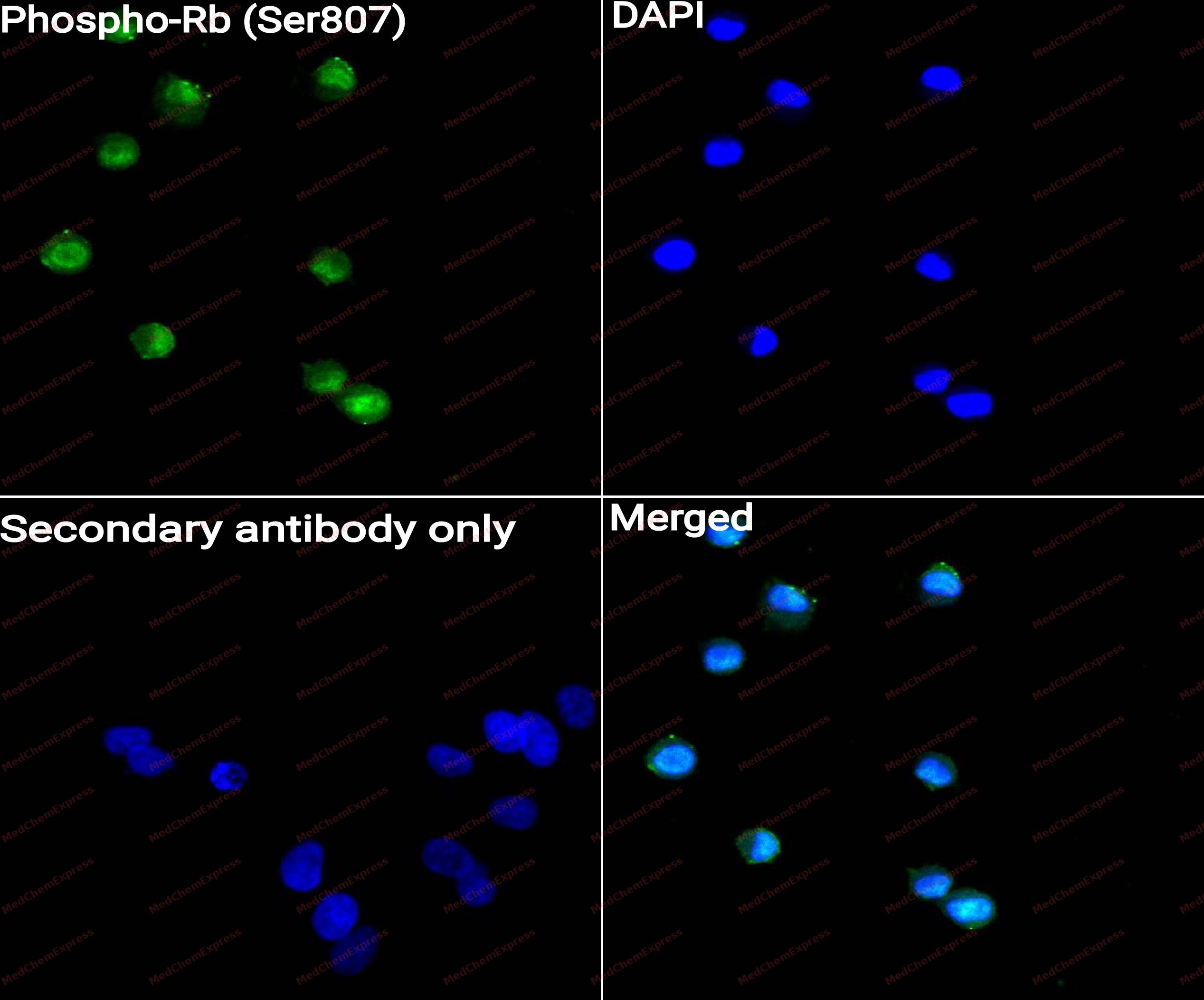 Immunocytochemistry analysis of L929 cells labeling Phospho-Rb (Ser807) with Phospho-Rb (Ser807) antibody (HY-P80476) at 1/50 dilution. Cells were fixed in 4% paraformaldehyde for 15 minutes at room temperature, permeabilized with 0.1% Triton X-100 for 10 minutes at room temperature, then blocked with QuickBlock™ Blocking Buffer for Immunol Staining for 10 min at room temperature. Cells were then incubated with Phospho-Rb (Ser807) antibody (HY-P80476) at 1/50 dilution in QuickBlock™ Blocking Buffer for Immunol Staining at 4 ℃. Alexa Fluor® 488-conjugated AffiniPure Goat Anti-Rabbit IgG H&L(HY-P8002, Green) was used as the secondary antibody at 1/1,000 dilution. PBS instead of the primary antibody was used as the secondary antibody only control. The Nuclear counterstain was DAPI (Blue).
 -
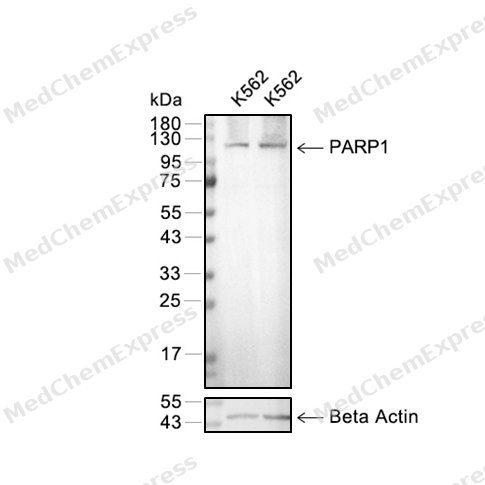 Western blot analysis of extracts from K562 (lane 2(20μg) and K562 (lane 3(40μg)using Phospho-Rb (HY-P80476) Rabbit mAb. Proteins were transferred to a PVDF membrane and blocked with 5% BSA in TBST for 2 hour at room temperature. The primary antibody (HY-P80476, 1/1000) and Loading control antibody (Beta Actin, HY-P80438, 1/3000) was used in 5% BSA in TBST at 4°C overnight. Goat Anti-Mouse/Rabbit IgG-HRP Secondary Antibody (HY-P8004/HY-P8001, 1/10,000) was used for 1 hour at room temperature.
 -
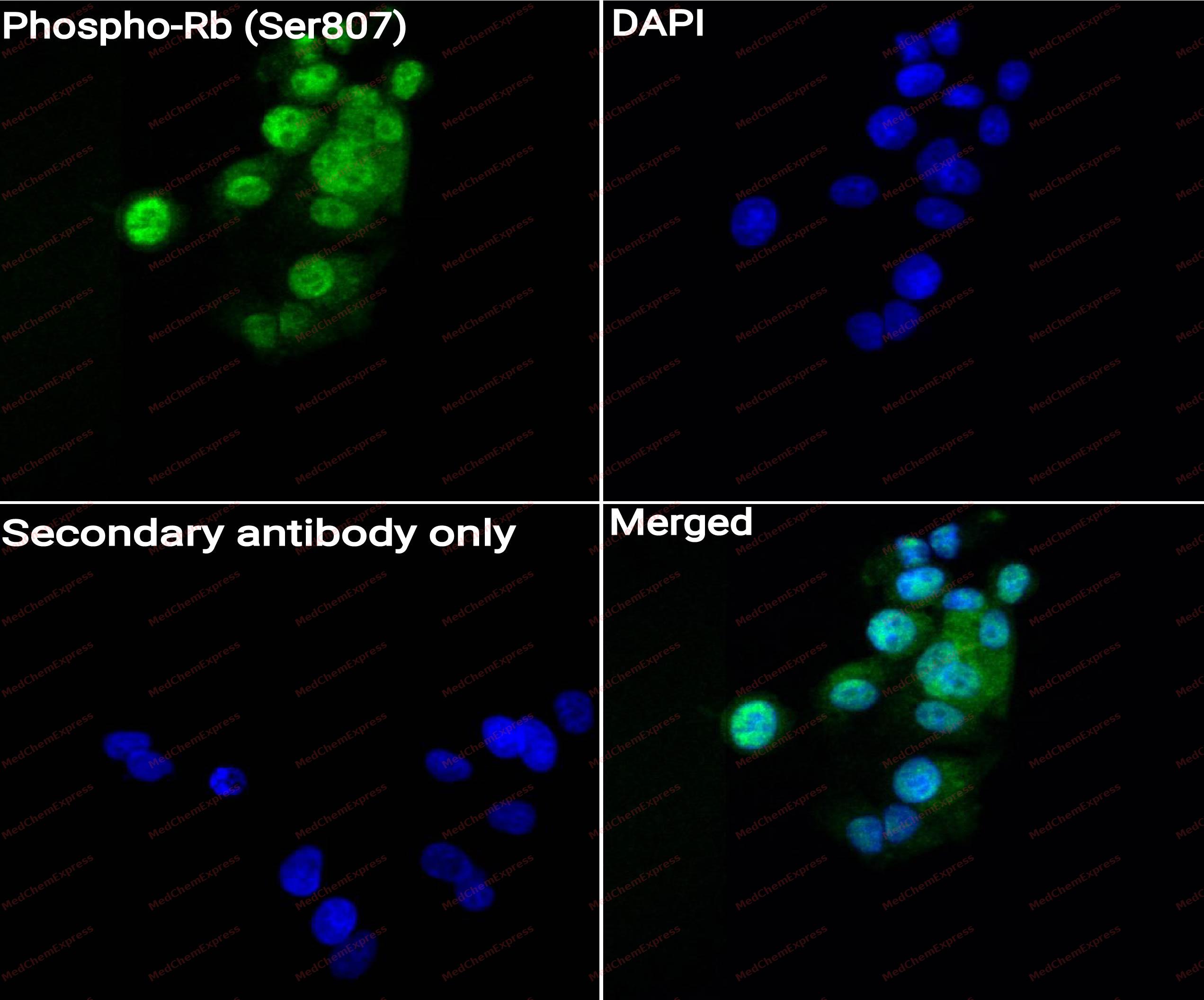 Immunocytochemistry analysis of MCF-7 cells labeling Phospho-Rb (Ser807) with Phospho-Rb (Ser807) antibody (HY-P80476) at 1/50 dilution. Cells were fixed in 4% paraformaldehyde for 15 minutes at room temperature, permeabilized with 0.1% Triton X-100 for 10 minutes at room temperature, then blocked with QuickBlock™ Blocking Buffer for Immunol Staining for 10 min at room temperature. Cells were then incubated with Phospho-Rb (Ser807) antibody (HY-P80476) at 1/50 dilution in QuickBlock™ Blocking Buffer for Immunol Staining at 4 ℃. Alexa Fluor® 488-conjugated AffiniPure Goat Anti-Rabbit IgG H&L(HY-P8002, Green) was used as the secondary antibody at 1/1,000 dilution. PBS instead of the primary antibody was used as the secondary antibody only control. The Nuclear counterstain was DAPI (Blue).


Background
-
Function
Rb is a Tumor suppressor that is a key regulator of the G1/S transition of the cell cycle. The hypophosphorylated form binds transcription regulators of the E2F family, preventing transcription of E2F-responsive genes. Both physically blocks E2Fs transactivating domain and recruits chromatin-modifying enzymes that actively repress transcription. Cyclin and CDK-dependent phosphorylation of RB1 induces its dissociation from E2Fs, thereby activating transcription of E2F responsive genes and triggering entry into S phase. RB1 also promotes the G0-G1 transition upon phosphorylation and activation by CDK3/cyclin-C. Directly involved in heterochromatin formation by maintaining overall chromatin structure and, in particular, that of constitutive heterochromatin by stabilizing histone methylation. Recruits and targets histone methyltransferases SUV39H1, KMT5B and KMT5C, leading to epigenetic transcriptional repression. Controls histone H4 'Lys-20' trimethylation. Inhibits the intrinsic kinase activity of TAF1. Mediates transcriptional repression by SMARCA4/BRG1 by recruiting a histone deacetylase (HDAC) complex to the c-FOS promoter. In resting neurons, transcription of the c-FOS promoter is inhibited by BRG1-dependent recruitment of a phospho-RB1-HDAC1 repressor complex. Upon calcium influx, RB1 is dephosphorylated by calcineurin, which leads to release of the repressor complex; (Microbial infection) In case of viral infections, interactions with SV40 large T antigen, HPV E7 protein or adenovirus E1A protein induce the disassembly of RB1-E2F1 complex thereby disrupting RB1's activity[1][2].
-
Subcellular Localization
Nucleus; Cytoplasm
-
Expression
Tissue_specificity:Expressed in the retina. Expressed in foreskin keratinocytes (protein level) (PubMed: 20940255) -
Subunit
The hypophosphorylated form interacts with and sequesters the E2F1 transcription factor, thereby inhibiting E2F1 transcription (PubMed:20940255, PubMed:8336704). Interacts with heterodimeric E2F/DP transcription factor complexes containing TFDP1 and either E2F1, E2F3, E2F4 or E2F5, or TFDP2 and E2F4. Interacts (when hyperphosphorylated and hypophosphorylated) with PKP3; the interaction inhibits RB1 interaction with and repression of the transcription factor E2F1, potentially via sequestering RB1 to the cytoplasm (By similarity). The unphosphorylated form interacts with EID1, ARID3B, KDM5A, SUV39H1, MJD2A/JHDM3A and THOC1. Interacts with the N-terminal domain of TAF1. Interacts with SNW1, ATAD5, AATF, DNMT1, LIN9, LMNA, KMT5B, KMT5C, PELP1, UHRF2 and TMPO-alpha. Interacts with GRIP1 and UBR4. Interacts with ARID4A and KDM5B. Interacts with E4F1 and LIMD1. Interacts with SMARCA4/BRG1 and HDAC1 (By similarity). Interacts with PSMA3 and USP4. Interacts (when methylated at Lys-860) with L3MBTL1. Interacts with CHEK2; phosphorylates RB1. Interacts with CDK1 and CDK2 (By similarity). Interacts with PRMT2. Interacts with CEBPA (PubMed:15107404). P-TEFB complex interacts with RB1; promotes phosphorylation of RB1 (PubMed:12037672). Interacts with RBBP9; the interaction disrupts RB1 binding to E2F1 (By similarity). Interacts with KAT2B/PCAF and EP300/P300 (By similarity). Interacts with PAX5 (PubMed:10197586). Interacts (phosphorylated and unphosphorylated) with BLCAP (PubMed:26986503). May interact with NDC80
-
SwissProt ID
-
Synonyms
Retinoblastoma-associated protein, p105-Rb, p110-RB1, pRb, pp110, Rb, RB1
-
Research Field
Signal Transduction
Documentation
-
Data Sheet (261 KB)
-
SDS (251 KB)
- English - EN (251 KB)
- Français - FR (251 KB)
- Deutsch - DE (251 KB)
- Norwegian - NO (251 KB)
- Español - ES (251 KB)
- Swedish - SV (251 KB)
- Italian - IT (251 KB)
- Korean - KR (251 KB)
- Portuguese - PT (251 KB)
-
User Guide for Antibodies (1077 KB)
References
[1]. Harbour JW, et al. Cdk phosphorylation triggers sequential intramolecular interactions that progressively block Rb functions as cells move through G1. Cell. 1999 Sep 17;98(6):859-69. [Content Brief]
[2]. Ren S, et al. Cyclin C/cdk3 promotes Rb-dependent G0 exit. Cell. 2004 Apr 16;117(2):239-51. [Content Brief]

 Powered by Bioz
