



TCP 1 alpha Antibody (YA2238)
(Synonyms: TCP1; CCT1; CCTA; T-complex protein 1 subunit alpha; TCP-1-alpha; CCT-alpha)
Based on 1 Customer Validation
TCP 1 alpha Antibody (YA2238) is a Rabbit-derived and non-conjugated IgG monoclonal antibody, targeting to TCP 1 alpha.
-
Host:
Rabbit
-
Isotype:
IgG
-
Application:
WB, IHC-P, ICC/IF, IP
-
Reactivity :
Human
-
Formulation:
Supplied in rabbit IgG in 50 mM Tris-Glycine, pH 7.4, 0.15 M NaCl, 40% glycerol, 0.01% sodium azide and 0.05% BSA.
-
Conjugation:
Non-conjugated


To place orders, for customer services and technical support, please contact: MedChemExpress USA
Tel: 609-228-6898 E-mail: [email protected] [email protected]





Applications
| Application |
WB

WB: Western Blot
|
IHC-P
IHC-P: Immunohistochemistry-Paraffin
|
ICC/IF
ICC/IF: Immunocytochemistry/
Immunofluorescence |
IP
IP: Immunoprecipitation
|
|---|---|---|---|---|
| Dilution Ratio | 1:1000-1:2000 | 1:100-1:200 | 1:50-1:200 | 1:50 |


Product Details
TCP 1 alpha Antibody (YA2238) is a Rabbit-derived and non-conjugated IgG monoclonal antibody, targeting to TCP 1 alpha.
-
Host Rabbit

-
Clonality Recombinant,Monoclonal

-
Species ReactivityHuman
-
Observed Molecular WeightObserved band size: 60 kDa
Note: Due to possible protein modifications or aggregation, the molecular weight should be confirmed by actual measurement, and the predicted value is for reference only.

-
Calculated Molecular Weight Predicted band size: 60 kDa

A synthesized peptide derived from human TCP1 alpha aa1-55/556.
Endogenous
Affinity Chromatography
Non-conjugated
Unmodified
IgG


Product Properties
-
Appearance
Solution
-
Formulation
Supplied in rabbit IgG in 50 mM Tris-Glycine, pH 7.4, 0.15 M NaCl, 40% glycerol, 0.01% sodium azide and 0.05% BSA.
-
Concentration
Batch-dependent, Please check the COA for the concentration of each lot. Check Lot Concentration
-
Storage & Stability
Stored at -20°C for 1 year. Avoid repeated freeze / thaw cycles.
-
Shipping
Shipping with blue ice.


Verification Images
-
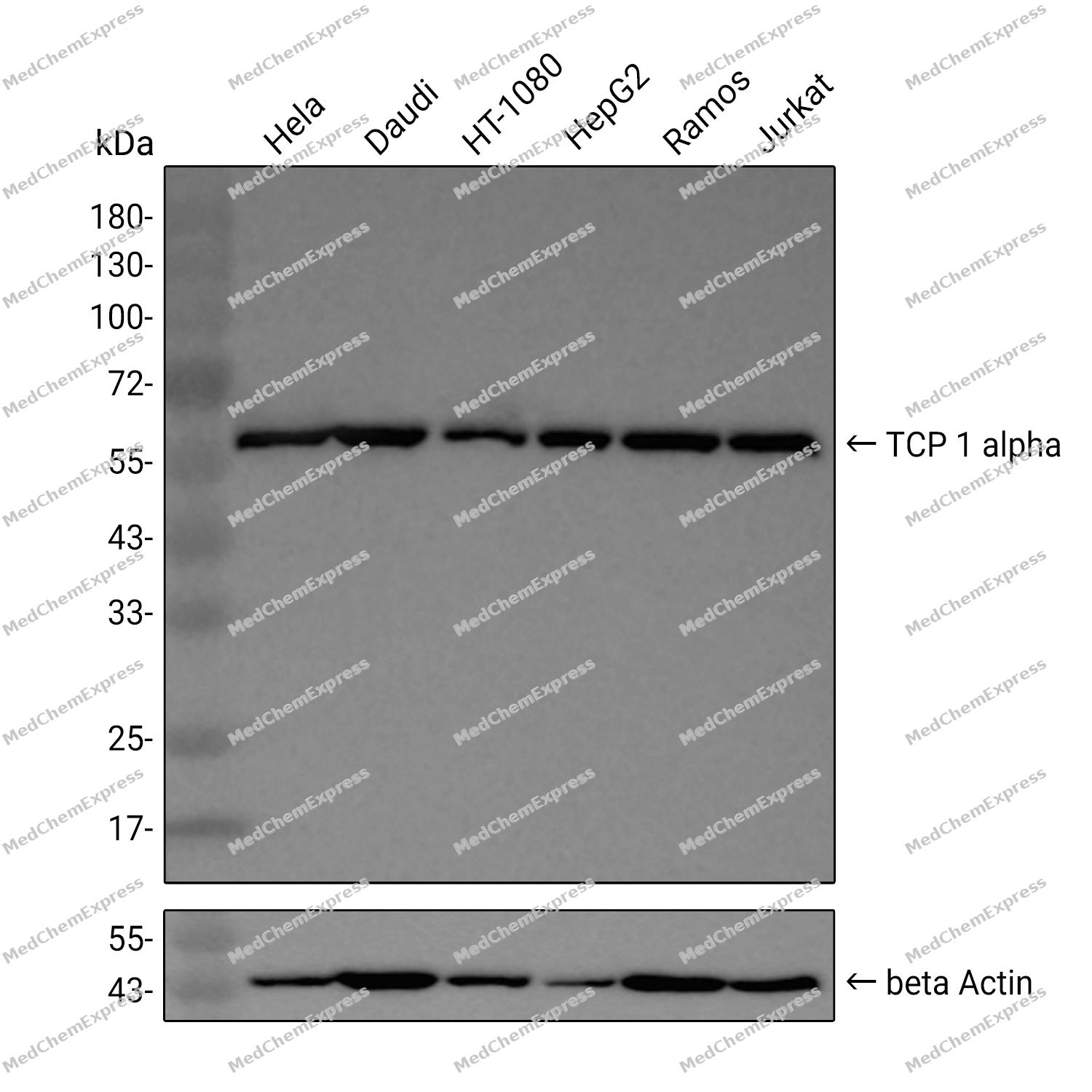 Western blot analysis was performed on extracts from Hela (lane 1, 15 μg), Daudi (lane 2, 15 μg), HT-1080 (lane 3, 15 μg), HepG2 (lane 4, 15 μg), Ramos (lane 5, 15 μg), and Jurkat (lane 6, 15 μg) using TCP 1 alpha Rabbit mAb. Proteins were transferred to a PVDF membrane and blocked with 5% non - fat milk in TBST at 4°C overnight. The primary antibody (1:1000 dilution) and the loading control antibody (beta-Actin(HRP), HY-P80993, 1:5000 dilution) was incubated in 5% non-fat milk in TBST for 1 hour at 37°C. Goat Anti - Rabbit IgG - HRP Secondary Antibody (1:20000 dilution) was then applied for 40 minutes at 37°C.
 -
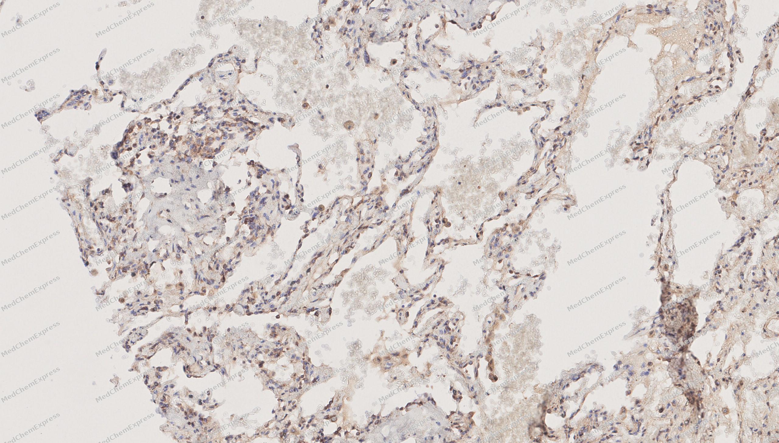 Immunohistochemical analysis of paraffin-embedded human lungs tissue using TCP 1 alpha antibody was performed. The section was pretreated using high-temperature mediated EDTA antigen retrieval buffer (pH 9.0), for 20 minutes. The tissues were incubated with primary antibody (HY-P82493, 1:600 dilution) at room temperature for 20 minutes. Detection was performed using an HRP conjugated compact polymer system. DAB was used as the chromogen. The tissues were counterstained with hematoxylin and mounted with neutral balsam mounting medium.
 -
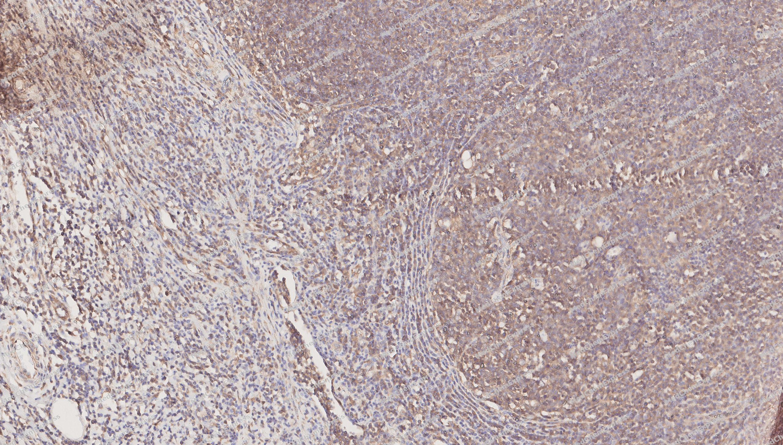 Immunohistochemical analysis of paraffin-embedded human appendix tissue using TCP 1 alpha antibody was performed. The section was pretreated using high-temperature mediated EDTA antigen retrieval buffer (pH 9.0), for 20 minutes. The tissues were incubated with primary antibody (HY-P82493, 1:600 dilution) at room temperature for 20 minutes. Detection was performed using an HRP conjugated compact polymer system. DAB was used as the chromogen. The tissues were counterstained with hematoxylin and mounted with neutral balsam mounting medium.
 -
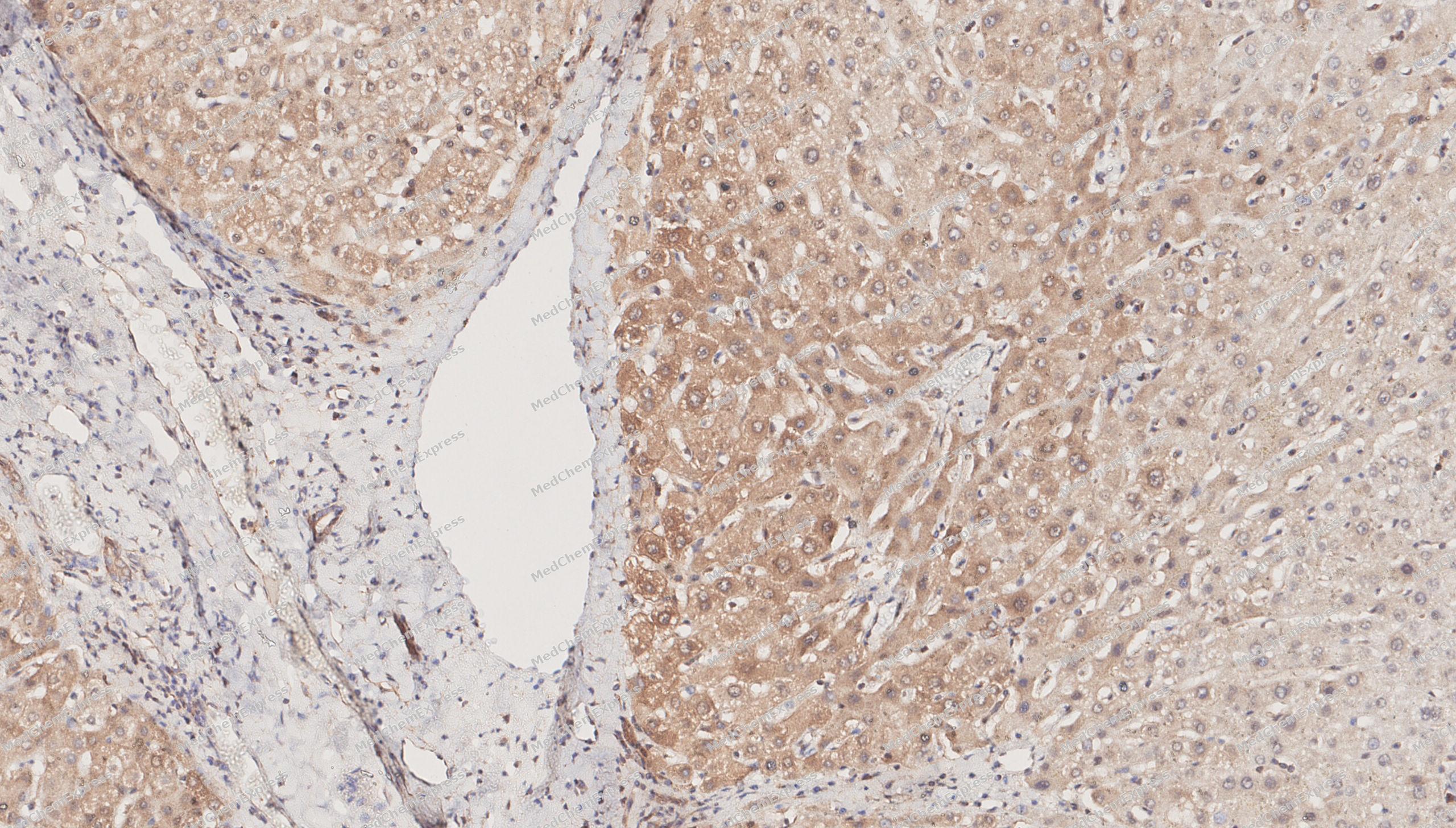 Immunohistochemical analysis of paraffin-embedded human Liver tissue using TCP 1 alpha antibody was performed. The section was pretreated using high-temperature mediated EDTA antigen retrieval buffer (pH 9.0), for 20 minutes. The tissues were incubated with primary antibody (HY-P82493, 1:600 dilution) at room temperature for 20 minutes. Detection was performed using an HRP conjugated compact polymer system. DAB was used as the chromogen. The tissues were counterstained with hematoxylin and mounted with neutral balsam mounting medium.
 -
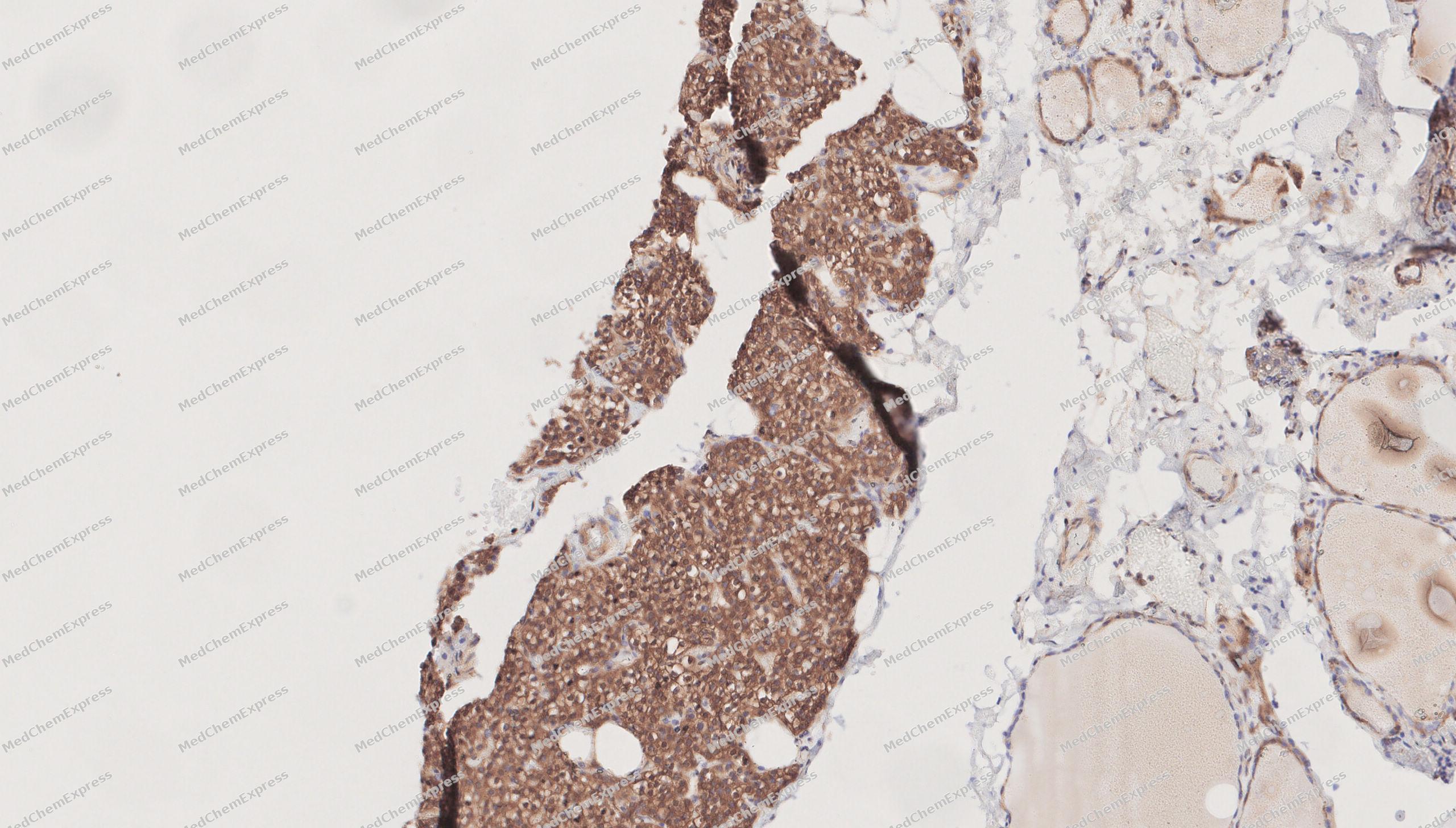 Immunohistochemical analysis of paraffin-embedded human thyroid tissue using TCP 1 alpha antibody was performed. The section was pretreated using high-temperature mediated EDTA antigen retrieval buffer (pH 9.0), for 20 minutes. The tissues were incubated with primary antibody (HY-P82493, 1:600 dilution) at room temperature for 20 minutes. Detection was performed using an HRP conjugated compact polymer system. DAB was used as the chromogen. The tissues were counterstained with hematoxylin and mounted with neutral balsam mounting medium.
 -
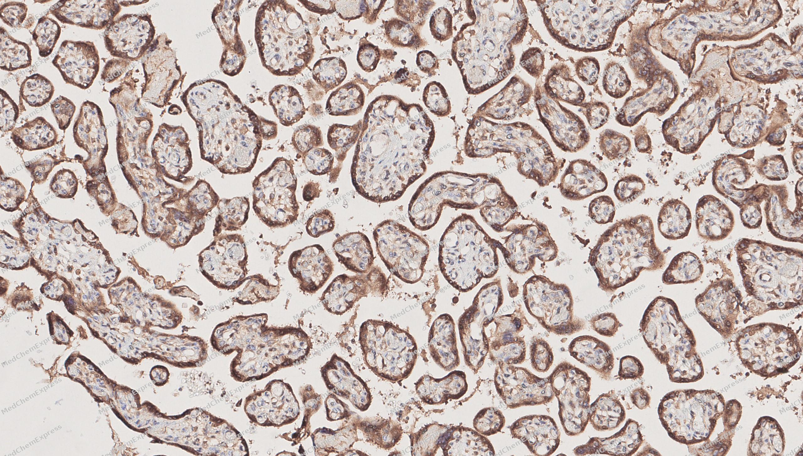 Immunohistochemical analysis of paraffin-embedded human placenta tissue using TCP 1 alpha antibody was performed. The section was pretreated using high-temperature mediated EDTA antigen retrieval buffer (pH 9.0), for 20 minutes. The tissues were incubated with primary antibody (HY-P82493, 1:600 dilution) at room temperature for 20 minutes. Detection was performed using an HRP conjugated compact polymer system. DAB was used as the chromogen. The tissues were counterstained with hematoxylin and mounted with neutral balsam mounting medium.
 -
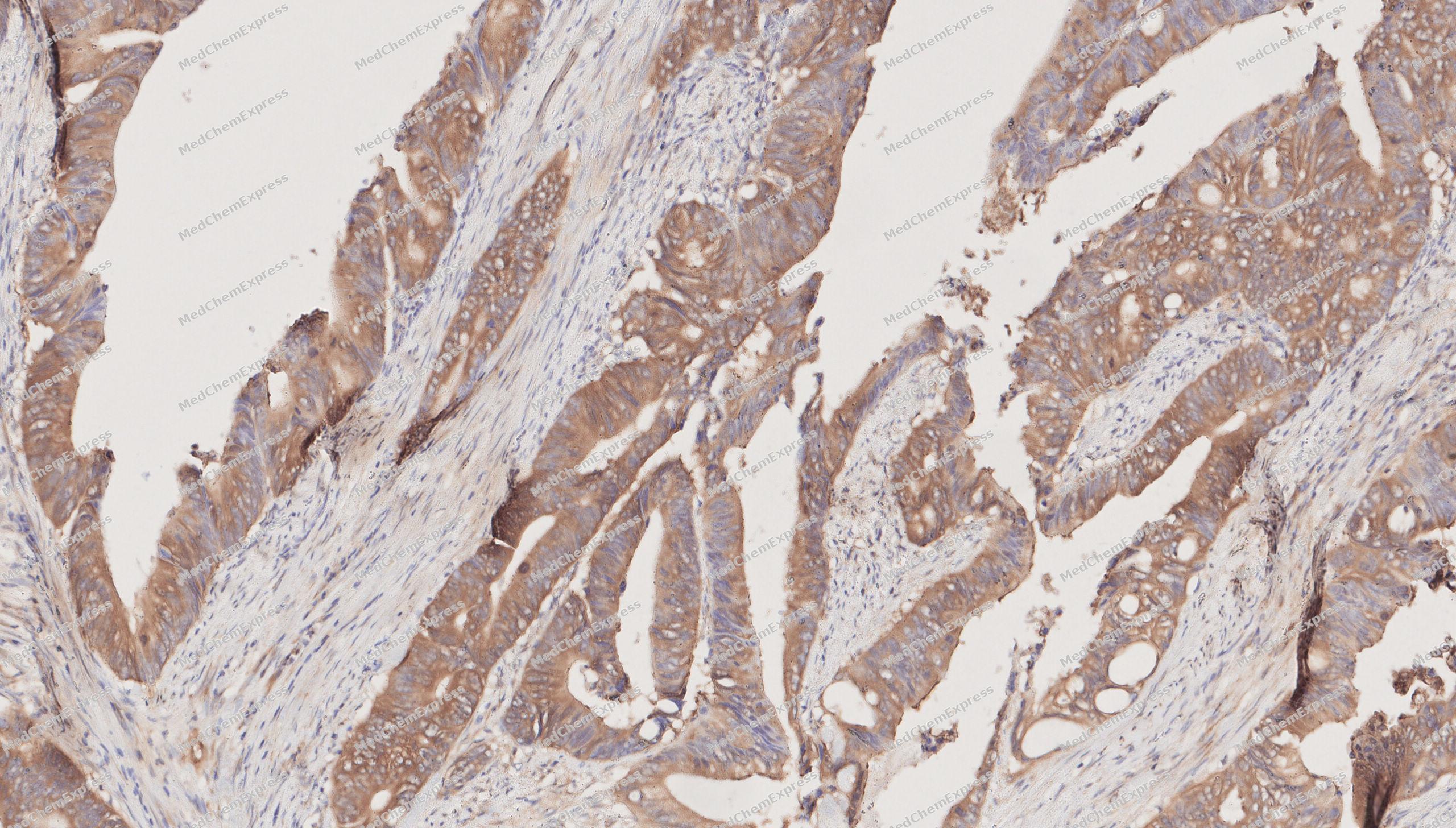 Immunohistochemical analysis of paraffin-embedded human Colorectal cancer tissue using TCP 1 alpha antibody was performed. The section was pretreated using high-temperature mediated EDTA antigen retrieval buffer (pH 9.0), for 20 minutes. The tissues were incubated with primary antibody (HY-P82493, 1:600 dilution) at room temperature for 20 minutes. Detection was performed using an HRP conjugated compact polymer system. DAB was used as the chromogen. The tissues were counterstained with hematoxylin and mounted with neutral balsam mounting medium.
 -
 Immunocytochemistry analysis of Hela cells labeling TCP 1 alpha with TCP 1 alpha Antibody (HY-P82493) at 1/100 dilution. Cells were fixed in 4% paraformaldehyde for 15 minutes at room temperature, permeabilized with 0.1% Triton X-100 in PBS for 15 minutes at room temperature, then blocked with quick block buffer for 10 minutes at room temperature. Cells were then incubated with TCP 1 alpha Antibody (HY-P82493) at 1/100 dilution in quick block buffer overnight at 4 ℃. AF488-conjugated Goat Anti-Rabbit IgG H&L (HY-P8002, Green) was used as the secondary antibody at 1/1,000 dilution. PBS instead of the primary antibody was used as the secondary antibody only control. The Nuclear counterstain was DAPI (Blue).
 -
 Immunocytochemistry analysis of Hela cells labeling TCP 1 alpha with TCP 1 alpha Antibody (HY-P82493) at 1/200 dilution. Cells were fixed in 4% paraformaldehyde for 15 minutes at room temperature, permeabilized with 0.1% Triton X-100 in PBS for 15 minutes at room temperature, then blocked with quick block buffer for 10 minutes at room temperature. Cells were then incubated with TCP 1 alpha Antibody (HY-P82493) at 1/200 dilution in quick block buffer overnight at 4 ℃. AF488-conjugated Goat Anti-Rabbit IgG H&L (HY-P8002, Green) was used as the secondary antibody at 1/1,000 dilution. PBS instead of the primary antibody was used as the secondary antibody only control. The Nuclear counterstain was DAPI (Blue).


Background
-
Function
TCP 1 alpha component of the chaperonin-containing T-complex (TRiC), a molecular chaperone complex that assists the folding of actin, tubulin and other proteins upon ATP hydrolysis. The TRiC complex mediates the folding of WRAP53/TCAB1, thereby regulating telomere maintenance. As part of the TRiC complex may play a role in the assembly of BBSome, a complex involved in ciliogenesis regulating transports vesicles to the cilia[1][2][3][4][5].
-
Subcellular Localization
Cytoplasm, cytosol; Cytoplasm, cytoskeleton, microtubule organizing center, centrosome
-
Subunit
Component of the chaperonin-containing T-complex (TRiC), a hexadecamer composed of two identical back-to-back stacked rings enclosing a protein folding chamber (PubMed:1630492, PubMed:20080638, PubMed:25467444, PubMed:36493755, PubMed:35449234, PubMed:37193829). Each ring is made up of eight different subunits: TCP1/CCT1, CCT2, CCT3, CCT4, CCT5, CCT6A/CCT6, CCT7, CCT8 (PubMed:36493755, PubMed:35449234, PubMed:37193829). Interacts with PACRG (PubMed:14532270). Interacts with GBA1 (PubMed:21098288). Interacts with DLEC1 (PubMed:33144677)
-
SwissProt ID
-
Synonyms
TCP1; CCT1; CCTA; T-complex protein 1 subunit alpha; TCP-1-alpha; CCT-alpha
-
Research Field
Signal Transduction


Documentation
-
Data Sheet (261 KB)
-
SDS (251 KB)
- English - EN (251 KB)
- Français - FR (251 KB)
- Deutsch - DE (251 KB)
- Norwegian - NO (251 KB)
- Español - ES (251 KB)
- Swedish - SV (251 KB)
- Italian - IT (251 KB)
- Korean - KR (251 KB)
- Portuguese - PT (251 KB)
-
User Guide for Antibodies (1077 KB)
References
[1]. Freund A, et al. Proteostatic control of telomerase function through TRiC-mediated folding of TCAB1. Cell. 2014 Dec 4;159(6):1389-403. [Content Brief]
[2]. Gestaut D, et al. Structural visualization of the tubulin folding pathway directed by human chaperonin TRiC/CCT. Cell. 2022 Dec 8;185(25):4770-4787.e20. [Content Brief]
[3]. Kelly JJ, et al. Snapshots of actin and tubulin folding inside the TRiC chaperonin. Nat Struct Mol Biol. 2022 May;29(5):420-429. [Content Brief]
[4]. Liu C, et al. Pathway and mechanism of tubulin folding mediated by TRiC/CCT along its ATPase cycle revealed using cryo-EM. Commun Biol. 2023 May 16;6(1):531. [Content Brief]
[5]. Seo S, et al. BBS6, BBS10, and BBS12 form a complex with CCT/TRiC family chaperonins and mediate BBSome assembly. Proc Natl Acad Sci U S A. 2010 Jan 26;107(4):1488-93. [Content Brief]

 Powered by Bioz
