



TERF1 Antibody (YA3239)
(Synonyms: TERF1; PIN2; TRBF1; TRF1)
Based on 1 Customer Validation
TERF1 Antibody (YA3239) is a Rabbit-derived and non-conjugated IgG monoclonal antibody, targeting to TERF1.
-
Host:
Rabbit
-
Isotype:
IgG
-
Application:
WB, ICC/IF
-
Reactivity :
Human, Mouse
-
Formulation:
Supplied in 10mM PBS, pH 7.4, 150mM sodium chloride, 0.05% BSA, 0.02% sodium azide and 50% glycerol.
-
Conjugation:
Non-conjugated


To place orders, for customer services and technical support, please contact: MedChemExpress USA
Tel: 609-228-6898 E-mail: [email protected] [email protected]





Applications
| Application |
WB

WB: Western Blot
|
ICC/IF
ICC/IF: Immunocytochemistry/
Immunofluorescence |
|---|---|---|
| Dilution Ratio | 1:500-1:1000 | 1:50-1:200 |


Product Details
TERF1 Antibody (YA3239) is a Rabbit-derived and non-conjugated IgG monoclonal antibody, targeting to TERF1.
-
Host Rabbit

-
Clonality Recombinant,Monoclonal

-
Species ReactivityHuman, Mouse
-
Observed Molecular WeightObserved band size: 50 kDa
Note: Due to possible protein modifications or aggregation, the molecular weight should be confirmed by actual measurement, and the predicted value is for reference only.

-
Calculated Molecular Weight Predicted band size: 50 kDa

A synthesized peptide derived from human TRF1 aa70-135.
Endogenous
Affinity Chromatography
Non-conjugated
Unmodified
IgG


Product Properties
-
Appearance
Liquid
-
Formulation
Supplied in 10mM PBS, pH 7.4, 150mM sodium chloride, 0.05% BSA, 0.02% sodium azide and 50% glycerol.
-
Concentration
Batch-dependent, Please check the COA for the concentration of each lot. Check Lot Concentration
-
Storage & Stability
Stored at -20°C for 1 year. Avoid repeated freeze / thaw cycles.
-
Shipping
Shipping with blue ice.


Verification Images
-
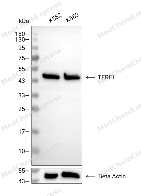 Western blot analysis of extracts from K562 (lane 2(20μg), K562 (lane 3(40μg), using TERF1 Antibody. Proteins were transferred to a PVDF membrane and blocked with 5% BSA in TBST for 2 hour at room temperature. The primary antibody and Loading control antibody (Beta Actin, HY-P80438, 1/3000) was used in 5% BSA in TBST at 4°C overnight. Goat Anti-Mouse/Rabbit IgG-HRP Secondary Antibody (HY-P8004/HY-P8001, 1/10,000) was used for 1 hour at room temperature.
 -
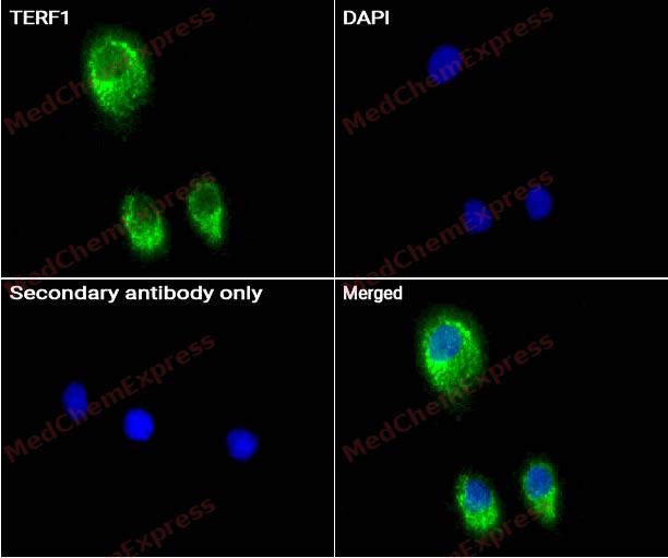 Immunocytochemistry analysis of HepG2 cells labeling TERF1 Antibody (HY-P83494) at 1/50 dilution. Cells were fixed in 4% paraformaldehyde for 15 minutes at room temperature, permeabilized with 0.1% Triton X-100 for 10 minutes at room temperature, then blocked with BSA for Immunol Staining for 10 min at room temperature. Cells were then incubated with TERF1 Antibody (HY-P83494) at 1/50 dilution in BSA for Immunol Staining at 4 ℃ overnight. AF488-conjugated AffiniPure Goat Anti-Rabbit IgG H&L(HY-P8002, Green) was used as the secondary antibody at 1/1,000 dilution. PBS instead of the primary antibody was used as the secondary antibody only control. The Nuclear counterstain was DAPI (Blue).
 -
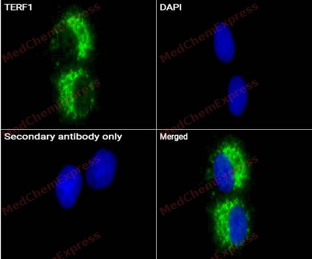 Immunocytochemistry analysis of HepG2 cells labeling TERF1 Antibody (HY-P83494) at 1/100 dilution. Cells were fixed in 4% paraformaldehyde for 15 minutes at room temperature, permeabilized with 0.1% Triton X-100 for 10 minutes at room temperature, then blocked with BSA for Immunol Staining for 10 min at room temperature. Cells were then incubated with TERF1 Antibody (HY-P83494) at 1/100 dilution in BSA for Immunol Staining at 4 ℃ overnight. AF488-conjugated AffiniPure Goat Anti-Rabbit IgG H&L(HY-P8002, Green) was used as the secondary antibody at 1/1,000 dilution. PBS instead of the primary antibody was used as the secondary antibody only control. The Nuclear counterstain was DAPI (Blue).


Background
-
Function
TERF1 binds the telomeric double-stranded 5'-TTAGGG-3' repeat and negatively regulates telomere length. Involved in the regulation of the mitotic spindle. Component of the shelterin complex (telosome) that is involved in the regulation of telomere length and protection. Shelterin associates with arrays of double-stranded 5'-TTAGGG-3' repeats added by telomerase and protects chromosome ends; without its protective activity, telomeres are no longer hidden from the DNA damage surveillance and chromosome ends are inappropriately processed by DNA repair pathways
-
Subcellular Localization
Nucleus; Cytoplasm, cytoskeleton, spindle; Chromosome, telomere
-
Expression
Tissue_specificity:Highly expressed and ubiquitous. Isoform Pin2 predominates
Induction:Expression is tightly regulated during the cell cycle; levels are low in G1 and S phase and increase during G2 phase and mitosis -
Isoforms & Post-Translational Modification
P54274 has 2 isomers: P54274-1: 50246 Da (predicted); P54274-2: 48239 Da (predicted).
Phosphorylated preferentially on Ser-219 in an ATM-dependent manner in response to ionizing DNA damage;ADP-ribosylation by TNKS1 or TNKS2 diminishes its ability to bind to telomeric DNA;Ubiquitinated by RLIM/RNF12, leading to its degradation by the proteasome. Ubiquitinated by a SCF (SKP1-CUL1-F-box protein) ubiquitin-protein ligase complex, leading to its degradation by the proteasome -
Subunit
Homodimer; can contain both isoforms. Found in a complex with POT1; TINF2 and TNKS1. Interacts with ATM, TINF2, TNKS1, TNKS2, PINX1, NEK2 and MAPRE1. Component of the shelterin complex (telosome) composed of TERF1, TERF2, TINF2, TERF2IP ACD and POT1. Interacts with RLIM (via N-terminus). Interacts with FBXO4. Interaction with TINF2 protects against interaction with FBXO4 and subsequent polyubiquitination and proteasomal degradation. Interacts with GNL3L; this interaction promotes homodimerization. Interacts with TIN2. Interacts with RTEL1. Interactions with GNL3L and TIN2 are mutually exclusive. Interacts with CCDC79/TERB1 (By similarity). Interacts with TRIOBP isoform 1; mediates TERF1 localization to the centrosome (PubMed:24692559)
-
SwissProt ID
-
Synonyms
TERF1; PIN2; TRBF1; TRF1
-
Research Field
Epigenetics and Nuclear Signaling


Documentation
-
Data Sheet (234 KB)
-
SDS (251 KB)
- English - EN (251 KB)
- Français - FR (251 KB)
- Deutsch - DE (251 KB)
- Norwegian - NO (251 KB)
- Español - ES (251 KB)
- Swedish - SV (251 KB)
- Italian - IT (251 KB)
- Korean - KR (251 KB)
- Portuguese - PT (251 KB)
-
User Guide for Antibodies (1077 KB)

 Powered by Bioz
