



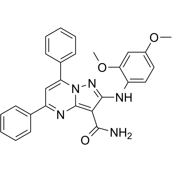
CDK1-IN-1

Based on 1 Customer Validation
CDK1-IN-1 is a potent CDK1 inhibitor (CDK1/CycB IC50=161.2 nM) with potential antiproliferative activity and selectivity for cancer tissues. CDK1-IN-1 induces apoptosis in p53 dependent manner through the intrinsic apoptotic pathway. CDK1-IN-1 is a potential targeted antitumor agent.
For research use only. We do not sell to patients.
- Purity : 99.33%
- CAS No.: 2761858-59-5
- Formula: C27H23N5O3
- Molecular Weight:465.50
-
Storage:Powder -20°C, 3 years , 4°C, 2 years ; In solvent -80°C, 6 months , -20°C, 1 month

To place orders, for customer services and technical support, please contact: MedChemExpress USA
Tel: 609-228-6898 E-mail: [email protected] [email protected]


-
 Biological Activity
-
Chemical Information
-
Solvent & Solubility
- Protocols
- Purity & Documentation
- References
-
 Help & FAQs




Biological Activity
Description
IC50 & Target
|
CDK1/cycB 161.2 nM (IC50) |
Cellular Effect
|
Cell Line

|
Type | Value | Description | References |
|---|---|---|---|---|
| HCT-116 | IC50 |
6.28 μM
Compound: 181a
|
Cytotoxicity against human HCT-116 cells by MTT assay
Cytotoxicity against human HCT-116 cells by MTT assay
|
[PMID: 36325400] |
| WI-38 | IC50 |
17.7 μM
Compound: 181a
|
Cytotoxicity against human WI-38 cells by MTT assay
Cytotoxicity against human WI-38 cells by MTT assay
|
[PMID: 36325400] |
In Vitro
CDK1-IN-1 (compound 7a) (10 µM, 48 hours) has the high antiproliferative activity with a mean percentage of growth inhibition of 48.5 % over NCI cancer cell lines, and caused more than 40% growth inhibition in 36 cancer cell lines from different cancer subtypes[1].
CDK1-IN-1 (0.1-100 μM, 48 hours) has better selectivity for cancer cells over normal cell lines (IC50 of 17.7 and 6.28 μM in WI-38 and HCT-116 cells, respectively; SI=2.8)[1].
CDK1-IN-1 (17.7 μM in WI-38 and 6.28 μM in HCT-116; 48 hours) has a superior activity on cancerous cells than normal cells in inducing apoptosis and arresting the cell cycle at the G2/M phase[1].
CDK1-IN-1 (17.7 μM in WI-38 and 6.28 μM in HCT-116; 48 hours) can cause cell death mainly through apoptosis rather than necrosis and confirmed that its activity is more selective to cancerous cells than normal cells[1].
CDK1-IN-1 (6.28 μM; 48 hours) induce apoptosis in p53 dependent manner through the intrinsic apoptotic pathway in HCT-116 cells[1].
MedChemExpress (MCE) has not independently confirmed the accuracy of these methods. They are for reference only.
-
Cell Line:60 human cancer cell lines (LOX IMVI, IGROV1, A498, COLO205 et al.)[1]
-
Concentration:10 µM
-
Incubation Time:48 hours
-
Result:Had the high antiproliferative activity with a mean percentage of growth inhibition of 48.5 % over NCI cancer cell lines, and caused more than 40% growth inhibition in 36 cancer cell lines from different cancer subtypes.
-
Cell Line:HCT-116 and WI-38 cells[1]
-
Concentration:0.1-100 μM
-
Incubation Time:48 hours
-
Result:Had better selectivity for cancer cells over normal cell lines (IC50 of 17.7 and 6.28 μM in WI-38 and HCT-116 cells, respectively; SI=2.8).
-
Cell Line:HCT-116 and WI-38 cells[1]
-
Concentration:17.7 μM in WI-38 and 6.28 μM in HCT-116
-
Incubation Time:48 hours
-
Result:Exerted a superior activity on cancerous cells than normal cells in inducing apoptosis and arresting the cell cycle at the G2/M phase.


Chemical Information
-
CAS No. 2761858-59-5
-
Appearance Solid
-
Molecular Weight 465.50
-
Formula C27H23N5O3
-
Color Yellow to orange
-
SMILES
O=C(C1=C2N=C(C3=CC=CC=C3)C=C(C4=CC=CC=C4)N2N=C1NC5=CC=C(OC)C=C5OC)N
-
Shipping
Room temperature in continental US; may vary elsewhere.
-
Storage
Powder -20°C 3 years 4°C 2 years In solvent -80°C 6 months -20°C 1 month


Solvent & Solubility
In Vitro:
DMSO : 25 mg/mL (53.71 mM; ultrasonic and warming and heat to 60°C; Hygroscopic DMSO has a significant impact on the solubility of product, please use newly opened DMSO)
Please refer to the solubility information to select the appropriate solvent. Once prepared, please aliquot and store the solution to prevent product inactivation from repeated freeze-thaw cycles.
Storage method and period of stock solution: -80°C, 6 months; -20°C, 1 month. When stored at -80°C, please use it within 6 months. When stored at -20°C, please use it within 1 month.
Please refer to the solubility information to select the appropriate solvent. Once prepared, please aliquot and store the solution to prevent product inactivation from repeated freeze-thaw cycles.
Storage method and period of stock solution: -80°C, 6 months; -20°C, 1 month. When stored at -80°C, please use it within 6 months. When stored at -20°C, please use it within 1 month.
Concentration (start) × Volume (start) = Concentration (final) × Volume (final)
In Vivo:
Select the appropriate dissolution method based on your experimental animal and administration route.
- For the following dissolution methods, please ensure to first prepare a clear stock solution using an In Vitro approach and then sequentially add co-solvents:
- To ensure reliable experimental results, the clarified stock solution can be appropriately stored based on storage conditions. As for the working solution for In Vivo experiments, it is recommended to prepare freshly and use it on the same day.
- The percentages shown for the solvents indicate their volumetric ratio in the final prepared solution. If precipitation or phase separation occurs during preparation, heat and/or sonication can be used to aid dissolution.
In Vivo Dissolution Calculator
Please enter the basic information of animal experiments:
-
-
-
-
Recommended: Prepare an additional quantity of animals to account for potential losses during experiments.
Please enter your animal formula composition:
-
%DMSO +
Recommended: Keep the proportion of DMSO in working solution below 2% if your animal is weak.
-
%+
-
+%Tween-80 + +
-
%Saline +
The co-solvents required include: DMSO, . All of co-solvents are available by MedChemExpress (MCE). , Tween 80. All of co-solvents are available by MedChemExpress (MCE).
Working solution concentration: 0.22 mg/mL
Method for preparing stock solution: mg drug dissolved in μL DMSO. Stock solution concentration: mg/mL.
1. Take μL DMSO stock solution;
2. Add μL .
μL , mix evenly;
3. Then add μL Tween 80, mix evenly;
4. Then add μL
Please ensure that the stock solution in the first step is dissolved to a clear state, and add co-solvents in sequence. You can use ultrasonic heating (ultrasonic cleaner, recommended frequency 20-40 kHz), vortexing, etc. to assist dissolution.


Purity & Documentation
-
Data Sheet (280 KB)
-
SDS (251 KB)
- English - EN (251 KB)
- Français - FR (251 KB)
- Deutsch - DE (251 KB)
- Norwegian - NO (251 KB)
- Español - ES (251 KB)
- Swedish - SV (251 KB)
- Italian - IT (251 KB)
- Korean - KR (251 KB)
- Portuguese - PT (251 KB)
-
Handling Instructions (2659 KB)
References
[1]. Elgiushy HR, et al. Identification of a promising hit from a new series of pyrazolo[1,5-a]pyrimidine based compounds as a potential anticancer agent with potent CDK1 inhibitory and pro-apoptotic properties through a multistep in vitro assessment [published online ahead of print, 2022 Jan 29]. Bioorg Chem. 2022;120:105646. [Content Brief]
[2]. Gangadevi S, et al. Kobophenol A Inhibits Binding of Host ACE2 Receptor with Spike RBD Domain of SARS-CoV-2, a Lead Compound for Blocking COVID-19. J Phys Chem Lett. 2021;12(7):1793-1802. [Content Brief]


Complete Stock Solution Preparation Table
Please refer to the solubility information to select the appropriate solvent. Once prepared, please aliquot and store the solution to prevent product inactivation from repeated freeze-thaw cycles.
Storage method and period of stock solution: -80°C, 6 months; -20°C, 1 month. When stored at -80°C, please use it within 6 months. When stored at -20°C, please use it within 1 month.
| Optional Solvent | Concentration Solvent Mass | 1 mg | 5 mg | 10 mg | 25 mg |
|---|---|---|---|---|---|
| DMSO | 1 mM | 2.1482 mL | 10.7411 mL | 21.4823 mL | 53.7057 mL |
| 5 mM | 0.4296 mL | 2.1482 mL | 4.2965 mL | 10.7411 mL | |
| 10 mM | 0.2148 mL | 1.0741 mL | 2.1482 mL | 5.3706 mL | |
| 15 mM | 0.1432 mL | 0.7161 mL | 1.4322 mL | 3.5804 mL | |
| 20 mM | 0.1074 mL | 0.5371 mL | 1.0741 mL | 2.6853 mL | |
| 25 mM | 0.0859 mL | 0.4296 mL | 0.8593 mL | 2.1482 mL | |
| 30 mM | 0.0716 mL | 0.3580 mL | 0.7161 mL | 1.7902 mL | |
| 40 mM | 0.0537 mL | 0.2685 mL | 0.5371 mL | 1.3426 mL | |
| 50 mM | 0.0430 mL | 0.2148 mL | 0.4296 mL | 1.0741 mL |


CDK1-IN-1 Related Classifications
HY-115924 Related Classifications
 Powered by Bioz