



CDK9-IN-19
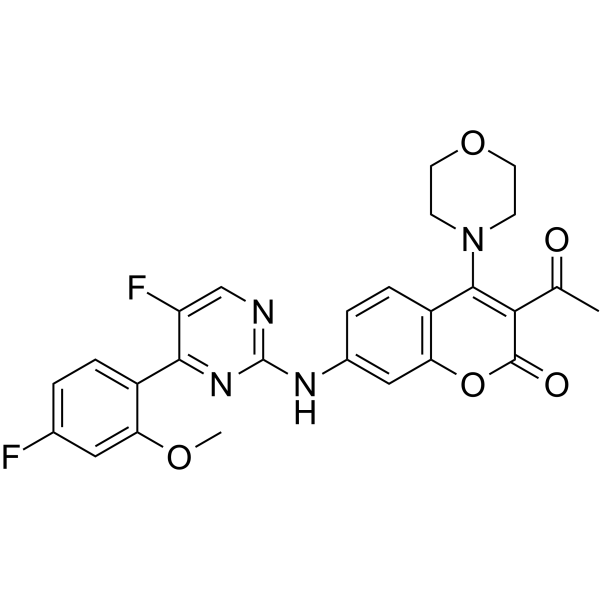
CDK9-IN-19 is a highly potent and selective CDK9 inhibitor with an IC50 value of 2.0 nM. CDK9-IN-19 has excellent cellular antiproliferative activity, moderate pharmacokinetic property and low hERG inhibition. CDK9-IN-19 significantly induces tumour growth inhibition in an MV4-11 xenograft mice model. CDK9-IN-19 can be used for researching acute myeloid leukaemia (AML).
Nos produits utilisent uniquement pour la recherche. Nous ne vendons pas aux patients.
- CAS No.: 2479306-60-8
- Formule: C26H22F2N4O5
- Masse moléculaire:508.47
-
Stockage:
Please store the product under the recommended conditions in the Certificate of Analysis.

To place orders, for customer services and technical support, please contact: MedChemExpress USA
Tel: 609-228-6898 E-mail: [email protected] [email protected]


-
 Activité biologique
-
Chemical Information
- Protocole
- Pureté et documentation
- Références
-
 Help & FAQs




Activité biologique
Description
IC50 & Target
[1]|
CDK9/cyclinT1 2 nM (IC50) |
Cellular Effect
|
Cell Line

|
Type | Value | Description | References |
|---|---|---|---|---|
| A-375 | IC50 |
0.09 μM
Compound: 30i
|
Antiproliferative activity against human A-375 cells assessed as inhibition of cell growth incubated for 72 hrs by Celltiter-Glo luminescent assay
Antiproliferative activity against human A-375 cells assessed as inhibition of cell growth incubated for 72 hrs by Celltiter-Glo luminescent assay
|
[PMID: 32447197] |
| A549 | IC50 |
0.47 μM
Compound: 30i
|
Antiproliferative activity against human A549 cells assessed as inhibition of cell growth incubated for 72 hrs by Celltiter-Glo luminescent assay
Antiproliferative activity against human A549 cells assessed as inhibition of cell growth incubated for 72 hrs by Celltiter-Glo luminescent assay
|
[PMID: 32447197] |
| BXPC-3 | IC50 |
0.08 μM
Compound: 30i
|
Antiproliferative activity against human BxPC3 cells assessed as inhibition of cell growth incubated for 72 hrs by Celltiter-Glo luminescent assay
Antiproliferative activity against human BxPC3 cells assessed as inhibition of cell growth incubated for 72 hrs by Celltiter-Glo luminescent assay
|
[PMID: 32447197] |
| HCT-116 | IC50 |
0.28 μM
Compound: 30i
|
Antiproliferative activity against human HCT-116 cells assessed as inhibition of cell growth incubated for 72 hrs by Celltiter-Glo luminescent assay
Antiproliferative activity against human HCT-116 cells assessed as inhibition of cell growth incubated for 72 hrs by Celltiter-Glo luminescent assay
|
[PMID: 32447197] |
| HepG2 | IC50 |
0.1 μM
Compound: 30i
|
Antiproliferative activity against human HepG2 cells assessed as inhibition of cell growth incubated for 72 hrs by Celltiter-Glo luminescent assay
Antiproliferative activity against human HepG2 cells assessed as inhibition of cell growth incubated for 72 hrs by Celltiter-Glo luminescent assay
|
[PMID: 32447197] |
| MDA-MB-231 | IC50 |
0.23 μM
Compound: 30i
|
Antiproliferative activity against human MDA-MB-231 cells assessed as inhibition of cell growth incubated for 72 hrs by Celltiter-Glo luminescent assay
Antiproliferative activity against human MDA-MB-231 cells assessed as inhibition of cell growth incubated for 72 hrs by Celltiter-Glo luminescent assay
|
[PMID: 32447197] |
| MIA PaCa-2 | IC50 |
0.46 μM
Compound: 30i
|
Antiproliferative activity against human MIA PaCa-2 cells assessed as inhibition of cell growth incubated for 72 hrs by Celltiter-Glo luminescent assay
Antiproliferative activity against human MIA PaCa-2 cells assessed as inhibition of cell growth incubated for 72 hrs by Celltiter-Glo luminescent assay
|
[PMID: 32447197] |
| MOLM-13 | IC50 |
0.26 μM
Compound: 30i
|
Antiproliferative activity against human MOLM-13 cells assessed as inhibition of cell growth incubated for 72 hrs by Celltiter-Glo luminescent assay
Antiproliferative activity against human MOLM-13 cells assessed as inhibition of cell growth incubated for 72 hrs by Celltiter-Glo luminescent assay
|
[PMID: 32447197] |
| MOLM-13 | IC50 |
0.26 μM
Compound: 5
|
Growth inhibition of human MOLM-13 cells
Growth inhibition of human MOLM-13 cells
|
[PMID: 34415148] |
| MV4-11 | IC50 |
0.09 μM
Compound: 30i
|
Antiproliferative activity against human MV4-11 cells assessed as inhibition of cell growth incubated for 72 hrs by Celltiter-Glo luminescent assay
Antiproliferative activity against human MV4-11 cells assessed as inhibition of cell growth incubated for 72 hrs by Celltiter-Glo luminescent assay
|
[PMID: 32447197] |
| MV4-11 | IC50 |
0.09 μM
Compound: 5
|
Growth inhibition of human MV4-11 cells incubated after 72 hrs by CTG- luminescence assay
Growth inhibition of human MV4-11 cells incubated after 72 hrs by CTG- luminescence assay
|
[PMID: 34415148] |
| NCI-H460 | IC50 |
0.64 μM
Compound: 30i
|
Antiproliferative activity against human NCI-H460 cells assessed as inhibition of cell growth incubated for 72 hrs by Celltiter-Glo luminescent assay
Antiproliferative activity against human NCI-H460 cells assessed as inhibition of cell growth incubated for 72 hrs by Celltiter-Glo luminescent assay
|
[PMID: 32447197] |
| NCI-N87 | IC50 |
0.6 μM
Compound: 30i
|
Antiproliferative activity against human NCI-N87 cells assessed as inhibition of cell growth incubated for 72 hrs by Celltiter-Glo luminescent assay
Antiproliferative activity against human NCI-N87 cells assessed as inhibition of cell growth incubated for 72 hrs by Celltiter-Glo luminescent assay
|
[PMID: 32447197] |
| PANC-1 | IC50 |
0.3 μM
Compound: 30i
|
Antiproliferative activity against human PANC-1 cells assessed as inhibition of cell growth incubated for 72 hrs by Celltiter-Glo luminescent assay
Antiproliferative activity against human PANC-1 cells assessed as inhibition of cell growth incubated for 72 hrs by Celltiter-Glo luminescent assay
|
[PMID: 32447197] |
| SW-620 | IC50 |
0.43 μM
Compound: 30i
|
Antiproliferative activity against human SW-620 cells assessed as inhibition of cell growth incubated for 72 hrs by Celltiter-Glo luminescent assay
Antiproliferative activity against human SW-620 cells assessed as inhibition of cell growth incubated for 72 hrs by Celltiter-Glo luminescent assay
|
[PMID: 32447197] |
In Vitro
CDK9-IN-19 (compound 30i) has antiproliferative activity against a panel of human tumor cell lines with IC50 ranks of 0.08~0.64 μM (for example: Hep G2, HCT-116, SW620, A549, MV-4-11, et al.)[1].
CDK9-IN-19 has low hERG inhibitory activity (IC50=10080 nM) and over 5000-fold selectivity for CDK9[1].
CDK9-IN-19 (0.1-0.8 μM; 24 h) reduces the expression levels of Mcl-1 and c-Myc[1].
MedChemExpress (MCE) has not independently confirmed the accuracy of these methods. They are for reference only.
-
Cell Line:MV4-11
-
Concentration:0.1, 0.2, 0.4 and 0.8 μM
-
Incubation Time:24 h
-
Result:Clearly reduced the expression levels of Mcl-1 and c-Myc at 0.1 μM, and completely reduced at 0.2-0.8 μM.
In Vivo
Pharmacokinetic Parameters of CDK9-IN-19 in ICR mice[1].
| PO (30 mg/kg) | IV (2 mg/kg) | |
| T1/2 (h) | NR | 0.23 |
| Tmax (h) | 0.25 | - |
| C0 (ng/mL) | - | 3060 |
| Cmax (ng/mL) | 665 | - |
| AUC0-t (ng/mL·h) | 1200 | 560 |
| AUC0-∞ (ng/mL·h) | NR | 561 |
| CL (L/h/kg) | - | 3.56 |
| VdSS (L/kg) | - | 0.67 |
| F (%) | 14.3 | - |
MedChemExpress (MCE) has not independently confirmed the accuracy of these methods. They are for reference only.
-
Animal Model:Female BALB/c nude mice (injected with MV4-11)[1]
-
Dosage:10, 20 or 40 mg/kg
-
Administration:IV, for 32 days
-
Result:Significantly suppressed the tumour progression and tumour growth inhibition (TGI) values up to 100% from days 14-32 at 40 mg/kg.


Chemical Information
-
CAS No. 2479306-60-8
-
Masse moléculaire 508.47
-
Formule C26H22F2N4O5
-
SMILES
O=C1C(C(C)=O)=C(N2CCOCC2)C3=C(O1)C=C(NC4=NC=C(F)C(C5=CC=C(F)C=C5OC)=N4)C=C3
-
Livraison
Room temperature in continental US; may vary elsewhere.
-
Stockage
Please store the product under the recommended conditions in the Certificate of Analysis.


Pureté et documentation
Références

Calculators
Concentration (start) × Volume (start) = Concentration (final) × Volume (final)


CDK9-IN-19 Related Classifications
HY-150562 Related Classifications
 Powered by Bioz