



Phospho-Tau (Ser396) Antibody (YA140)
(Synonyms: MAPTL, MTBT1, TAU, MAPT, Microtubule-associated protein tau, Neurofibrillary tangle protein, Paired helical filament-tau, PHF-tau)
Based on 1 Customer Validation
Phospho-Tau (Ser396) Antibody (YA140) is a Rabbit-derived and non-conjugated IgG monoclonal antibody, targeting to Phospho-Tau (Ser396).
-
Host:
Rabbit
-
Isotype:
IgG
-
Application:
WB, IHC-P, IHC-F, ICC/IF, IF-Tissue
-
Reactivity :
Human, Mouse, Rat, Monkey, Pig
-
Formulation:
Supplied in 1*TBS (pH7.4), 0.05% BSA and 40% Glycerol. Preservative: 0.05% Sodium Azide.
-
Conjugation:
Non-conjugated


To place orders, for customer services and technical support, please contact: MedChemExpress USA
Tel: 609-228-6898 E-mail: [email protected] [email protected]





Applications
| Application |
WB

WB: Western Blot
|
ICC/IF
ICC/IF: Immunocytochemistry/
Immunofluorescence |
IHC-P
IHC-P: Immunohistochemistry-Paraffin
|
IHC-F
IHC-F: Immunohistochemistry-Frozen
|
IF-Tissue
IF-Tissue: Immunofluorescence-Tissue
|
|---|---|---|---|---|---|
| Dilution Ratio | 1:500-1:1000 | 1:100-1:500 | 1:50-1:200 | 1:200-1:500 | 1:100-1:500 |


Product Details
Phospho-Tau (Ser396) Antibody (YA140) is a Rabbit-derived and non-conjugated IgG monoclonal antibody, targeting to Phospho-Tau (Ser396).
-
Host Rabbit

-
Clonality Recombinant,Monoclonal

-
Species ReactivityHuman, Mouse, Rat, Monkey, Pig
-
Observed Molecular WeightObserved band size: 70/130 kDa
Note: Due to possible protein modifications or aggregation, the molecular weight should be confirmed by actual measurement, and the predicted value is for reference only.

-
Calculated Molecular Weight Predicted band size: 79 kDa

Entrez Gene: 4137 Human ; 17762 Mouse ;
SwissProt: P10636 Human ; P10637 Mouse ; P19332 Rat
OMIM: 600274 Human
Synthetic phosphopeptide corresponding to residues surrounding Ser396 of Human Tau.The exact sequence is proprietary to MCE.
Endogenous
Protein A affinity purified.
Non-conjugated
Phosphorylated
IgG


Product Properties
-
Appearance
Solution
-
Formulation
Supplied in 1*TBS (pH7.4), 0.05% BSA and 40% Glycerol. Preservative: 0.05% Sodium Azide.
-
Concentration
Batch-dependent, Please check the COA for the concentration of each lot. Check Lot Concentration
-
Storage & Stability
Stored at -20°C for 1 year. Avoid repeated freeze / thaw cycles.
-
Shipping
Shipping with blue ice.


Verification Images
-
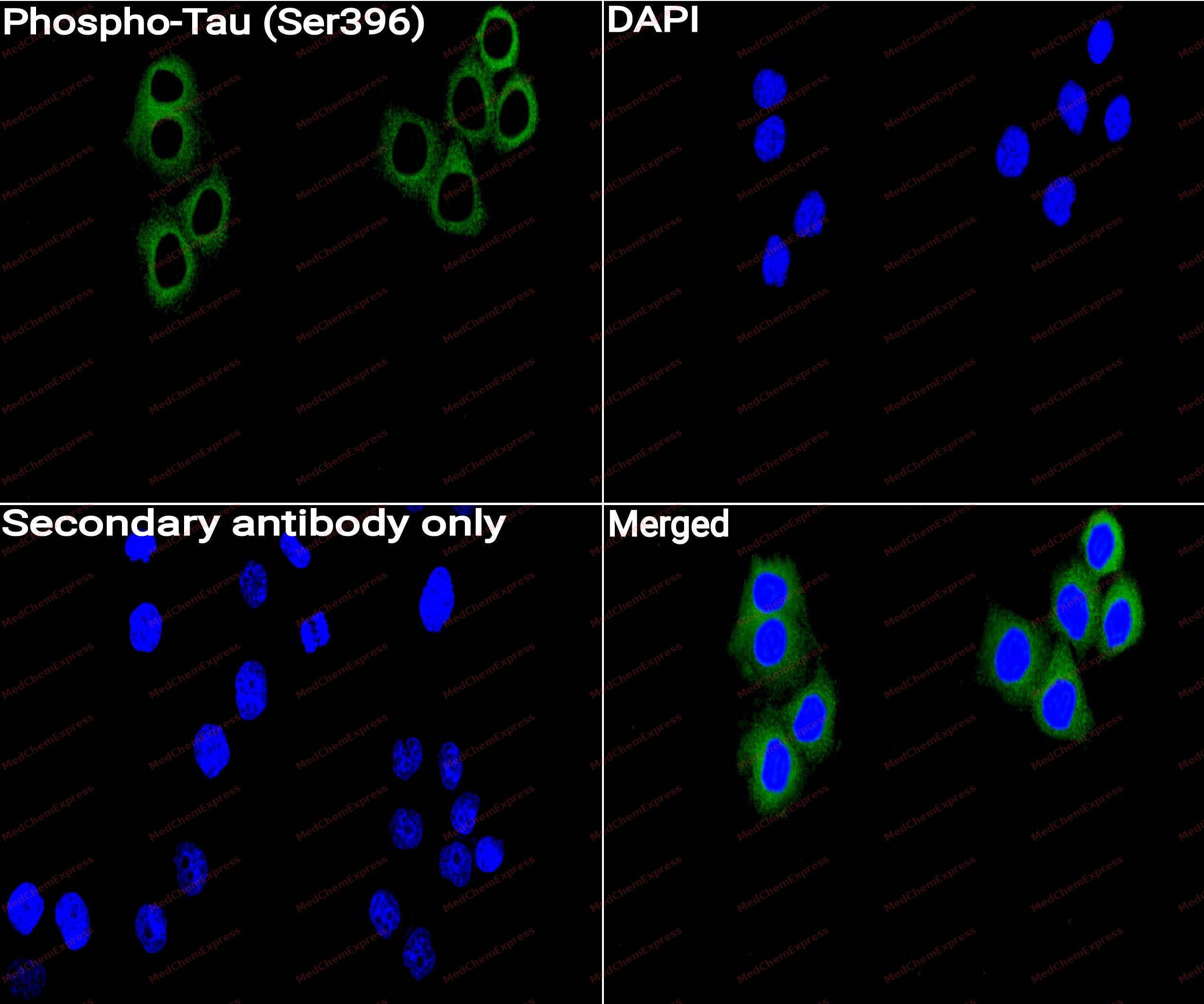 Immunocytochemistry analysis of Hela cells labeling Phospho-Tau (Ser396) with Phospho-Tau (Ser396) Antibody (HY-P80480) at 1/100 dilution. Cells were fixed in 4% paraformaldehyde for 15 minutes at room temperature, permeabilized with 0.1% Triton X-100 for 10 minutes at room temperature, then blocked with QuickBlock™ Blocking Buffer for Immunol Staining for 10 min at room temperature. Cells were then incubated with Phospho-Tau (Ser396) Antibody (HY-P80480)at 1/100 dilution in QuickBlock™ Blocking Buffer for Immunol Staining at 4 ℃. Alexa Fluor® 488-conjugated AffiniPure Goat Anti-Rabbit IgG H&L(HY-P8002,Green) was used as the secondary antibody at 1/1,000 dilution. PBS instead of the primary antibody was used as the secondary antibody only control. The Nuclear counterstain was DAPI (Blue).
 -
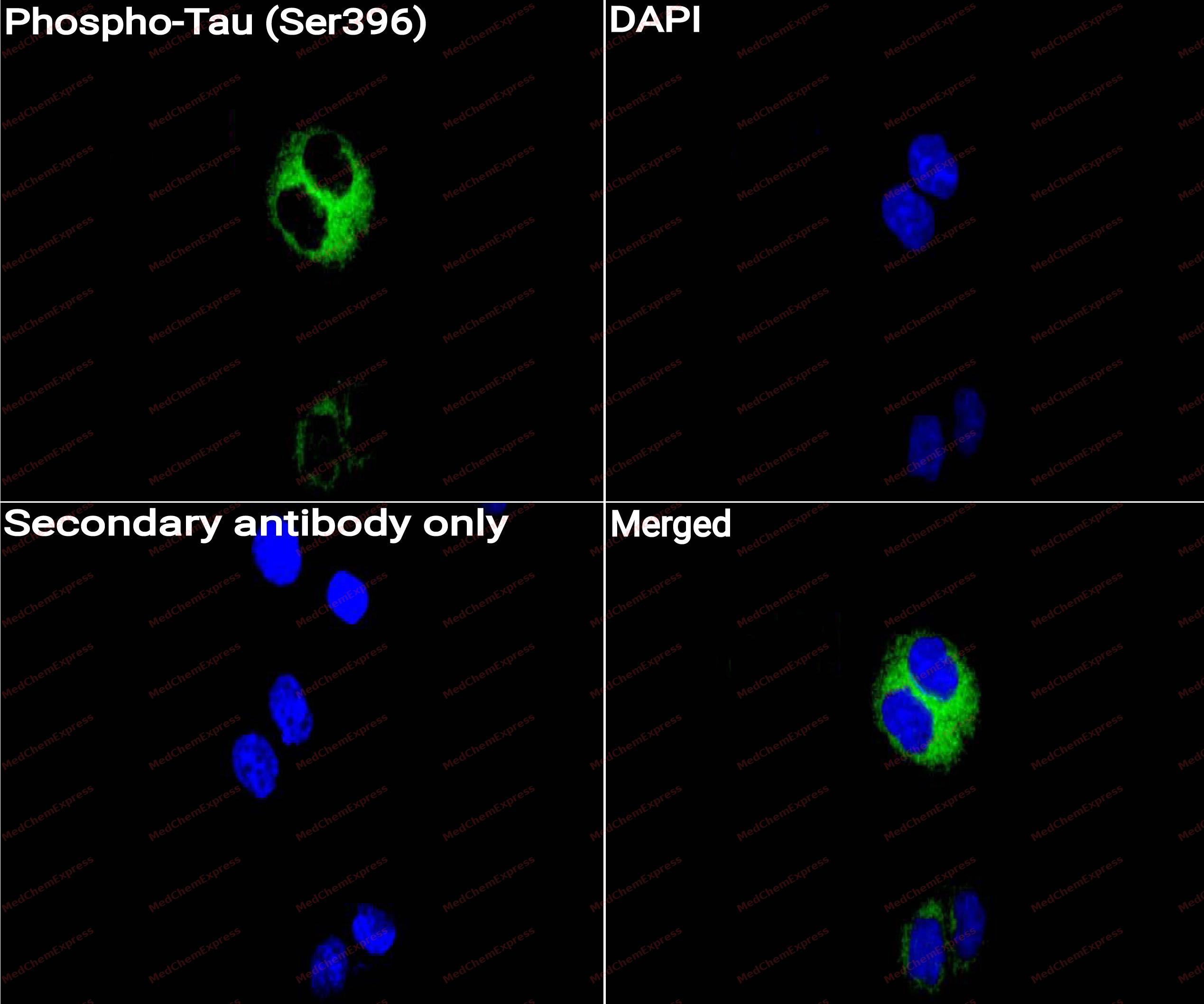 Immunocytochemistry analysis of Hela cells labeling Phospho-Tau (Ser396) with Phospho-Tau (Ser396) Antibody (HY-P80480) at 1/50 dilution. Cells were fixed in 4% paraformaldehyde for 15 minutes at room temperature, permeabilized with 0.1% Triton X-100 for 10 minutes at room temperature, then blocked with QuickBlock™ Blocking Buffer for Immunol Staining for 10 min at room temperature. Cells were then incubated with Phospho-Tau (Ser396) Antibody (HY-P80480) at 1/50 dilution in QuickBlock™ Blocking Buffer for Immunol Staining at 4 ℃. Alexa Fluor® 488-conjugated AffiniPure Goat Anti-Rabbit IgG H&L(HY-P8002, Green) was used as the secondary antibody at 1/1,000 dilution. PBS instead of the primary antibody was used as the secondary antibody only control. The Nuclear counterstain was DAPI (Blue).
 -
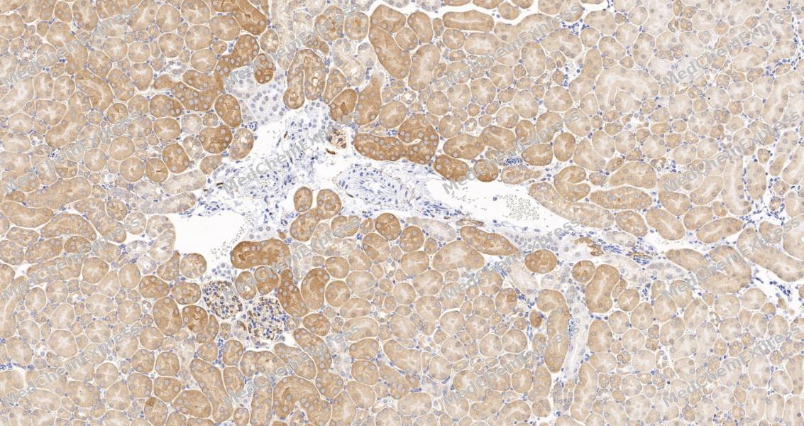 Immunohistochemical analysis of paraffin-embedded mouse kidney tissue using Phospho-Tau Antibody. The section was pre-treated using heat mediated antigen retrieval with Tris-EDTA buffer (pH 9.0) for 8 minutes. The tissues were blocked in QuickBlock for 20 minutes at room temperature, washed with ddH2O and PBS, and then probed with the primary antibody at 1/100 dilution in 4℃ overnight. The detection was performed using an HRP conjugated compact polymer system. DAB was used as the chromogen. Tissues were counterstained with hematoxylin and mounted with DPX.
 -
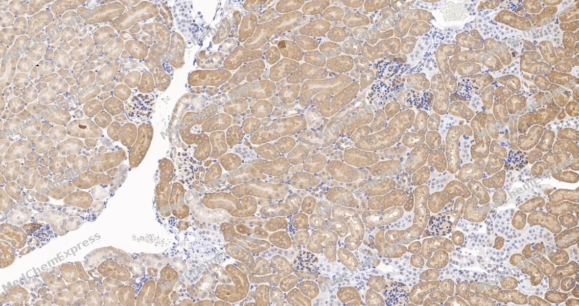 Immunohistochemical analysis of paraffin-embedded mouse kidney tissue using Phospho-Tau Antibody. The section was pre-treated using heat mediated antigen retrieval with Tris-EDTA buffer (pH 9.0) for 8 minutes. The tissues were blocked in QuickBlock for 20 minutes at room temperature, washed with ddH2O and PBS, and then probed with the primary antibody at 1/100 dilution in 4℃ overnight. The detection was performed using an HRP conjugated compact polymer system. DAB was used as the chromogen. Tissues were counterstained with hematoxylin and mounted with DPX.


Background
-
Function
Tau promotes microtubule assembly and stability, and might be involved in the establishment and maintenance of neuronal polarity. The C-terminus binds axonal microtubules while the N-terminus binds neural plasma membrane components, suggesting that tau functions as a linker protein between both. Axonal polarity is predetermined by TAU/MAPT localization (in the neuronal cell) in the domain of the cell body defined by the centrosome. The short isoforms allow plasticity of the cytoskeleton whereas the longer isoforms may preferentially play a role in its stabilization[1][2].
-
Subcellular Localization
Cytoplasm, cytosol; Cell membrane; Peripheral membrane protein; Cytoplasmic side; Cytoplasm, cytoskeleton; Cell projection, axon; Cell projection, dendrite; Secreted
-
Expression
Tissue_specificity:It is expressed in neurons. The PNS-tau subtype is expressed in the peripheral nervous system, while other subtypes are expressed in the central nervous system. -
Subunit
Interacts with MARK1, MARK2, MARK3 and MARK4 (PubMed:23666762). Interacts with PSMC2 through SQSTM1 (By similarity). Interacts with SQSTM1 when polyubiquitinated (PubMed:15953362). Interacts with FKBP4 (By similarity). Binds to CSNK1D (PubMed:14761950). Interacts with SGK1 (PubMed:16982696). Interacts with EPM2A; the interaction dephosphorylates MAPT at Ser-396 (PubMed:19542233). Interacts with PIN1 (PubMed:11313338). Interacts with LRRK2 (PubMed:26014385). Interacts with LRP1, leading to endocytosis; this interaction is reduced in the presence of LRPAP1/RAP (PubMed:32296178)
-
SwissProt ID
-
Synonyms
MAPTL, MTBT1, TAU, MAPT, Microtubule-associated protein tau, Neurofibrillary tangle protein, Paired helical filament-tau, PHF-tau
-
Research Field
Neuroscience


Documentation
-
Data Sheet (260 KB)
-
SDS (252 KB)
- English - EN (252 KB)
- Français - FR (252 KB)
- Deutsch - DE (252 KB)
- Norwegian - NO (252 KB)
- Español - ES (252 KB)
- Swedish - SV (252 KB)
- Italian - IT (252 KB)
- Korean - KR (252 KB)
- Portuguese - PT (252 KB)
-
User Guide for Antibodies (1077 KB)
References
[1]. Yoshida H, et al. Phosphorylation of microtubule-associated protein tau by AMPK-related kinases. J Neurochem. 2012 Jan;120(1):165-76. [Content Brief]
[2]. Sandberg A, et al. Fibrillation and molecular characteristics are coherent with clinical and pathological features of 4-repeat tauopathy caused by MAPT variant G273R. Neurobiol Dis. 2020 Dec;146:105079. [Content Brief]

 Powered by Bioz
