



L-Ascorbic acid-13C6-1

Based on 1 Customer Validation
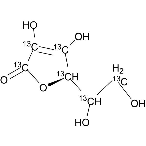
L-Ascorbic acid-13C6-1 (L-Ascorbate-1; Vitamin C-13C6-1) is a 13C labeled L-Ascorbic acid (HY-B0166). L-Ascorbic acid (L-Ascorbate), an electron donor, is an endogenous antioxidant agent. L-Ascorbic acid inhibits selectively Cav3.2 channels with an IC50 of 6.5 μM. L-Ascorbic acid is also a collagen deposition enhancer and an elastogenesis inhibitor. L-Ascorbic acid exhibits anti-cancer effects through the generation of reactive oxygen species (ROS) and selective damage to cancer cells.
For research use only. We do not sell to patients.
- Purity : 99.37%
- CAS No.: 1331939-77-5
- Formula: 13C6H8O6
- Molecular Weight:182.08
-
Storage:
-20°C, protect from light, stored under nitrogen
* In solvent : -80°C, 6 months; -20°C, 1 month (protect from light, stored under nitrogen)

To place orders, for customer services and technical support, please contact: MedChemExpress USA
Tel: 609-228-6898 E-mail: [email protected] [email protected]


-
 Biological Activity
-
Chemical Information
-
Solvent & Solubility
- Protocols
- Purity & Documentation
- References
-
 Help & FAQs




Biological Activity
Description
In Vitro
Stable heavy isotopes of hydrogen, carbon, and other elements have been incorporated into drug molecules, largely as tracers for quantitation during the drug development process. Deuteration has gained attention because of its potential to affect the pharmacokinetic and metabolic profiles of drugs[1].
MedChemExpress (MCE) has not independently confirmed the accuracy of these methods. They are for reference only.
Application
1. This compound can be used as a tracer
2. This compound can be used as an internal standard for quantitative analysis by NMR, GC-MS, or LC-MS.


Chemical Information
-
CAS No. 1331939-77-5
-
Unlabeled CAS 50-81-7
-
Appearance Solid
-
Molecular Weight 182.08
-
Formula 13C6H8O6
-
Color White to off-white
-
SMILES
O[13CH2][13CH]([13C@@H]1[13C](O)=[13C]([13C](O1)=O)O)O
-
Synonyms
L-Ascorbate-1; Vitamin C-13C6-1
-
Shipping
Room temperature in continental US; may vary elsewhere.
-
Storage
-20°C, protect from light, stored under nitrogen
* In solvent : -80°C, 6 months; -20°C, 1 month (protect from light, stored under nitrogen)


Solvent & Solubility
In Vitro:
DMSO : 100 mg/mL (549.21 mM; Need ultrasonic and warming; Hygroscopic DMSO has a significant impact on the solubility of product, please use newly opened DMSO)
Please refer to the solubility information to select the appropriate solvent. Once prepared, please aliquot and store the solution to prevent product inactivation from repeated freeze-thaw cycles.
Storage method and period of stock solution: -80°C, 6 months; -20°C, 1 month (protect from light, stored under nitrogen). When stored at -80°C, please use it within 6 months. When stored at -20°C, please use it within 1 month.
Please refer to the solubility information to select the appropriate solvent. Once prepared, please aliquot and store the solution to prevent product inactivation from repeated freeze-thaw cycles.
Storage method and period of stock solution: -80°C, 6 months; -20°C, 1 month (protect from light, stored under nitrogen). When stored at -80°C, please use it within 6 months. When stored at -20°C, please use it within 1 month.
Concentration (start) × Volume (start) = Concentration (final) × Volume (final)


Purity & Documentation
-
Data Sheet (277 KB)
-
SDS (393 KB)
- English - EN (393 KB)
- Français - FR (393 KB)
- Deutsch - DE (393 KB)
- Norwegian - NO (393 KB)
- Español - ES (393 KB)
- Swedish - SV (393 KB)
- Italian - IT (393 KB)
- Korean - KR (393 KB)
- Portuguese - PT (393 KB)
-
Handling Instructions (2659 KB)
References
[1]. Russak EM, et al. Impact of Deuterium Substitution on the Pharmacokinetics of Pharmaceuticals. Ann Pharmacother. 2019 Feb;53(2):211-246. [Content Brief]
[2]. Sebastian J Padayatty, et al. Vitamin C as an antioxidant: evaluation of its role in disease prevention. J Am Coll Nutr. 2003 Feb;22(1):18-35. [Content Brief]
[3]. Sungrae Cho, et al. Hormetic dose response to L-ascorbic acid as an anti-cancer drug in colorectal cancer cell lines according to SVCT-2 expression. Sci Rep. 2018 Jul 27;8(1):11372. [Content Brief]
[4]. Satyanarayana Sreemantula, et al. Influence of antioxidant (L- ascorbic acid) on tolbutamide induced hypoglycaemia/antihyperglycaemia in normal and diabetic rats. BMC Endocr Disord. 2005 Mar 3;5(1):2. [Content Brief]
[5]. Michael T Nelson, et al. Molecular mechanisms of subtype-specific inhibition of neuronal T-type calcium channels by ascorbate. J Neurosci. 2007 Nov 14;27(46):12577-83. [Content Brief]
[6]. Aleksander Hinek, et al. Sodium L-ascorbate enhances elastic fibers deposition by fibroblasts from normal and pathologic human skin. J Dermatol Sci. 2014 Sep;75(3):173-82. [Content Brief]


Complete Stock Solution Preparation Table
Please refer to the solubility information to select the appropriate solvent. Once prepared, please aliquot and store the solution to prevent product inactivation from repeated freeze-thaw cycles.
Storage method and period of stock solution: -80°C, 6 months; -20°C, 1 month (protect from light, stored under nitrogen). When stored at -80°C, please use it within 6 months. When stored at -20°C, please use it within 1 month.
| Optional Solvent | Concentration Solvent Mass | 1 mg | 5 mg | 10 mg | 25 mg |
|---|---|---|---|---|---|
| DMSO | 1 mM | 5.4921 mL | 27.4605 mL | 54.9209 mL | 137.3023 mL |
| 5 mM | 1.0984 mL | 5.4921 mL | 10.9842 mL | 27.4605 mL | |
| 10 mM | 0.5492 mL | 2.7460 mL | 5.4921 mL | 13.7302 mL | |
| 15 mM | 0.3661 mL | 1.8307 mL | 3.6614 mL | 9.1535 mL | |
| 20 mM | 0.2746 mL | 1.3730 mL | 2.7460 mL | 6.8651 mL | |
| 25 mM | 0.2197 mL | 1.0984 mL | 2.1968 mL | 5.4921 mL | |
| 30 mM | 0.1831 mL | 0.9153 mL | 1.8307 mL | 4.5767 mL | |
| 40 mM | 0.1373 mL | 0.6865 mL | 1.3730 mL | 3.4326 mL | |
| 50 mM | 0.1098 mL | 0.5492 mL | 1.0984 mL | 2.7460 mL | |
| 60 mM | 0.0915 mL | 0.4577 mL | 0.9153 mL | 2.2884 mL | |
| 80 mM | 0.0687 mL | 0.3433 mL | 0.6865 mL | 1.7163 mL | |
| 100 mM | 0.0549 mL | 0.2746 mL | 0.5492 mL | 1.3730 mL |


L-Ascorbic acid-13C6-1 Related Classifications
HY-B0166S8 Related Classifications
 Powered by Bioz