



Semaphorin-3C/SEMA3C Protein, Human (HEK293, C-His)
Based on 1 publication(s) in Google Scholar
Semaphorin-3C (SEMA3C) protein plays a crucial role in developmental processes by binding to plexin family members, particularly PLXND1, orchestrating intricate cellular signaling pathways. Its indispensable contribution to normal cardiovascular development during embryogenesis and its function as an attractant for growing axons underscore SEMA3C's significance in axon growth and guidance. Semaphorin-3C/SEMA3C Protein, Human (HEK293, C-His) is the recombinant human-derived Semaphorin-3C/SEMA3C, expressed by HEK293, with C-6*His labeled tag.
- Species: Human
- Source: HEK293
-
Storage:Stored at -20°C for 2 years from date of receipt. After reconstitution, it is stable at 4°C for 1 week or -20°C for longer (with carrier protein). It is recommended to freeze aliquots at -20°C or -80°C for extended storage.


To place orders, for customer services and technical support, please contact: MedChemExpress USA
Tel: 609-228-6898 E-mail: [email protected] [email protected]


-
 Biological Activity
-
Technical Parameters
-
Product Properties
- Documentation
- Help & FAQs




Biological Activity
Description
Semaphorin-3C (SEMA3C) protein plays a crucial role in developmental processes by binding to plexin family members, particularly PLXND1, orchestrating intricate cellular signaling pathways. Its indispensable contribution to normal cardiovascular development during embryogenesis and its function as an attractant for growing axons underscore SEMA3C's significance in axon growth and guidance. Semaphorin-3C/SEMA3C Protein, Human (HEK293, C-His) is the recombinant human-derived Semaphorin-3C/SEMA3C, expressed by HEK293, with C-6*His labeled tag.
Background
Semaphorin-3C (SEMA3C) protein serves as a pivotal regulator in developmental processes by binding to members of the plexin family. Crucially, SEMA3C is indispensable for normal cardiovascular development in embryogenesis. Its functionality extends to acting as an attractant for growing axons, thereby playing a significant role in axon growth and guidance. In molecular interactions, SEMA3C forms a specific association with PLXND1, highlighting its involvement in intricate cellular signaling pathways that contribute to the orchestration of developmental events.
Verified Bioactivity
Measured by the ability of the immobilized protein to support the adhesion of SVEC4-10 mouse vascular endothelial cells to Fibronectin. Recombinant Human Semaphorin 3C immobilized at 10 μg/mL, with 2 ng/mL rhFN-1, 100 μL/well, will induce approximately 40%-80% adhesion.
MCE Validation Data
-
Purity - SDS-PAGE
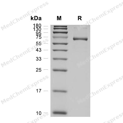
Purity - SDS-PAGE


Publications (1)
-
Journal Impact Factor
-
Most Recent
-
Am J Pathol
Semaphorin 3C/Plexin D1 Interaction Regulates Collagen Metabolism in Keloid Fibroblasts via the Transforming Growth Factor-β1 Signaling Pathway. [Abstract]2026 May;196(5):1169-1187. PMID: 41833636


Technical Parameters
-
Species Human
-
Source HEK293
-
Tag C-6*His
-
Accession
Q99985-1 (G21-S738)
-
Gene ID10512
-
Protein Length
Partial
-
Synonyms
SEMA3C; Semaphorin-3C; Prev. SEMAE; Uncharacterized Protein SEMA3C; SemE; Semaphorin E; Sema Domain, Immunoglobulin Domain (Ig), Short Basic Domain, Secreted, (Semaphorin) 3C; Semaphorin-E; Semaphorin 3C; Sema E
-
AA Sequence
GSSQPQARVYLTFDELRETKTSEYFSLSHHPLDYRILLMDEDQDRIYVGSKDHILSLNINNISQEALSVFWPASTIKVEECKMAGKDPTHGCGNFVRVIQTFNRTHLYVCGSGAFSPVCTYLNRGRRSEDQVFMIDSKCESGKGRCSFNPNVNTVSVMINEELFSGMYIDFMGTDAAIFRSLTKRNAVRTDQHNSKWLSEPMFVDAHVIPDGTDPNDAKVYFFFKEKLTDNNRSTKQIHSMIARICPNDTGGLRSLVNKWTTFLKARLVCSVTDEDGPETHFDELEDVFLLETDNPRTTLVYGIFTTSSSVFKGSAVCVYHLSDIQTVFNGPFAHKEGPNHQLISYQGRIPYPRPGTCPGGAFTPNMRTTKEFPDDVVTFIRNHPLMYNSIYPIHKRPLIVRIGTDYKYTKIAVDRVNAADGRYHVLFLGTDRGTVQKVVVLPTNNSVSGELILEELEVFKNHAPITTMKISSKKQQLYVSSNEGVSQVSLHRCHIYGTACADCCLARDPYCAWDGHSCSRFYPTGKRRSRRQDVRHGNPLTQCRGFNLKAYRNAAEIVQYGVKNNTTFLECAPKSPQASIKWLLQKDKDRRKEVKLNERIIATSQGLLIRSVQGSDQGLYHCIATENSFKQTIAKINFKVLDSEMVAVVTDKWSPWTWASSVRALPFHPKDIMGAFSHSEMQMINQYCKDTRQQHQQGDESQKMRGDYGKLKALINS
-
Predicted Molecular Mass
81.4 kDa
-
Molecular Weight
Approximately 65-72 kDa, based on SDS-PAGE under reducing conditions.
-
Glycosylation
Yes
-
Purity
≥ 95%, as determined by reducing SDS-PAGE.


Product Properties
Lyophilized powder.
Lyophilized from a 0.22 μm filtered solution of PBS, pH 6.5, 8% trehalose.
<1 EU/μg, determined by LAL method.
It is not recommended to reconstitute to a concentration less than 100 μg/mL in ddH2O.
Stored at -20°C for 2 years from date of receipt. After reconstitution, it is stable at 4°C for 1 week or -20°C for longer (with carrier protein). It is recommended to freeze aliquots at -20°C or -80°C for extended storage.
Room temperature in continental US; may vary elsewhere.


Documentation
-
Data Sheet (240 KB)
-
SDS (251 KB)
- English - EN (251 KB)
- Français - FR (251 KB)
- Deutsch - DE (251 KB)
- Norwegian - NO (251 KB)
- Español - ES (251 KB)
- Swedish - SV (251 KB)
- Italian - IT (251 KB)
- Korean - KR (251 KB)
- Portuguese - PT (251 KB)
-
Handling Instructions (2659 KB)

Calculators
Concentration (start) × Volume (start) = Concentration (final) × Volume (final)


 Powered by Bioz

