



TGF beta 2/TGFB2 Protein, Canine (HEK293, His)

Based on 1 Customer Validation
Transforming growth factor-beta 2 (TGF-β2), an extracellular glycosylated protein, is a member of the TGF-β superfamily. TGFβ2 controls key physiological processes including cell migration, proliferation and differentiation via signalling through type I and type II receptors (TGFβR1 and TGFβR2). TGF-β2 is an immune suppressor involved in the development of immune tolerance, and also regulates embryonic development. TGF beta 2/TGFB2 Protein, Canine (HEK293, His) is produced in HEK293 cells with a C-Terminal His-tag. It and consists of 414 amino acids (M1-S414).
- Species: Canine
- Source: HEK293
-
Storage:Stored at -20°C for 2 years from date of receipt. After reconstitution, it is stable at 4°C for 1 week or -20°C for longer (with carrier protein). It is recommended to freeze aliquots at -20°C or -80°C for extended storage.


To place orders, for customer services and technical support, please contact: MedChemExpress USA
Tel: 609-228-6898 E-mail: [email protected] [email protected]


-
 Biological Activity
-
Technical Parameters
-
Product Properties
- Documentation
- References
- Help & FAQs




Biological Activity
Description
Transforming growth factor-beta 2 (TGF-β2), an extracellular glycosylated protein, is a member of the TGF-β superfamily. TGFβ2 controls key physiological processes including cell migration, proliferation and differentiation via signalling through type I and type II receptors (TGFβR1 and TGFβR2). TGF-β2 is an immune suppressor involved in the development of immune tolerance, and also regulates embryonic development[1][2]. TGF beta 2/TGFB2 Protein, Canine (HEK293, His) is produced in HEK293 cells with a C-Terminal His-tag. It and consists of 414 amino acids (M1-S414).
Background
In mammals, three different isoforms of TGF-β are described (TGF-β1, TGF-β2 and TGF-β3; transforming growth factor beta) to regulate apoptosis, proliferation, differentiation, migration and invasion processes utilising overlapping but not redundant mechanisms. All three isoforms are expressed in the liver, but their expression is differentially distributed among liver cell types. TGF-β2 expression in different liver cell types and is also associated with developmental defects and fibrotic diseases in mice[1][2][3].
The sequence of amino acids in TGF-β2 proteins from different species is very stable, which leads to the conclusion that in the process of evolution, TGF-β2 has been only slightly altered, and that both in humans and in animals, its function is similar.
TGFβ2 is a transforming growth factor beta (TGFB) family cytokine, with members of this cytokine family playing broad regulatory roles and controlling key physiological processes including cell migration, proliferation and differentiation via signalling through type I and type II receptors (TGFβR1 and TGFβR2), with signals propagating via the downstream regulatory SMAD proteins. This TGFβ/SMAD pathway is frequently dysregulated in human cancer. TGFβ cytokines are capable of suppressing T cell growth in response to IL‐2. The degree of TGFβ2 expression correlated with the expression of several different markers of immune cell subsets within tumours. In addition, TGF-β2 regulates embryonic development and, therefore not surprisingly, global Tgfb2 null mice exhibit a wide range of developmental defects and perinatal mortality[1][2][3].
TGF-β2 is an immune suppressor involved in the development of immune tolerance, and recombinant TGF-β2 incubation is more potent than TGF-β1 or TGF-β3 in suppressing macrophage inflammatory responses. TGF-β2 is shown to correlate with bad prognosis in intrahepatic CCAs and hepatocellular carcinoma. Mechanistically, canonical Smad signalling as well as crosstalk with Yap, Hippo, Wnt and β-catenin signalling have been demonstrated in the liver and other organs[1][2][3].
In Vitro
Recombinant human TGF-β2 (1.25, 2.5, 5, 1, and 2 ng/mL; for 24 h) increases extracellular matrix (ECM) protein synthesis and secretion in optic nerve head (ONH) astrocytes and lamina cribrosa (LC) cells. TGF-β2 induces phosphorylation of canonical signaling proteins Smad2/3 but does not alter phosphorylation of non-canonical signaling proteins ERK1/2, p38, and JNK1/2 proteins in ONH astrocytes and LC cells[4].
In Vivo
Recombinant human TGF-β2 (12 μg/dog; for 28 days; porous-coated titanium implant) increases in fixation strength and stiffness of the implant in a canine model[5].
MCE Validation Data
-
Purity - SDS-PAGE
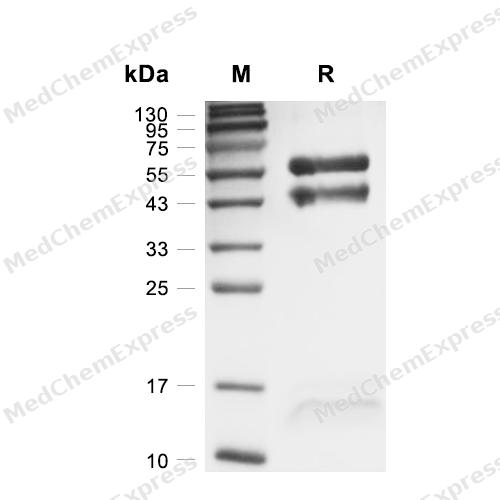
Purity - SDS-PAGE


Technical Parameters
-
Species Canine
-
Source HEK293
-
Tag C-6*His
-
Accession
XP_858677.1 (L21-S414)
-
Molecular Construction
-
N-term
-
TGFB2 (L21-S414)
Accession # XP_858677.1 -
His
-
C-term
-
-
Protein Length
Full Length of Mature Protein
-
Synonyms
TGFB2; Transforming Growth Factor, Beta 2; Transforming Growth Factor Beta 2; BSC-1 Cell Growth Inhibitor; Glioblastoma-Derived T-Cell Suppressor Factor; TGF-Beta2; Transforming Growth Factor Beta-2 Proprotein; Polyergin; Prepro-Transforming Growth Factor
-
AA Sequence
LSTCSTLDMDQFMRKRIEAIRGQILSKLKLTSPPEDYPEPEEVPPEVISIYNSTRDLLQEKASRRAAACERERSDEEYYAKEVYKIDMPPFFPSENAIPPTFYRPYFRIVRFDVSAMEKNASNLVKAEFRVFRLQNPKARVPEQRIELYQILKSKDLTSPTQRYIDSKVVKTRAEGEWLSFDVTDAVHEWLHHKDRNLGFKISLHCPCCTFVPSNNYIIPNKSEELEARFAGIDGTSTYTSGDQKTIKSTRKKNSGKTPHLLLMLLPSYRLESQQSNRRKKRALDAAYCFRNVQDNCCLRPLYIDFKRDLGWKWIHEPKGYNANFCAGACPYLWSSDTQHSRVLSLYNTINPEASASPCCVSQDLEPLTILYYIGKTPKIEQLSNMIVKSCKCS
-
Predicted Molecular Mass
45.5 kDa
-
Molecular Weight
Approximately 13-16 kDa & 40-60 kDa, based on SDS-PAGE under reducing conditions.
-
Purity
≥ 95%, as determined by reducing SDS-PAGE.


Product Properties
Lyophilized powder.
Lyophilized from a 0.22 μm filtered solution of PBS, pH 7.4.
<1 EU/μg, determined by LAL method.
It is not recommended to reconstitute to a concentration less than 100 μg/mL in ddH2O. For long term storage it is recommended to add a carrier protein (0.1% BSA, 5% HSA, 10% FBS or 5% Trehalose).
Stored at -20°C for 2 years from date of receipt. After reconstitution, it is stable at 4°C for 1 week or -20°C for longer (with carrier protein). It is recommended to freeze aliquots at -20°C or -80°C for extended storage.
Room temperature in continental US; may vary elsewhere.


Documentation
-
Data Sheet (267 KB)
-
SDS (252 KB)
- English - EN (252 KB)
- Français - FR (252 KB)
- Deutsch - DE (252 KB)
- Norwegian - NO (252 KB)
- Español - ES (252 KB)
- Swedish - SV (252 KB)
- Italian - IT (252 KB)
- Korean - KR (252 KB)
- Portuguese - PT (252 KB)
-
Handling Instructions (2659 KB)


References
[1]. Hirokazu Takahashi, et al. TGF-β2 is an exercise-induced adipokine that regulates glucose and fatty acid metabolism. Nat Metab. 2019 Feb;1(2):291-303. [Content Brief]
[2]. Zunqiang Xiao, et al. TGFβ2 is a prognostic-related biomarker and correlated with immune infiltrates in gastric cancer. J Cell Mol Med. 2020 Jul;24(13):7151-7162. [Content Brief]
[3]. Anne Dropmann, et al. TGF-β2 silencing to target biliary-derived liver diseases. Gut. 2020 Sep;69(9):1677-1690. [Content Brief]
[4]. Gulab S Zode, et al. Transforming growth factor-β2 increases extracellular matrix proteins in optic nerve head cells via activation of the Smad signaling pathway. Mol Vis. 2011;17:1745-58. [Content Brief]
[5]. D R Sumner, et al. Additive enhancement of implant fixation following combined treatment with rhTGF-beta2 and rhBMP-2 in a canine model. J Bone Joint Surg Am. 2006 Apr;88(4):806-17. [Content Brief]

Calculators
Concentration (start) × Volume (start) = Concentration (final) × Volume (final)


 Powered by Bioz

