



7DG

Based on 1 Customer Validation
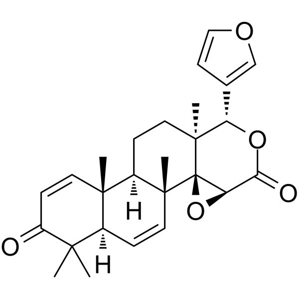
7DG (7-Desacetoxy-6,7-dehydrogedunin) is a PKR inhibitor, P2X7 purinergic receptor inhibitor, and skin-lightening agent. 7DG binds outside the ATP-catalytic domain of PKR, blocks the kinase activity-independent protein-protein interactions of PKR, inhibits the phosphorylation and activity of PKR, disrupts ASC assembly and caspase-1 activation, and suppresses the activation of the NLRP1 inflammasome. 7DG inhibits pyroptosis, suppresses the ATP-P2X7 signaling pathway, and abolishes ATP-induced increases in the expression levels of MITF, tyrosinase, PMEL/gp100, and melanin content. 7DG exerts skin-lightening effects in cultured skin in vitro. 7DG can be used in research related to chronic obstructive pulmonary disease, gout, type 2 diabetes, Alzheimer's disease, and hyperpigmentary skin disorders.
Nur für Forschungszwecke. Wir verkaufen nicht an Patienten.
- Reinheit : 98%
- CAS. Nr.: 26927-01-5
- Formel: C26H30O5
- Molecular Weight:422.51
-
Speicherung:Powder -20°C, 3 years , 4°C, 2 years ; In solvent -80°C, 6 months , -20°C, 1 month

To place orders, for customer services and technical support, please contact: MedChemExpress USA
Tel: 609-228-6898 E-mail: [email protected] [email protected]


-
 Biologische Aktivität
-
Chemical Information
- Protokoll
- Reinheit & Dokumentation
- Verweise
-
 Help & FAQs


Alle P2X Receptor Isoform-spezifische Produkte anzeigen
MoreAlle Caspase Isoform-spezifische Produkte anzeigen
More

Biologische Aktivität
Beschreibung
IC50 & Target
protein kinase R (PKR)[1]
In Vitro
7DG (0.001-10 μM; administered within up to 1 h after LT exposure) reversibly protects J774 macrophages from lethal toxin (LT)-induced pyroptotic cell death, with an IC50 of 5 μM, and remains active when added within 1 hour after LT exposure[1].
7DG (10 μM, 2 h) inhibits LT-induced caspase-1 activation in J774 macrophages by targeting upstream steps of caspase-1 activation, without directly inhibiting the enzymatic activity of caspase-1[1].
7DG (20 μM; 2 h) inhibits LPS (HY-D1056)-induced IκBβ degradation in J774 macrophages, and suppresses NLRP3 inflammasome-mediated caspase-1 activation in J774 macrophages treated with LPS + nigericin[1].
7DG (1 h) inhibits the assembly of NLRP1 and NLRP3 inflammasomes in immortalized mouse macrophages with constitutive NLRP3 expression by targeting the kinase-independent function of PKR[1].
7DG directly targets PKR in macrophages, inhibits PKR phosphorylation, and protects macrophages from LT-induced NLRP1 inflammasome-mediated pyroptosis[2].
7DG (5.6-50 μM; 30 min) inhibits P2X7 activity in HOS-P2X7 cells[3].
Incubation with 10 μM 7DG for 4 days (within the concentration range of 0.1-10 μM over 4 days) significantly reduces the viability of PHEM cells[3].
7DG (0.5-2 μM; 1 h pre-incubation, followed by 3 days of co-treatment with 100 μM ATP) potently inhibits ATP (HY-B2176)-induced P2X7 activity in PHEMs[3].
7DG (0.5-2 μM; 1 h pre-incubation followed by 72 h co-treatment with 100 μM ATP) inhibits ATP-induced melanogenesis in PHEMs by reducing the expression of melanogenesis-related proteins, tyrosinase activity, and melanin content[3].
7DG (0.5-2 μM; 4-72 h) reduces melanogenesis in PHEMs without ATP stimulation[3].
7DG (2 μM, daily for 9 consecutive days, with UVB irradiation supplemented on days 5 to 7) inhibits UVB-induced pigmentation in organ-cultured human skin after 9 days of treatment[3].
MedChemExpress (MCE) has not independently confirmed the accuracy of these methods. They are for reference only.
-
Cell Line:J774A.1 BALB/c murine macrophages (J774)
-
Concentration:0.001, 0.01, 0.1, 1, 10 μM
-
Incubation Time:up to 1 h after LT exposure
-
Result:Completely protected J774 macrophages from LT-induced death at 10 μM, with an IC50 of 5 μM.
Retained protective activity when added up to 1 hour after LT exposure, but not at later time points.
Lost all protective activity after washout, indicating it acts as a reversible inhibitor.
-
Cell Line:J774 macrophages
-
Concentration:20 μM
-
Incubation Time:2 h (pre-treatment); 30-40 min (LPS exposure)
-
Result:Prevented LPS-induced IκBβ degradation in J774 macrophages, similar to the proteasome inhibitor Epoxo, which blocks IκBβ degradation.


Chemical Information
-
CAS. Nr. 26927-01-5
-
Appearance Solid
-
Molecular Weight 422.51
-
Formel C26H30O5
-
Color White to off-white
-
SMILES
O=C1O[C@@H](C2=COC=C2)[C@]3(C)CC[C@@]4([H])[C@](C=C[C@@]5([H])C(C)(C)C(C=C[C@@]54C)=O)(C)[C@@]36[C@@H]1O6
-
Synonyms
7-Desacetoxy-6,7-dehydrogedunin
-
Versand
Room temperature in continental US; may vary elsewhere.
-
Speicherung
Powder -20°C 3 years 4°C 2 years In solvent -80°C 6 months -20°C 1 month


Reinheit & Dokumentation
-
Data Sheet (281 KB)
-
SDS (392 KB)
- English - EN (392 KB)
- Français - FR (392 KB)
- Deutsch - DE (392 KB)
- Norwegian - NO (392 KB)
- Español - ES (392 KB)
- Swedish - SV (392 KB)
- Italian - IT (392 KB)
- Korean - KR (392 KB)
- Portuguese - PT (392 KB)
-
Handling Instructions (2659 KB)
Verweise
[1]. Hett EC, et al. Chemical genetics reveals a kinase-independent role for protein kinase R in pyroptosis. Nat Chem Biol. 2013;9(6):398-405. [Content Brief]
[2]. Chukwurah E, et al. A tale of two proteins: PACT and PKR and their roles in inflammation. FEBS J. 2021;288(22):6365-6391. [Content Brief]
[3]. Park S, et al. 7-desacetoxy-6,7-dehydrogedunin discovered by high-throughput screening system suppresses melanogenesis through ATP-P2X7 signaling inhibition. J Dermatol Sci. 2022;108(3):157-166. [Content Brief]

Calculators
Konzentration (Stammlösung) × Volumen (Stammlösung) = Konzentration (Ziellösung) × Volumen (Ziellösung)


7DG Related Classifications
HY-124857 Related Classifications
 Powered by Bioz