


Belinostat
Based on 38 publication(s) in Google Scholar
Belinostat (PXD101; PX105684) is a potent HDAC inhibitor with an IC50 of 27 nM in HeLa cell extracts.
For research use only. We do not sell to patients.
- Purity: 99.94%
- CAS No.: 866323-14-0
- Formula: C15H14N2O4S
- Molecular Weight:318.35
-
Storage:Powder -20°C, 3 years , 4°C, 2 years ; In solvent -80°C, 2 years , -20°C, 1 year

To place orders, for customer services and technical support, please contact: MedChemExpress USA
Tel: 609-228-6898 E-mail: [email protected] [email protected]


-
 Biological Activity
-
Chemical Information
-
Solvent & Solubility
- Protocol
- Purity & Documentation
- References
-
 Help & FAQs
-


Cell Cycle/DNA Damage Compound Library
HY-L004
-
Epigenetics Compound Library
HY-L005
-
FDA-Approved Drug Library
HY-L022
-
Histone Modification Research Compound Library
HY-L024
-
Anti-Cancer Compound Library
HY-L025
-
Autophagy Compound Library
HY-L029
-
Anti-Aging Compound Library
HY-L034
-
Drug Repurposing Compound Library
HY-L035
-
Covalent Screening Library
HY-L036
-
Reprogramming Compound Library
HY-L039
-
Oxygen Sensing Compound Library
HY-L045
-
Anti-COVID-19 Compound Library
HY-L052
-
Orally Active Compound Library
HY-L061
-
FDA Approved & Pharmacopeial Drug Library
HY-L066
-
Anti-Breast Cancer Compound Library
HY-L074
-
Drug-Induced Liver Injury (DILI) Compound Library
HY-L076
-
Anti-Pancreatic Cancer Compound Library
HY-L077
-
Anti-Blood Cancer Compound Library
HY-L079
-
Targeted Therapy Drug Library
HY-L080
-
Anti-Liver Cancer Compound Library
HY-L101
-
Rare Diseases Drug Library
HY-L102
-
Chemotherapy Drug Library
HY-L112
-
FDA-Approved Anticancer Drug Library
HY-L122
-
Anti-Prostate Cancer Compound Library
HY-L124
-
Anti-Pulmonary Fibrosis Compound Library
HY-L125
-
Osteogenesis Compound Library
HY-L131
-
Off-patent Drug Library
HY-L141
-
Cell Death Library
HY-L162
-
Anti-Hematopathy Compound Library
HY-L171
-
Anti-Ovarian Cancer Compound Library
HY-L173
-
Multi-Target Compound Library
HY-L176
-
Bioactive Compound Library Max
HY-L181
-
MCE Bioactive Compound Library
HY-L001V
-
Covalent Screening Library Plus
HY-L036P
-
Drug Repurposing Compound Library Plus
HY-L035P
-
FDA-Approved Drug Library Plus
HY-L022P
-
FDA-Approved Drug Library Mini
HY-L022M
-
Bioactive Compound Library
HY-L001
-
Anti-Brain Cancer Compound Library
HY-L188
-
High-Throughput Bioactive Compound Library
HY-L205
-
Anti-Cancer Approved Drug Library
HY-L213
-
High-Efficiency Gene Editing Compound Library
HY-L244
-
Lactylation Compound Library
HY-L249


Publications Citing Use of MedChemExpress (MCE) Belinostat
More- Signal Transduct Target Ther. 2025 Oct 14;10(1):341. [Abstract]
- Adv Mater. 2025 Oct 10:e05951. [Abstract]
- Cancer Res. 2022 Feb 15;82(4):681-694. [Abstract]
- Nat Commun. 2023 Apr 13;14(1):2095. [Abstract]
- Nat Commun. 2017 Sep 5;8(1):435. [Abstract]
- J Exp Clin Cancer Res. 2023 Oct 7;42(1):260. [Abstract]
- Pharmacol Res. 2022 Feb:176:105969. [Abstract]
- Cell Death Dis. 2019 May 24;10(6):400. [Abstract]
- Cell Death Dis. 2018 Jan 26;9(2):129. [Abstract]
- NPJ Breast Cancer. 2023 Aug 11;9(1):66. [Abstract]
- Blood Adv. 2025 Jul 2:bloodadvances.2024015322. [Abstract]
- Cell Rep. 2022 Sep 20;40(12):111396. [Abstract]
- Cell Syst. 2018 Apr 25;6(4):424-443.e7. [Abstract]
- JCI Insight. 2025 Oct 9:e195385. [Abstract]
- Mol Ther Nucleic Acids. 2025 Dec 15.
- Cells. 2026 Apr 10;15(8):673. [Abstract]
- Commun Biol. 2026 Jun 1. [Abstract]
- Eur J Pharmacol. 2025 Oct 15:1005:178066. [Abstract]
- Pharmaceuticals (Basel). 2021 Nov 30;14(12):1244. [Abstract]
- Cancers (Basel). 2022 Mar 19;14(6):1575. [Abstract]
- Rheumatology (Oxford). 2025 Aug 13:keaf437. [Abstract]
- Bioengineering (Basel). 2025 Oct 19;12(10):1121. [Abstract]
- Viruses. 2020 Jun 3;12(6):609. [Abstract]
- Int J Parasitol Drugs Drug Resist. 2026 May 17;31:100649.
- Exp Cell Res. 2020 Aug 1;393(1):112054. [Abstract]
- Int Immunol. 2017 Dec 18;29(10):457-469. [Abstract]
- Invest New Drugs. 2016 Oct;34(5):552-64. [Abstract]
- Saudi Med J. 2024 Feb;45(2):121-127. [Abstract]
- bioRxiv. 2026 Mar 14.
- Res Sq. 2025 Sep 23.
- bioRxiv. 2025 Aug 25.
- Patent. US20250152731A1.
- Patent. US20250161243A1.
- Patent. US20220362183A1.
- Annals of Medical Research. 2021;28(5):941-5.
- bioRxiv. 2021 Jan 5.
- Textbook of Personalized Medicine. 2020 Dec 6.
- Patent. US20200218874A1
 Customer Validation & Images

-
Cell Proliferation/Viability Assay
-
In Vivo Efficacy Study
-
In Vivo Imaging
-
WB
-
IF


Biological Activity
|
HDAC6 82 nM (IC50) |
HDAC 27 nM (IC50, Hela cell) |
|
Cell Line
|
Type | Value | Description | References |
|---|---|---|---|---|
| 143B | GI50 |
3.3 μM
Compound: 54; PDX-101
|
Antiproliferative activity against human 143B cells
Antiproliferative activity against human 143B cells
|
[PMID: 37875056] |
| A2780 | IC50 |
0.67 μM
Compound: PXD101
|
Antiproliferative activity against human A2780 cells after 96 hrs by celltiter 96 assay
Antiproliferative activity against human A2780 cells after 96 hrs by celltiter 96 assay
|
[PMID: 21634430] |
| A549 | GI50 |
0.78 μM
Compound: PXD101
|
Antiproliferative activity against human A549 cells after 48 hrs by SRB assay
Antiproliferative activity against human A549 cells after 48 hrs by SRB assay
|
[PMID: 27344487] |
| A549 | GI50 |
0.23 μM
Compound: 3; PXD101
|
Growth inhibition of human A549 cells after 48 hrs by sulforhodamine B assay
Growth inhibition of human A549 cells after 48 hrs by sulforhodamine B assay
|
[PMID: 28395150] |
| A549 | IC50 |
0.077 μM
Compound: 2
|
Antiproliferative activity against human A549 cells after 72 hrs by resazurin dye based fluorescence assay
Antiproliferative activity against human A549 cells after 72 hrs by resazurin dye based fluorescence assay
|
[PMID: 29456804] |
| ASPC1 | GI50 |
0.65 μM
Compound: 3; PXD101
|
Growth inhibition of human AsPC1 cells after 48 hrs by sulforhodamine B assay
Growth inhibition of human AsPC1 cells after 48 hrs by sulforhodamine B assay
|
[PMID: 28395150] |
| COLO 205 | IC50 |
0.7 μM
Compound: PXD101
|
Antiproliferative activity against human COLO205 cells after 96 hrs by celltiter 96 assay
Antiproliferative activity against human COLO205 cells after 96 hrs by celltiter 96 assay
|
[PMID: 21634430] |
| HCT-116 | IC50 |
0.6 μM
Compound: PXD101
|
Antiproliferative activity against human HCT116 cells after 96 hrs by celltiter 96 assay
Antiproliferative activity against human HCT116 cells after 96 hrs by celltiter 96 assay
|
[PMID: 21634430] |
| HCT-116 | IC50 |
160 nM
Compound: 2, PXD101
|
Antiproliferative activity against human HCT116 cells assessed as growth inhibition
Antiproliferative activity against human HCT116 cells assessed as growth inhibition
|
[PMID: 21650221] |
| HCT-116 | IC50 |
0.16 μM
Compound: 2, PXD-101
|
Antiproliferative activity against human HCT116 cells
Antiproliferative activity against human HCT116 cells
|
[PMID: 21742496] |
| HCT-116 | GI50 |
0.13 μM
Compound: PXD101
|
Antiproliferative activity against human HCT116 cells after 48 hrs by SRB assay
Antiproliferative activity against human HCT116 cells after 48 hrs by SRB assay
|
[PMID: 27344487] |
| HEK293 | IC50 |
15 nM
Compound: PXD101
|
Inhibition of HDAC6 in HEK293 cells
Inhibition of HDAC6 in HEK293 cells
|
[PMID: 18308563] |
| HEK293 | IC50 |
18 nM
Compound: PXD101
|
Inhibition of HDAC1 in HEK293 cells
Inhibition of HDAC1 in HEK293 cells
|
[PMID: 18308563] |
| HEK293 | IC50 |
46 nM
Compound: PXD101
|
Inhibition of HDAC3 in HEK293 cells
Inhibition of HDAC3 in HEK293 cells
|
[PMID: 18308563] |
| HEK293 | IC50 |
1.4 μM
Compound: 33
|
Cytotoxicity against HEK293 cells after 48 hrs by resazurin assay
Cytotoxicity against HEK293 cells after 48 hrs by resazurin assay
|
[PMID: 28241112] |
| HEK293 | IC50 |
1420 nM
Compound: Belinostat
|
Cytotoxicity against HEK293 cells after 48 hrs by resazurin dye based assay
Cytotoxicity against HEK293 cells after 48 hrs by resazurin dye based assay
|
[PMID: 30245402] |
| HEL | IC50 |
0.1 μM
Compound: PXD101
|
Antiproliferative activity against human HEL cells after 48 hrs by MTT assay
Antiproliferative activity against human HEL cells after 48 hrs by MTT assay
|
[PMID: 29533873] |
| HEL | IC50 |
<0.78125 μM
Compound: PXD101
|
Antiproliferative activity against human HEL cells after 48 hrs by MTT assay
Antiproliferative activity against human HEL cells after 48 hrs by MTT assay
|
[PMID: 30879863] |
| HeLa | IC50 |
28 nM
Compound: 17a, PXD101
|
Inhibition of HDAC from human HeLa cells
Inhibition of HDAC from human HeLa cells
|
[PMID: 18247554] |
| HeLa | IC50 |
27 nM
Compound: PXD101
|
Inhibition of HDAC in human HeLa cells using Fluor de Lys as substrate by fluorescence assay
Inhibition of HDAC in human HeLa cells using Fluor de Lys as substrate by fluorescence assay
|
[PMID: 23639537] |
| HeLa | IC50 |
26.4 nM
Compound: 8, PXD101
|
Inhibition of HDAC in human HeLa cells nuclear extracts incubated for 30 mins by fluorescent assay
Inhibition of HDAC in human HeLa cells nuclear extracts incubated for 30 mins by fluorescent assay
|
[PMID: 25113875] |
| HeLa | IC50 |
81 nM
Compound: 3; PXD101
|
Inhibition of HDAC in human HeLa cell nuclear extract using Ac-Lys(Ac)-pNA as substrate after 30 mins by fluorescence assay
Inhibition of HDAC in human HeLa cell nuclear extract using Ac-Lys(Ac)-pNA as substrate after 30 mins by fluorescence assay
|
[PMID: 28395150] |
| HeLa | IC50 |
0.087 μM
Compound: 2
|
Antiproliferative activity against human HeLa cells after 72 hrs by resazurin dye based fluorescence assay
Antiproliferative activity against human HeLa cells after 72 hrs by resazurin dye based fluorescence assay
|
[PMID: 29456804] |
| HeLa | IC50 |
0.51 μM
Compound: PXD101
|
Antiproliferative activity against human HeLa cells after 48 hrs by MTT assay
Antiproliferative activity against human HeLa cells after 48 hrs by MTT assay
|
[PMID: 29533873] |
| HeLa | IC50 |
0.087 μM
Compound: PXD101
|
Inhibition of HDAC in human HeLa nuclear extract using Boc-Lys(Ac)-AMC as substrate preincubated for 5 mins followed by substrate addition and measured after 30 mins by fluorescence assay
Inhibition of HDAC in human HeLa nuclear extract using Boc-Lys(Ac)-AMC as substrate preincubated for 5 mins followed by substrate addition and measured after 30 mins by fluorescence assay
|
[PMID: 30879863] |
| HeLa | IC50 |
1.07 μM
Compound: PXD101
|
Antiproliferative activity against human HeLa cells after 48 hrs by MTT assay
Antiproliferative activity against human HeLa cells after 48 hrs by MTT assay
|
[PMID: 30879863] |
| HeLa | IC50 |
189 nM
Compound: PXD101
|
Inhibition Class 1 histone deacetylase in human HeLa nuclear extracts using Fluor-de- Lys-green substrate by fluorescence assay
Inhibition Class 1 histone deacetylase in human HeLa nuclear extracts using Fluor-de- Lys-green substrate by fluorescence assay
|
[PMID: 31400937] |
| HeLa | IC50 |
10 nM
Compound: 25; PXD-101
|
Inhibition of HDAC in human HeLa cell extracts
Inhibition of HDAC in human HeLa cell extracts
|
[PMID: 31536895] |
| HL-60 | GI50 |
1.09 μM
Compound: PXD101
|
Antiproliferative activity against human HL60 cells after 48 hrs by SRB assay
Antiproliferative activity against human HL60 cells after 48 hrs by SRB assay
|
[PMID: 27344487] |
| HOS | GI50 |
3.3 μM
Compound: 54; PDX-101
|
Antiproliferative activity against human HOS cells
Antiproliferative activity against human HOS cells
|
[PMID: 37875056] |
| HT-29 | IC50 |
0.86 μM
Compound: PXD101
|
Antiproliferative activity against human HT-29 cells after 48 hrs by MTT assay
Antiproliferative activity against human HT-29 cells after 48 hrs by MTT assay
|
[PMID: 30879863] |
| Huh-7 | CC50 |
0.68 μM
Compound: 7, PXD101
|
Cytotoxicity against human HuH7 cells assessed as inhibition of cell viability after 3 days by CellTiter 96 assay
Cytotoxicity against human HuH7 cells assessed as inhibition of cell viability after 3 days by CellTiter 96 assay
|
[PMID: 25490700] |
| Huh-7 | EC50 |
0.12 μM
Compound: 7, PXD101
|
Antiviral activity against HCV genotype 1b infected in human Huh7 cells after 3 days by luciferase reporter gene assay
Antiviral activity against HCV genotype 1b infected in human Huh7 cells after 3 days by luciferase reporter gene assay
|
[PMID: 25490700] |
| Huh-7 | CC50 |
0.65 μM
Compound: Belinostat
|
Cytotoxicity against human HuH7 cells assessed as reduction in cell viability incubated for 3 days by MTT assay
Cytotoxicity against human HuH7 cells assessed as reduction in cell viability incubated for 3 days by MTT assay
|
[PMID: 31201063] |
| Jurkat | IC50 |
0.07 μM
Compound: PXD101
|
Antiproliferative activity against human Jurkat cells after 48 hrs by MTT assay
Antiproliferative activity against human Jurkat cells after 48 hrs by MTT assay
|
[PMID: 29533873] |
| Jurkat | GI50 |
0.11 μM
Compound: 2
|
Growth inhibition of human Jurkat cells
Growth inhibition of human Jurkat cells
|
[PMID: 37429084] |
| K562 | IC50 |
1.1 μM
Compound: PXD101
|
Antiproliferative activity against human K562 cells after 48 hrs by MTT assay
Antiproliferative activity against human K562 cells after 48 hrs by MTT assay
|
[PMID: 29533873] |
| K562 | IC50 |
1.07 μM
Compound: PXD101
|
Antiproliferative activity against human K562 cells after 48 hrs by MTT assay
Antiproliferative activity against human K562 cells after 48 hrs by MTT assay
|
[PMID: 30879863] |
| MCF7 | IC50 |
0.096 μM
Compound: 2
|
Antiproliferative activity against human MCF7 cells after 72 hrs by resazurin dye based fluorescence assay
Antiproliferative activity against human MCF7 cells after 72 hrs by resazurin dye based fluorescence assay
|
[PMID: 29456804] |
| MDA-MB-231 | GI50 |
0.28 μM
Compound: 3; PXD101
|
Growth inhibition of human MDA-MB-231 cells after 48 hrs by sulforhodamine B assay
Growth inhibition of human MDA-MB-231 cells after 48 hrs by sulforhodamine B assay
|
[PMID: 28395150] |
| MDA-MB-231 | IC50 |
0.062 μM
Compound: 2
|
Antiproliferative activity against human MDA-MB-231 cells after 72 hrs by resazurin dye based fluorescence assay
Antiproliferative activity against human MDA-MB-231 cells after 72 hrs by resazurin dye based fluorescence assay
|
[PMID: 29456804] |
| MOLT-4 | IC50 |
0.14 μM
Compound: PXD101
|
Antiproliferative activity against human MOLT4 cells after 48 hrs by MTT assay
Antiproliferative activity against human MOLT4 cells after 48 hrs by MTT assay
|
[PMID: 29533873] |
| MOLT-4 | GI50 |
0.12 μM
Compound: 2
|
Growth inhibition of human MOLT-4 cells measured after 24 hrs by MTT assay
Growth inhibition of human MOLT-4 cells measured after 24 hrs by MTT assay
|
[PMID: 37429084] |
| NCI-H1299 | IC50 |
460 nM
Compound: 2, PXD101
|
Antiproliferative activity against human H1299 cells
Antiproliferative activity against human H1299 cells
|
[PMID: 21650221] |
| NCI-H23 | IC50 |
2.53 μM
Compound: 54; PDX-101
|
Antiproliferative activity against human NCI-H23 cells incubated for 48 hrs by colorimetric method
Antiproliferative activity against human NCI-H23 cells incubated for 48 hrs by colorimetric method
|
[PMID: 37875056] |
| NFF | IC50 |
1.4 μM
Compound: 33
|
Cytotoxicity against human NFF cells after 72 hrs by SRB assay
Cytotoxicity against human NFF cells after 72 hrs by SRB assay
|
[PMID: 28241112] |
| NFF | IC50 |
1420 nM
Compound: Belinostat
|
Cytotoxicity against human NFF cells after 72 hrs by sulforhodamine B assay
Cytotoxicity against human NFF cells after 72 hrs by sulforhodamine B assay
|
[PMID: 30245402] |
| NFF | IC50 |
2.37 μM
Compound: Belinostat
|
Cytotoxicity against human NFF cells incubated for 72 hrs by SRB assay
Cytotoxicity against human NFF cells incubated for 72 hrs by SRB assay
|
[PMID: 39208744] |
| PC-3 | IC50 |
0.45 μM
Compound: PXD101
|
Antiproliferative activity against human PC3 cells after 96 hrs by celltiter 96 assay
Antiproliferative activity against human PC3 cells after 96 hrs by celltiter 96 assay
|
[PMID: 21634430] |
| PC-3 | GI50 |
0.39 μM
Compound: PXD101
|
Antiproliferative activity against human PC3 cells after 48 hrs by SRB assay
Antiproliferative activity against human PC3 cells after 48 hrs by SRB assay
|
[PMID: 27344487] |
| PC-3 | GI50 |
0.31 μM
Compound: 3; PXD101
|
Growth inhibition of human PC3 cells after 48 hrs by sulforhodamine B assay
Growth inhibition of human PC3 cells after 48 hrs by sulforhodamine B assay
|
[PMID: 28395150] |
| PC-3 | IC50 |
1.3 μM
Compound: PXD101
|
Antiproliferative activity against human PC3 cells after 48 hrs by MTT assay
Antiproliferative activity against human PC3 cells after 48 hrs by MTT assay
|
[PMID: 29533873] |
| PC-3 | IC50 |
1.32 μM
Compound: PXD101
|
Antiproliferative activity against human PC3 cells after 48 hrs by MTT assay
Antiproliferative activity against human PC3 cells after 48 hrs by MTT assay
|
[PMID: 30879863] |
| PC-3 | IC50 |
2.53 μM
Compound: 54; PDX-101
|
Antiproliferative activity against human PC-3 cells incubated for 48 hrs by colorimetric method
Antiproliferative activity against human PC-3 cells incubated for 48 hrs by colorimetric method
|
[PMID: 37875056] |
| RAW264.7 | IC50 |
2.2 μM
Compound: 8, PXD101
|
Anti-inflammatory activity in LPS-stimulated mouse RAW264.7 cells assessed as suppression of nitric oxide production pre-incubated for 1 hr before LPS stimulation for 24 hrs by Griess reagent based assay
Anti-inflammatory activity in LPS-stimulated mouse RAW264.7 cells assessed as suppression of nitric oxide production pre-incubated for 1 hr before LPS stimulation for 24 hrs by Griess reagent based assay
|
[PMID: 25113875] |
| RAW264.7 | IC50 |
4.7 μM
Compound: 8, PXD101
|
Anti-inflammatory activity in LPS-stimulated mouse RAW264.7 cells assessed as suppression of TNFalpha production pre-incubated for 1 hr before LPS stimulation for 24 hrs by ELISA method
Anti-inflammatory activity in LPS-stimulated mouse RAW264.7 cells assessed as suppression of TNFalpha production pre-incubated for 1 hr before LPS stimulation for 24 hrs by ELISA method
|
[PMID: 25113875] |
| RAW264.7 | IC50 |
8.28 μM
Compound: 8, PXD101
|
Anti-inflammatory activity in LPS-stimulated mouse RAW264.7 cells assessed as suppression of PGE2 production pre-incubated for 1 hr before LPS stimulation for 24 hrs by enzyme immunoassay method
Anti-inflammatory activity in LPS-stimulated mouse RAW264.7 cells assessed as suppression of PGE2 production pre-incubated for 1 hr before LPS stimulation for 24 hrs by enzyme immunoassay method
|
[PMID: 25113875] |
| RAW264.7 | IC50 |
0.059 nM
Compound: 8, PXD101
|
Anti-inflammatory activity in LPS-stimulated mouse RAW264.7 cells assessed as suppression of IL6 production pre-incubated for 1 hr before LPS stimulation for 24 hrs by ELISA method
Anti-inflammatory activity in LPS-stimulated mouse RAW264.7 cells assessed as suppression of IL6 production pre-incubated for 1 hr before LPS stimulation for 24 hrs by ELISA method
|
[PMID: 25113875] |
| RAW264.7 | IC50 |
0.059 x 10⁻³ μM
Compound: 8, PXD101
|
Anti-inflammatory activity in LPS-stimulated mouse RAW264.7 cells assessed as suppression of IL6 production pre-incubated for 1 hr before LPS stimulation for 24 hrs by ELISA method
Anti-inflammatory activity in LPS-stimulated mouse RAW264.7 cells assessed as suppression of IL6 production pre-incubated for 1 hr before LPS stimulation for 24 hrs by ELISA method
|
[PMID: 25113875] |
| SAOS-2 | GI50 |
3.3 μM
Compound: 54; PDX-101
|
Antiproliferative activity against human SAOS-2 cells
Antiproliferative activity against human SAOS-2 cells
|
[PMID: 37875056] |
| SW-620 | IC50 |
2.53 μM
Compound: 54; PDX-101
|
Antiproliferative activity against human SW620 cells incubated for 48 hrs by colorimetric method
Antiproliferative activity against human SW620 cells incubated for 48 hrs by colorimetric method
|
[PMID: 37875056] |
| THP-1 | GI50 |
0.075 μM
Compound: 2
|
Growth inhibition of human THP-1 cells
Growth inhibition of human THP-1 cells
|
[PMID: 37429084] |
| THP-1 | IC50 |
0.4 μM
Compound: Belinostat
|
Inhibition of cell growth of human THP-1 cells
Inhibition of cell growth of human THP-1 cells
|
[PMID: 38236416] |
| U-937 | IC50 |
0.4 μM
Compound: PXD101
|
Antiproliferative activity against human U937 cells after 48 hrs by MTT assay
Antiproliferative activity against human U937 cells after 48 hrs by MTT assay
|
[PMID: 30879863] |
Belinostat (PXD101) induces a concentration-dependent (0.2-5 μM) increase in acetylation of histone H4 in tumor cell lines. Belinostat is cytotoxic in vitro in a number of tumor cell lines with IC50s in the range 0.2-3.4 μM as determined by a clonogenic assay and induces apoptosis. Belinostat inhibits the growth of a number of human tumor cell lines in vitro with IC50s determined by a clonogenic assay in the range 0.2-3.4 μM[1]. Belinostat (PXD101) is a potent histone deacetylase (HDAC) inhibitor, potently inhibits the enzymatic activity of purified recombinant HDAC6 (IC50 of 82 nM)[2].
MedChemExpress (MCE) has not independently confirmed the accuracy of these methods. They are for reference only.
MedChemExpress (MCE) has not independently confirmed the accuracy of these methods. They are for reference only.
| NCT Number | Sponsor | Condition | Start Date |
Phase
|
|---|---|---|---|---|
| NCT01329991 | Plexxikon| | 2011-05 | PHASE1 |


Chemical Information
-
CAS No. 866323-14-0
-
Appearance Solid
-
Molecular Weight 318.35
-
Formula C15H14N2O4S
-
Color White to off-white
-
SMILES
O=S(NC1=CC=CC=C1)(C2=CC(/C=C/C(NO)=O)=CC=C2)=O
-
Synonyms
PXD101; PX105684
-
Shipping
Room temperature in continental US; may vary elsewhere.
-
Storage
Powder -20°C 3 years 4°C 2 years In solvent -80°C 2 years -20°C 1 year


Publications (38)
-
Journal Impact Factor
-
Most Recent
-
Signal Transduct Target Ther
The noncanonical function of liver-type phosphofructokinase potentiates the efficacy of HDAC inhibitors in cancer. [Abstract]2025 Oct 14;10(1):341. PMID: 41083431
Belinostat purchased from MedChemExpress. Usage Cited in: Signal Transduct Target Ther. 2025 Oct 14;10(1):341. [Abstract]
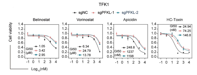
Cell viability of TFK1 cells expressing either a negative control sgRNA (sgNC) or sgRNAs targeting PFKL (sgPFKL-1 and sgPFKL-2) following treatment with specified concentrations of belinostat, vorinostat, apicidin or HC-toxin for 72 hours.
-
Adv Mater
Dual Targeting of m7G tRNA Modification and Histone Acetylation using Carrier-Free Nano-Epidrugs to Evoke Osteosarcoma Chemosensitization. [Abstract]2025 Oct 10:e05951. PMID: 41074238 -
Cancer Res
An In Vivo CRISPR Screen Identifies Stepwise Genetic Dependencies of Metastatic Progression. [Abstract]2022 Feb 15;82(4):681-694. PMID: 34916221
Belinostat purchased from MedChemExpress. Usage Cited in: Cancer Res. 2022 Feb 15;82(4):681-694. [Abstract]
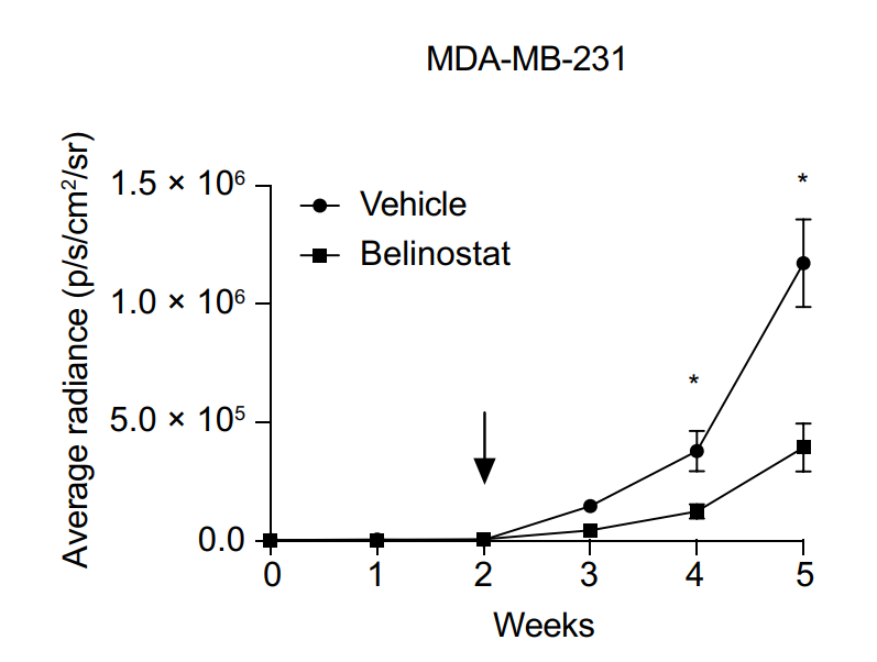
Plot showing the luminescence signal (ph/sec/cm2/sr) over time from NSG mice intracranially injected with either 2.0 × 10⁵ MDA-MB-231-GFP-Luc cells and treated either with vehicle (l-arginine (100 mg/mL) in ddH2O) or 80 mg/kg Belinostat for five consecutive days a week, for a total of three cycles.
Belinostat purchased from MedChemExpress. Usage Cited in: Cancer Res. 2022 Feb 15;82(4):681-694. [Abstract]
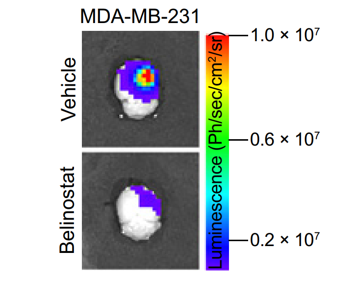
Representative images of the bioluminescence signal measured from brains of NSG mice intracranially injected with either MDA-MB-231-GFP-Luc cells and treated either with vehicle [l-arginine (100 mg/mL) in ddH2O] or 80 mg/kg belinostat.
-
Nat Commun
Histone 4 lysine 5/12 acetylation enables developmental plasticity of Pristionchus mouth form. [Abstract]2023 Apr 13;14(1):2095. PMID: 37055396 -
Nat Commun
Phenotype-driven precision oncology as a guide for clinical decisions one patient at a time. [Abstract]2017 Sep 5;8(1):435. PMID: 28874669 -
J Exp Clin Cancer Res
Comparative analysis of response to treatments and molecular features of tumor-derived organoids versus cell lines and PDX derived from the same ovarian clear cell carcinoma. [Abstract]2023 Oct 7;42(1):260. PMID: 37803448 -
Pharmacol Res
The histone deacetylase inhibitor belinostat ameliorates experimental autoimmune encephalomyelitis in mice by inhibiting TLR2/MyD88 and HDAC3/ NF-κB p65-mediated neuroinflammation. [Abstract]2022 Feb:176:105969. PMID: 34758400 -
Cell Death Dis
Targeting HDAC/OAZ1 axis with a novel inhibitor effectively reverses cisplatin resistance in non-small cell lung cancer. [Abstract]2019 May 24;10(6):400. PMID: 31127087 -
Cell Death Dis
Targeting EHMT2 reverses EGFR-TKI resistance in NSCLC by epigenetically regulating the PTEN/AKT signaling pathway. [Abstract]2018 Jan 26;9(2):129. PMID: 29374157
Belinostat purchased from MedChemExpress. Usage Cited in: Cell Death Dis. 2018 Jan 26;9(2):129. [Abstract]
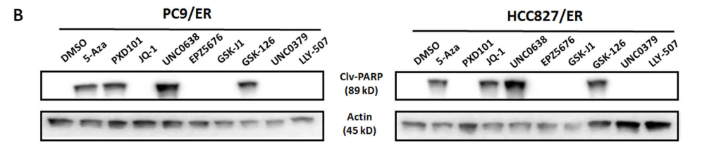
Belinostat (PDX101: 5/10 μM). PXD101The effects of treatment with the indicated epigenetic inhibitors on cleaved PARP (Clv-PARP) expression in both PC9/ER and HCC827/ER cells. β-actin is used as a loading control.
-
NPJ Breast Cancer
Epigenetically upregulating TROP2 and SLFN11 enhances therapeutic efficacy of TROP2 antibody drug conjugate sacitizumab govitecan. [Abstract]2023 Aug 11;9(1):66. PMID: 37567892 -
Blood Adv
BH3 mimetic drugs overcome the microenvironment-induced resistance to crizotinib in ALK+ anaplastic large cell lymphoma. [Abstract]2025 Jul 2:bloodadvances.2024015322. PMID: 40601898 -
Cell Rep
[Studies on ECMO. II. The effect of ECMO in respiratory failure and the trial of long-term ECMO; two experimental studies in dogs and goats]. [Abstract]2022 Sep 20;40(12):111396. PMID: 3613050 -
Cell Syst
A Library of Phosphoproteomic and Chromatin Signatures for Characterizing Cellular Responses to Drug Perturbations. [Abstract]2018 Apr 25;6(4):424-443.e7. PMID: 29655704 -
JCI Insight
Methylation-induced suppression of YAP/TAZ confers sensitivity to HDAC inhibitors in high grade IDH mutant gliomas. [Abstract]2025 Oct 9:e195385. PMID: 41066183 -
-
Cells
HDAC Inhibition Induces Transient Phenotypic Inertia in Dormant OCCC Spheroids by Derepression of Cell Cycle Genes. [Abstract]2026 Apr 10;15(8):673. PMID: 42041541 -
Commun Biol
CBX4 enhances acute monocytic leukemia development via HDAC-mediated suppression of Runx1. [Abstract]2026 Jun 1. PMID: 42225948 -
Eur J Pharmacol
Identification of novel SIRT1 up-regulators using a cell-based high-throughput screening assay. [Abstract]2025 Oct 15:1005:178066. PMID: 40818658 -
Pharmaceuticals (Basel)
Valproic Acid Prodrug Affects Selective Markers, Augments Doxorubicin Anticancer Activity and Attenuates Its Toxicity in a Murine Model of Aggressive Breast Cancer. [Abstract]2021 Nov 30;14(12):1244. PMID: 34959644 -
Cancers (Basel)
Identification of New Vulnerabilities in Conjunctival Melanoma Using Image-Based High Content Drug Screening. [Abstract]2022 Mar 19;14(6):1575. PMID: 35326726 -
Rheumatology (Oxford)
Autophagy inhibitors block pathogenic NET release in immune-mediated inflammatory disease without impairing host defence. [Abstract]2025 Aug 13:keaf437. PMID: 40802538 -
Bioengineering (Basel)
Precision Oncology for High-Grade Gliomas: A Tumor Organoid Model for Adjuvant Treatment Selection. [Abstract]2025 Oct 19;12(10):1121. PMID: 41155119 -
Viruses
Identification of Combinations of Protein Kinase C Activators and Histone Deacetylase Inhibitors That Potently Reactivate Latent HIV. [Abstract]2020 Jun 3;12(6):609. PMID: 32503121 -
-
Exp Cell Res
Network-based analysis with primary cells reveals drug response landscape of acute myeloid leukemia. [Abstract]2020 Aug 1;393(1):112054. PMID: 32376287 -
Int Immunol
Generation of allo-antigen-specific induced Treg stabilized by vitamin C treatment and its application for prevention of acute graft versus host disease model. [Abstract]2017 Dec 18;29(10):457-469. PMID: 29126272 -
Invest New Drugs
Molecular and cellular effects of a novel hydroxamate-based HDAC inhibitor - belinostat - in glioblastoma cell lines: a preliminary report. [Abstract]2016 Oct;34(5):552-64. PMID: 27468826
Belinostat purchased from MedChemExpress. Usage Cited in: Invest New Drugs. 2016 Oct;34(5):552-64. [Abstract]
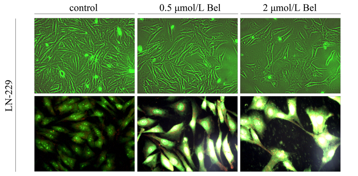
Phenotypic characteristics of LN-229 glioblastoma cells after 48 hours of treatment with Belinostat (Bel). Representative images are shown in the figure. Morphological changes induced by 0.5 and 2 μmol/L Bel treatment for 48 hours were assessed by phase contrast microscopy (100x magnification).
-
Saudi Med J
Investigation of the effect of belinostat on MCF-7 breast cancer stem cells via the Wnt, Notch, and Hedgehog signaling pathway. [Abstract]2024 Feb;45(2):121-127. PMID: 38309728 -
-
-
-
-
-
-
-
-
-


Solvent & Solubility
DMSO : 100 mg/mL (314.12 mM; Need ultrasonic; Hygroscopic DMSO has a significant impact on the solubility of product, please use newly opened DMSO)
Ethanol : 25 mg/mL (78.53 mM; ultrasonic and warming and heat to 60°C)
Please refer to the solubility information to select the appropriate solvent. Once prepared, please aliquot and store the solution to prevent product inactivation from repeated freeze-thaw cycles.
Storage method and period of stock solution: -80°C, 2 years; -20°C, 1 year. When stored at -80°C, please use it within 2 years. When stored at -20°C, please use it within 1 year.
Concentration (start) × Volume (start) = Concentration (final) × Volume (final)
Select the appropriate dissolution method based on your experimental animal and administration route.
- For the following dissolution methods, please ensure to first prepare a clear stock solution using an In Vitro approach and then sequentially add co-solvents:
- To ensure reliable experimental results, the clarified stock solution can be appropriately stored based on storage conditions. As for the working solution for In Vivo experiments, it is recommended to prepare freshly and use it on the same day.
- The percentages shown for the solvents indicate their volumetric ratio in the final prepared solution. If precipitation or phase separation occurs during preparation, heat and/or sonication can be used to aid dissolution.
Add each solvent one by one: 10% DMSO 90% (20% SBE-β-CD in Saline)
Solubility: ≥ 2.5 mg/mL (7.85 mM); Clear solution
This protocol yields a clear solution of ≥ 2.5 mg/mL (saturation unknown).
Taking 1 mL working solution as an example, add 100 μL DMSO stock solution (25.0 mg/mL) to 900 μL 20% SBE-β-CD in Saline, and mix evenly.
Preparation of 20% SBE-β-CD in Saline (4°C, storage for one week): 2 g SBE-β-CD powder is dissolved in 10 mL Saline, completely dissolve until clear.
Add each solvent one by one: 10% DMSO 40% PEG300 5% Tween-80 45% Saline
Solubility: ≥ 2.08 mg/mL (6.53 mM); Clear solution
This protocol yields a clear solution of ≥ 2.08 mg/mL (saturation unknown).
Taking 1 mL working solution as an example, add 100 μL DMSO stock solution (20.8 mg/mL) to 400 μL PEG300, and mix evenly; then add 50 μL Tween-80 and mix evenly; then add 450 μL Saline to adjust the volume to 1 mL.
Preparation of Saline: Dissolve 0.9 g sodium chloride in ddH₂O and dilute to 100 mL to obtain a clear Saline solution.
Please enter the basic information of animal experiments:
-
-
-
-
Recommended: Prepare an additional quantity of animals to account for potential losses during experiments.
Please enter your animal formula composition:
-
%DMSO +
Recommended: Keep the proportion of DMSO in working solution below 2% if your animal is weak.
-
%+
-
+%Tween-80 + +
-
%Saline +
The co-solvents required include: DMSO, . All of co-solvents are available by MedChemExpress (MCE). , Tween 80. All of co-solvents are available by MedChemExpress (MCE).
Working solution concentration: 0.22 mg/mL
Method for preparing stock solution: mg drug dissolved in μL DMSO. Stock solution concentration: mg/mL.
1. Take μL DMSO stock solution;
2. Add μL .
μL , mix evenly;
3. Then add μL Tween 80, mix evenly;
4. Then add μL
Please ensure that the stock solution in the first step is dissolved to a clear state, and add co-solvents in sequence. You can use ultrasonic heating (ultrasonic cleaner, recommended frequency 20-40 kHz), vortexing, etc. to assist dissolution.

Protocol
For activity assays, the reaction is carried out in a total volume of 150 μL of buffer [60 mM Tris (pH 7.4) containing 30% glycerol] containing 2 μL of cell extract and, where used, 2 μL of Belinostat. The reaction is started by the addition of 2 μL of [3H]labeled substrate (acetylated histone H4 peptide corresponding to the 20 NH2-terminal residues). Samples are incubated at 37°C for 45 min, and the reaction stopped by the addition of HCl and acetic acid (0.72 and 0.12 M final concentrations, respectively). Released [3H]acetate is extracted into 750 μL of ethyl acetate, and samples are centrifuged at 12,000× g for 5 min. The upper phase (600 μL) is transferred to 3 mL of scintillation fluid and counted[1].
MedChemExpress (MCE) has not independently confirmed the accuracy of these methods. They are for reference only.
The human ovarian cell line A2780 and Cisplatin (A2780/cp70) and Doxorubicin (2780AD) resistant derivatives are grown in RPMI 1640 supplemented with glutamine (2 mM) and FCS (10%). The human colon (HCT116 and HT29), melanoma (HS852), prostate (PC3), lung (CALU-3), and breast (MCF7) cell lines are grown in RPMI 1640 and the rest in DMEM supplemented as above. The human non-small cell lung cancer cell line WIL is grown in DMEM supplemented as above. Drug sensitivity is determined by a clonogenic assay. Briefly, cells are plated in 5 mL of medium at a density of 8×10⁴ cells/25 cm2 flask and allowed to attach and grow for 48 h. Cells are exposed to Belinostat (five concentrations from 0.016 to 10 μM) for 24 h. The medium is removed, and 1 mL of trypsin/EDTA is added to each flask. Once the cells have detached, 1 mL of medium is added, the cells are resuspended, and those from the control untreated flask are counted. Cells are diluted and plated into 6-cm Petri dishes (three per flask) at a density of 500-2000 cells/dish depending on the cell line. Cells from the drug-treated flasks are diluted and plated as for the control flasks. Dishes are incubated for 10-15 days at 37°C. Cells are washed with PBS, fixed in methanol, and stained with crystal violet, and colonies that contained ≥50 cells counted. Sensitivity is expressed as the IC50 (mean±SE of three experiments) defined as the concentration of drug required to reduce the number of colonies to 50% of that of the control untreated cells[1].
MedChemExpress (MCE) has not independently confirmed the accuracy of these methods. They are for reference only.
Mice[1]
For the human tumor xenograft studies, monolayer cultures are harvested with trypsin/EDTA (0.25%/1 mM in PBS) and resuspended in PBS. About 107 cells are injected s.c. into the right flank of athymic nude mice (CD1 nu/nu mice). After 10-15 days when the mean tumor diameter is ≥0.5 cm, animals are randomized into groups of six for experiments. Belinostat is dissolved in DMSO and then diluted in water to give a final concentration of DMSO of 10% and is administered i.p. at the times specified. This formulation gives sufficient solubility for doses of ≤ 40 mg/kg. Mice are weighed daily, and tumor volumes are estimated by caliper measurements assuming spherical geometry (volume=d3×π/6).
MedChemExpress (MCE) has not independently confirmed the accuracy of these methods. They are for reference only.


Purity & Documentation
-
Data Sheet (279 KB)
-
SDS (396 KB)
- English - EN (396 KB)
- Français - FR (396 KB)
- Deutsch - DE (396 KB)
- Norwegian - NO (396 KB)
- Español - ES (396 KB)
- Swedish - SV (396 KB)
- Italian - IT (396 KB)
- Korean - KR (396 KB)
- Portuguese - PT (396 KB)
-
Handling Instructions (2659 KB)
References
[1]. Plumb JA, et al. Pharmacodynamic response and inhibition of growth of human tumor xenografts by the novel histonedeacetylase inhibitor PXD101. Mol Cancer Ther. 2003 Aug;2(8):721-8. [Content Brief]
[2]. Qian X, et al. Activity of PXD101, a histone deacetylase inhibitor, in preclinical ovarian cancer studies. Mol Cancer Ther. 2006 Aug;5(8):2086-95. [Content Brief]
[3]. Chia S, et al. Phenotype-driven precision oncology as a guide for clinical decisions one patient at a time. Nat Commun. 2017 Sep 5;8(1):435. [Content Brief]


Complete Stock Solution Preparation Table
Please refer to the solubility information to select the appropriate solvent. Once prepared, please aliquot and store the solution to prevent product inactivation from repeated freeze-thaw cycles.
Storage method and period of stock solution: -80°C, 2 years; -20°C, 1 year. When stored at -80°C, please use it within 2 years. When stored at -20°C, please use it within 1 year.
| Optional Solvent | Concentration Solvent Mass | 1 mg | 5 mg | 10 mg | 25 mg |
|---|---|---|---|---|---|
| Ethanol / DMSO | 1 mM | 3.1412 mL | 15.7060 mL | 31.4120 mL | 78.5299 mL |
| 5 mM | 0.6282 mL | 3.1412 mL | 6.2824 mL | 15.7060 mL | |
| 10 mM | 0.3141 mL | 1.5706 mL | 3.1412 mL | 7.8530 mL | |
| 15 mM | 0.2094 mL | 1.0471 mL | 2.0941 mL | 5.2353 mL | |
| 20 mM | 0.1571 mL | 0.7853 mL | 1.5706 mL | 3.9265 mL | |
| 25 mM | 0.1256 mL | 0.6282 mL | 1.2565 mL | 3.1412 mL | |
| 30 mM | 0.1047 mL | 0.5235 mL | 1.0471 mL | 2.6177 mL | |
| 40 mM | 0.0785 mL | 0.3926 mL | 0.7853 mL | 1.9632 mL | |
| 50 mM | 0.0628 mL | 0.3141 mL | 0.6282 mL | 1.5706 mL | |
| 60 mM | 0.0524 mL | 0.2618 mL | 0.5235 mL | 1.3088 mL | |
| DMSO | 80 mM | 0.0393 mL | 0.1963 mL | 0.3926 mL | 0.9816 mL |
| 100 mM | 0.0314 mL | 0.1571 mL | 0.3141 mL | 0.7853 mL |


 Powered by Bioz