



Inobrodib
Based on 14 publication(s) in Google Scholar
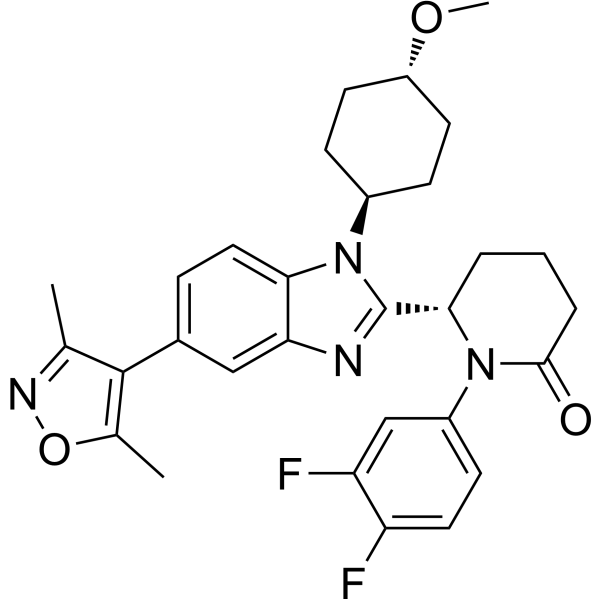
Inobrodib (CCS1477) is an orally active, potent, and selective inhibitor of the p300/CBP bromodomain. Inobrodib binds to p300 and CBP with Kd values of 1.3 and 1.7 nM, respectively, and with 170/130-fold selectivity compared with BRD4 with a Kd of 222 nM. CCS1477 inhibits cell proliferation in prostate cancer cell lines and decreases androgen receptor (AR)- and C-MYC-regulated gene expression.
For research use only. We do not sell to patients.
- Purity : 99.95%
- CAS No.: 2222941-37-7
- Formula: C30H32F2N4O3
- Molecular Weight:534.60
-
Storage:Powder -20°C, 3 years , 4°C, 2 years ; In solvent -80°C, 2 years , -20°C, 1 year

To place orders, for customer services and technical support, please contact: MedChemExpress USA
Tel: 609-228-6898 E-mail: [email protected] [email protected]


-
 Biological Activity
-
Chemical Information
-
Solvent & Solubility
- Protocols
- Purity & Documentation
- References
-
 Help & FAQs


-
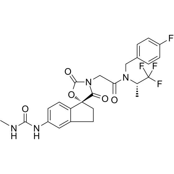
HY-107455
A-485p300/CBP Inhibitor -
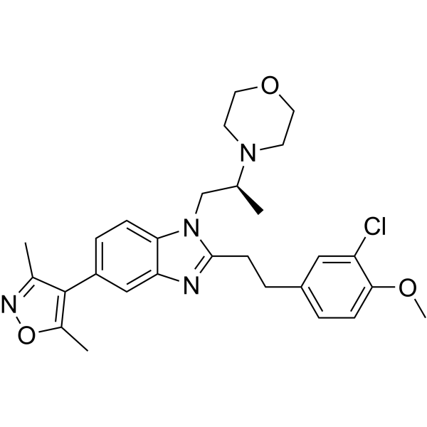
HY-15826
SGC-CBP30CBP/p300 Inhibitor -
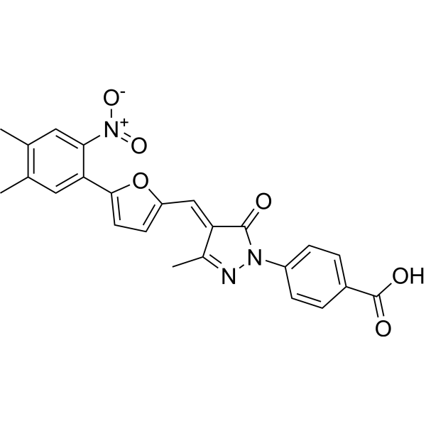
HY-13823
C646p300/CBP HAT Inhibitor -
HY-139664
GSK-3685032DNMT1 Inhibitor -

HY-15371
ForskolinAdenylate Cyclase Activator -
HY-108696
GNE-781CBP Inhibitor -
HY-13030
(+)-JQ-1BET Inhibitor -
HY-13803
TazemetostatEZH2 Inhibitor -
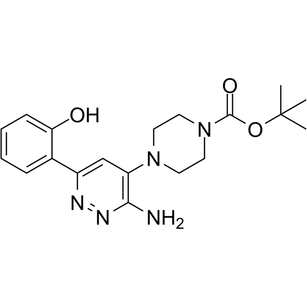
HY-145446
SGC-SMARCA-BRDVIIIPB1(5)/SMARCA2/4 Inhibitor -
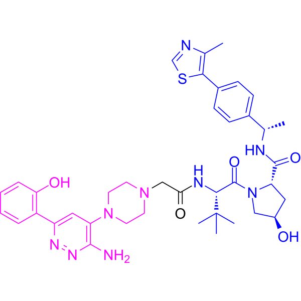
HY-145388
AU-15330Anticancer Reagent -
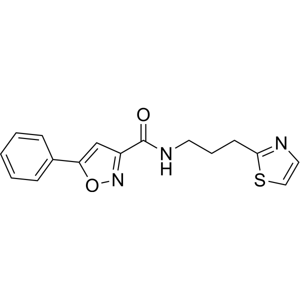
HY-141644
PY-60YAP Activator -
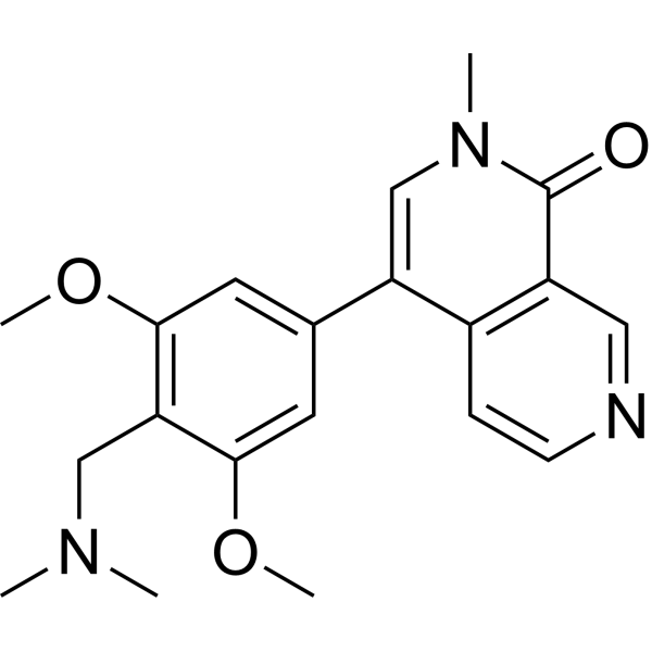
HY-100351
BI-7273BRD9 Inhibitor
Publications Citing Use of MedChemExpress (MCE) Inobrodib
More- Cell. 2026 Jun 11;189(12):3541-3552.e18. [Abstract]
- Blood. 2025 May 29;145(22):2614-2629. [Abstract]
- Cancer Res. 2026 Jul 17.
- Nat Commun. 2024 Jun 5;15(1):4770. [Abstract]
- Nucleic Acids Res. 2024 Jan 25;52(2):625-642. [Abstract]
- Adv Sci (Weinh). 2026 Mar 1:e17231. [Abstract]
- Cell Death Dis. 2025 Jun 3;16(1):432. [Abstract]
- J Med Chem. 2024 Apr 11;67(7):5275-5304. [Abstract]
- J Med Chem. 2024 Apr 11;67(7):5351-5372. [Abstract]
- Cell Mol Life Sci. 2024 Apr 2;81(1):160. [Abstract]
- Transl Res. 2026 Sep 6:297:112-128.
- EMBO Rep. 2025 Oct;26(19):4766-4793. [Abstract]
- Cancer Res Commun. 2025 Jan 1;5(1):24-38. [Abstract]
- bioRxiv. 2023 Mar 3.
 Customer Validation & Images

-
WB
-
RT-PCR
-
Cell Proliferation/Viability Assay
-
Cell Proliferation/Viability Assay
-
RT-PCR


Biological Activity
Description
IC50 & Target
|
CBP/p300 |
Cellular Effect
|
Cell Line

|
Type | Value | Description | References |
|---|---|---|---|---|
| CWR22R | IC50 |
0.42 μM
Compound: 4; CCS1477
|
Antiproliferative activity against human 22Rv1 cells assessed as cell growth inhibition by Celltiter-Glo assay
Antiproliferative activity against human 22Rv1 cells assessed as cell growth inhibition by Celltiter-Glo assay
|
[PMID: 34962793] |
| CWR22R | IC50 |
184 nM
Compound: 3; CCS1477
|
Antiproliferative activity against AR positive human 22Rv1 cells assessed inhibition of cell growth measured after 4 days by CellTiter-Glo Luminescent cell viability assay
Antiproliferative activity against AR positive human 22Rv1 cells assessed inhibition of cell growth measured after 4 days by CellTiter-Glo Luminescent cell viability assay
|
[PMID: 38530938] |
| CWR22R | IC50 |
184 nM
Compound: 5; CCS1477
|
Antiproliferative activity against human 22Rv1 cells assessed as cell growth inhibition incubated for 4 days by celtiter-glo luminescent assay
Antiproliferative activity against human 22Rv1 cells assessed as cell growth inhibition incubated for 4 days by celtiter-glo luminescent assay
|
[PMID: 38477974] |
| EOL1 | IC50 |
<3 μM
Compound: 4; CCS1477
|
Antiproliferative activity against human EOL1 cells
Antiproliferative activity against human EOL1 cells
|
[PMID: 38649304] |
| Hep 3B2 | IC50 |
1.928 μM
Compound: 4; CCS1477
|
Antiproliferative activity against human Hep3B cells assessed as inhibition of cell proliferation incubated for 6 days by CCK-8 assay
Antiproliferative activity against human Hep3B cells assessed as inhibition of cell proliferation incubated for 6 days by CCK-8 assay
|
[PMID: 39356741] |
| HepG2 | IC50 |
0.869 μM
Compound: 4; CCS1477
|
Antiproliferative activity against human HepG2 cells assessed as inhibition of cell proliferation incubated for 6 days by CCK-8 assay
Antiproliferative activity against human HepG2 cells assessed as inhibition of cell proliferation incubated for 6 days by CCK-8 assay
|
[PMID: 39356741] |
| KG-1 | IC50 |
<3 μM
Compound: 4; CCS1477
|
Antiproliferative activity against human KG-1 cells
Antiproliferative activity against human KG-1 cells
|
[PMID: 38649304] |
| KG-1 | IC50 |
17.04 μM
Compound: 4; CCS1477
|
Antiproliferative activity against human KG-1 cells assessed as inhibition of cell growth incubated for 72 hrs by celltiter glo luminescence analysis
Antiproliferative activity against human KG-1 cells assessed as inhibition of cell growth incubated for 72 hrs by celltiter glo luminescence analysis
|
[PMID: 38829718] |
| L02 | IC50 |
20 μM
Compound: 4; CCS1477
|
Cytotoxicity against human HL7702 cells assessed as inhibition of cell growth incubated for 72 hrs by celltiter glo luminescence analysis
Cytotoxicity against human HL7702 cells assessed as inhibition of cell growth incubated for 72 hrs by celltiter glo luminescence analysis
|
[PMID: 38829718] |
| LNCaP | IC50 |
0.06 μM
Compound: 4; CCS1477
|
Antiproliferative activity against human LNCaP cells assessed as cell growth inhibition by Celltiter-Glo assay
Antiproliferative activity against human LNCaP cells assessed as cell growth inhibition by Celltiter-Glo assay
|
[PMID: 34962793] |
| LNCaP | IC50 |
412 nM
Compound: 3; CCS1477
|
Antiproliferative activity against AR positive human LNCaP cells assessed inhibition of cell growth measured after 4 days by CellTiter-Glo Luminescent cell viability assay
Antiproliferative activity against AR positive human LNCaP cells assessed inhibition of cell growth measured after 4 days by CellTiter-Glo Luminescent cell viability assay
|
[PMID: 38530938] |
| LNCaP | IC50 |
412 nM
Compound: 5; CCS1477
|
Antiproliferative activity against human LNCaP cells assessed as cell growth inhibition incubated for 4 days by celtiter-glo luminescent assay
Antiproliferative activity against human LNCaP cells assessed as cell growth inhibition incubated for 4 days by celtiter-glo luminescent assay
|
[PMID: 38477974] |
| LNCaP C4-2B | IC50 |
1.65 μM
Compound: 4; CCS1477
|
Antiproliferative activity against human LNCaP C4-2B cells assessed as inhibition of cell growth by Celltiter-Glo assay
Antiproliferative activity against human LNCaP C4-2B cells assessed as inhibition of cell growth by Celltiter-Glo assay
|
[PMID: 34962793] |
| MOLM-13 | IC50 |
102.5 nM
Compound: 4; CCS1477
|
Antiproliferative activity against human MOLM-13 cells incubated for 72 hrs by Cell-Titer Glo assay
Antiproliferative activity against human MOLM-13 cells incubated for 72 hrs by Cell-Titer Glo assay
|
[PMID: 38649304] |
| MOLM-16 | IC50 |
0.37 μM
Compound: 4; CCS1477
|
Antiproliferative activity against human MOLM16 cells assessed as inhibition of cell growth incubated for 72 hrs by celltiter glo luminescence analysis
Antiproliferative activity against human MOLM16 cells assessed as inhibition of cell growth incubated for 72 hrs by celltiter glo luminescence analysis
|
[PMID: 38829718] |
| MOLM-16 | IC50 |
12.9 nM
Compound: 4; CCS1477
|
Antiproliferative activity against human MOLM16 cells incubated for 72 hrs by Cell-Titer Glo assay
Antiproliferative activity against human MOLM16 cells incubated for 72 hrs by Cell-Titer Glo assay
|
[PMID: 38649304] |
| MOLT-4 | IC50 |
0.247 μM
Compound: 4; CCS1477
|
Antiproliferative activity against human MOLT-4 cells assessed as inhibition of cell proliferation incubated for 6 days by CCK-8 assay
Antiproliferative activity against human MOLT-4 cells assessed as inhibition of cell proliferation incubated for 6 days by CCK-8 assay
|
[PMID: 39356741] |
| MV4-11 | IC50 |
0.014 μM
Compound: 4; CCS1477
|
Antiproliferative activity against human MV4-11 cells assessed as inhibition of cell growth incubated for 120 hrs by celltiter glo luminescence analysis
Antiproliferative activity against human MV4-11 cells assessed as inhibition of cell growth incubated for 120 hrs by celltiter glo luminescence analysis
|
[PMID: 38829718] |
| MV4-11 | IC50 |
20.4 nM
Compound: 4; CCS1477
|
Antiproliferative activity against human MV4-11 cells incubated for 120 hrs by Cell-Titer Glo assay
Antiproliferative activity against human MV4-11 cells incubated for 120 hrs by Cell-Titer Glo assay
|
[PMID: 38649304] |
| OPM-2 | IC50 |
30 nM
Compound: 4; CCS1477
|
Antiproliferative activity against human OPM-2 cells assessed as inhibition of cell proliferation incubated for 6 days by CCK8 assay
Antiproliferative activity against human OPM-2 cells assessed as inhibition of cell proliferation incubated for 6 days by CCK8 assay
|
[PMID: 39356741] |
| RPMI-8226 | IC50 |
0.082 μM
Compound: 4; CCS1477
|
Antiproliferative activity against human RPMI-8226 cells assessed as inhibition of cell proliferation incubated for 6 days by CCK-8 assay
Antiproliferative activity against human RPMI-8226 cells assessed as inhibition of cell proliferation incubated for 6 days by CCK-8 assay
|
[PMID: 39356741] |
| SK-HEP1 | IC50 |
4.679 μM
Compound: 1; CCS1477
|
Antiproliferative activity against human SK-HEP1 cells assessed as inhibition of cell growth incubated for 5 days by CCK-8 assay
Antiproliferative activity against human SK-HEP1 cells assessed as inhibition of cell growth incubated for 5 days by CCK-8 assay
|
[PMID: 38316017] |
| THP-1 | IC50 |
<3 μM
Compound: 4; CCS1477
|
Antiproliferative activity against human THP-1 cells
Antiproliferative activity against human THP-1 cells
|
[PMID: 38649304] |
| VCaP | IC50 |
195 nM
Compound: 5; CCS1477
|
Antiproliferative activity against human VCaP cells assessed as cell growth inhibition incubated for 4 days by celtiter-glo luminescent assay
Antiproliferative activity against human VCaP cells assessed as cell growth inhibition incubated for 4 days by celtiter-glo luminescent assay
|
[PMID: 38477974] |
| VCaP | IC50 |
53.6 nM
Compound: 3; CCS1477
|
Antiproliferative activity against AR positive human VCaP cells assessed inhibition of cell growth measured after 4 days by CellTiter-Glo Luminescent cell viability assay
Antiproliferative activity against AR positive human VCaP cells assessed inhibition of cell growth measured after 4 days by CellTiter-Glo Luminescent cell viability assay
|
[PMID: 38530938] |
In Vitro
Inobrodib binding to cellular histones in an in-cell BRET assay gives an IC50 of 19 nM for p300 and 1060 nM for BRD4. Inobrodib shows potent growth-inhibitory activity in VCaP, 22Rv1, and LNCaP95 (all IC50 < 100 nM) that express both AR-FL and AR-V7[1].
Inobrodib (0-3000 nM; 48 hours) reduces expression of AR-regulated genes (KLK2, KLK3, and TMPRSS2) in both 22Rv1 and LNCaP95 cells. Inobrodib also reduces C-MYC protein expression in both 22Rv1 and LNCaP95 cells and AR-V7 protein expression in 22Rv1 cells, without clear impact on AR-FL protein expression in 22Rv1 and LNCaP95 cells. Inobrodib reduces C-MYC mRNA and downstream AR and C-MYC signaling in 22Rv1 and C4-2 cells at 16 hours. Inobrodib regulates AR signaling by affecting the recruitment of CBP, p300, and AR-FL to known AR binding sites, and has the potential to abrogate persistent AR signaling in CRPC[1].
MedChemExpress (MCE) has not independently confirmed the accuracy of these methods. They are for reference only.
In Vivo
Inobrodib (20 mg/kg; oral gavage; daily for 8 days) decreases AR and AR-V7 signaling and inhibits growth in a patient- derived model of lethal prostate cancer (NOD/SCID gamma (NSG) male castrated mice)[1].
MedChemExpress (MCE) has not independently confirmed the accuracy of these methods. They are for reference only.
-
Animal Model:Noncastrated male athymic nude mice[1]
-
Dosage:10-30 mg/kg
-
Administration:Oral gavage; at 10 or 20 mg/kg daily (QD) or at 30 mg/kg every other day (QOD) for 28 days
-
Result:Affected tumor growth at 10 mg/kg daily, 20 mg/kg daily, and 30 mg/kg every other day.
- RT-PCR
- RNA extraction experimental
- BrdU Incorporation Assay
- Somatic Cell Culture
- CFSE Dye Dilution Proliferation Assay
- Dye-dilution cell tracking and proliferation staining
- CCK-8/WST-8 Cell Proliferation Assay
- Protocol for Cell Cycle
- Cell Counting-Based Growth Curve Assay
- Colony Formation (Clonogenic) Assay
- Real Time qPCR (Q-PCR)
- Cell Viability Determination by MTT Colorimetric Assay
- EdU Incorporation Assay (Click Chemistry-Based DNA Synthesis Measurement)
- Ki-67 Immunostaining Proliferation Assay
- PCNA Immunodetection Proliferation Assay
- Protocol for Cell Counting and Cell Density Analysis
- Research Protocol for Endocrine Diseases
- MTT Cell Proliferation Assay
Clinical Trial
| NCT Number | Sponsor | Condition | Start Date |
Phase
|
|---|---|---|---|---|
| NCT01329991 | Plexxikon| | 2011-05 | PHASE1 |


Chemical Information
-
CAS No. 2222941-37-7
-
Appearance Solid
-
Molecular Weight 534.60
-
Formula C30H32F2N4O3
-
Color White to off-white
-
SMILES
O=C1N(C2=CC=C(F)C(F)=C2)[C@H](C3=NC4=CC(C5=C(C)ON=C5C)=CC=C4N3[C@H]6CC[C@H](OC)CC6)CCC1
-
Synonyms
CCS1477
-
Shipping
Room temperature in continental US; may vary elsewhere.
-
Storage
Powder -20°C 3 years 4°C 2 years In solvent -80°C 2 years -20°C 1 year


Publications (14)
-
Journal Impact Factor
-
Most Recent
-
Cell
2026 Jun 11;189(12):3541-3552.e18. PMID: 41999746 -
Blood
Targeting Caseinolytic Mitochondrial Matrix Peptidase, a Novel Contributor to High-risk Behavior, in Multiple Myeloma. [Abstract]2025 May 29;145(22):2614-2629. PMID: 39912779 -
-
Nat Commun
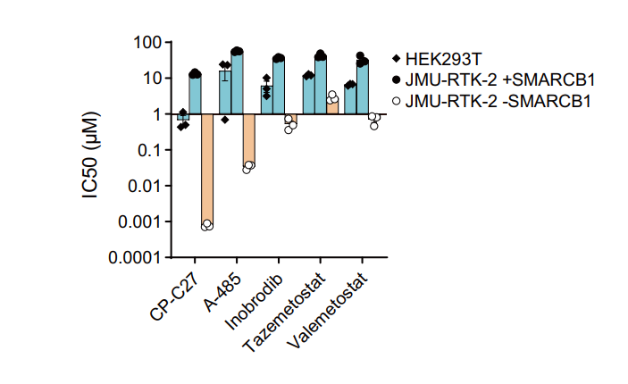
Targeting dependency on a paralog pair of CBP/p300 against de-repression of KREMEN2 in SMARCB1-deficient cancers. [Abstract]2024 Jun 5;15(1):4770. PMID: 38839769
Inobrodib purchased from MedChemExpress. Usage Cited in: Nat Commun. 2024 Jun 5;15(1):4770. [Abstract]
IC50 values of the CBP/p300 inhibitors CP-C27, A-485, and Inobrodib (6 days), the EZH2 inhibitor tazemetostat, and the EZH1/EZH2 inhibitor valemetostat in HEK293T, JMU-RTK-2 +SMARCB1, and JMU-RTK-2 -SMARCB1 cells.
-
Nucleic Acids Res
Chromatin accessibility and pioneer factor FOXA1 restrict glucocorticoid receptor action in prostate cancer. [Abstract]2024 Jan 25;52(2):625-642. PMID: 38015476
Inobrodib purchased from MedChemExpress. Usage Cited in: Nucleic Acids Res. 2024 Jan 25;52(2):625-642. [Abstract]
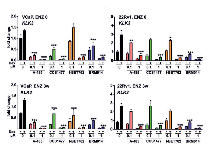
Bar graphs depict KLK3 gene expression analysis in VCaP ENZ 0, VCaP ENZ 3w, 22Rv1 ENZ 0, and 22Rv1 ENZ 3w cells treated with or without Dex in the presence or absence of 0.1 or 1 μM of indicated inhibitor (A-485, CCS1477, I-BET762, BRM014).
-
Adv Sci (Weinh)
BCR::ABL1-Induced Enhancer Reprogramming Uncovers Hypersensitivity of Ph+B-ALL Cells to Enhancer-Targeting Drugs. [Abstract]2026 Mar 1:e17231. PMID: 41764406 -
Cell Death Dis
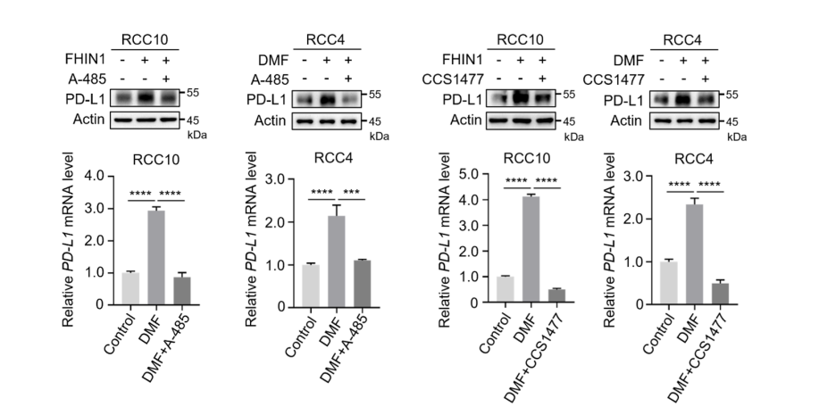
Oncometabolite fumarate facilitates PD-L1 expression and immune evasion in clear cell renal cell carcinoma. [Abstract]2025 Jun 3;16(1):432. PMID: 40461489
Inobrodib purchased from MedChemExpress. Usage Cited in: Cell Death Dis. 2025 Jun 3;16(1):432. [Abstract]
Western blot and qRT-PCR analyses were performed to evaluate the PD-L1 levels in RCC10 and RCC4 cells treated with DMF/FHIN1, both in the absence and presence of 20 μM A485 or 100 nM CCS1477, an inhibitor of p300.
-
J Med Chem
Discovery of CBPD-268 as an Exceptionally Potent and Orally Efficacious CBP/p300 PROTAC Degrader Capable of Achieving Tumor Regression. [Abstract]2024 Apr 11;67(7):5275-5304. PMID: 38477974 -
J Med Chem
Discovery of CBPD-409 as a Highly Potent, Selective, and Orally Efficacious CBP/p300 PROTAC Degrader for the Treatment of Advanced Prostate Cancer. [Abstract]2024 Apr 11;67(7):5351-5372. PMID: 38530938 -
Cell Mol Life Sci
EP300/CREBBP acetyltransferase inhibition limits steroid receptor and FOXA1 signaling in prostate cancer cells. [Abstract]2024 Apr 2;81(1):160. PMID: 38564048 -
-
EMBO Rep
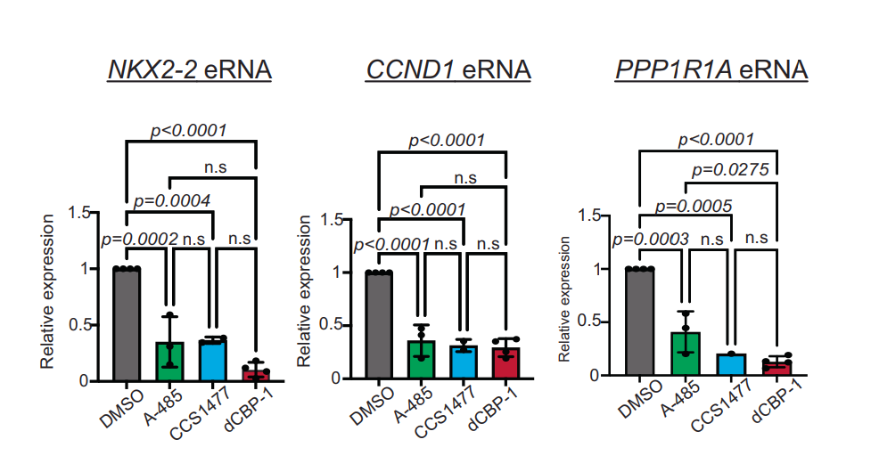
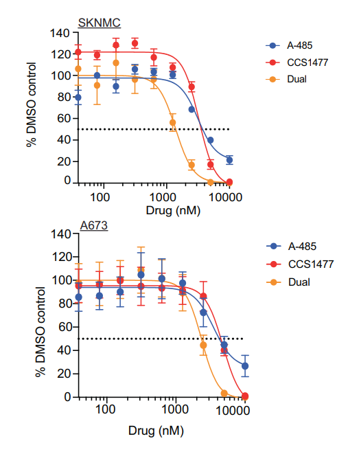
p300/CBP is an essential driver of pathogenic enhancer activity and gene expression in Ewing sarcoma. [Abstract]2025 Oct;26(19):4766-4793. PMID: 40890402
Inobrodib purchased from MedChemExpress. Usage Cited in: EMBO Rep. 2025 Oct;26(19):4766-4793. [Abstract]
RT-qPCR for eRNA of NKX2-2, CCND1 and PPP1R1A enhancers following 6 h treatment of A-485, CCS1477 (1 μM), and dCBP-1 treatment in SKNMC cells.
Inobrodib purchased from MedChemExpress. Usage Cited in: EMBO Rep. 2025 Oct;26(19):4766-4793. [Abstract]
Cell viability of SKNMC and A673 cells treated with A-485, CCS1477 (1 μM) and Dual combination for 6 days measured by CellTiter-Glo.
-
Cancer Res Commun
Efficacy of CBP/p300 Dual Inhibitors against Derepression of KREMEN2 in cBAF-Deficient Cancers. [Abstract]2025 Jan 1;5(1):24-38. PMID: 39625239 -


Solvent & Solubility
In Vitro:
DMSO : 100 mg/mL (187.06 mM; Need ultrasonic; Hygroscopic DMSO has a significant impact on the solubility of product, please use newly opened DMSO)
Please refer to the solubility information to select the appropriate solvent. Once prepared, please aliquot and store the solution to prevent product inactivation from repeated freeze-thaw cycles.
Storage method and period of stock solution: -80°C, 2 years; -20°C, 1 year. When stored at -80°C, please use it within 2 years. When stored at -20°C, please use it within 1 year.
Please refer to the solubility information to select the appropriate solvent. Once prepared, please aliquot and store the solution to prevent product inactivation from repeated freeze-thaw cycles.
Storage method and period of stock solution: -80°C, 2 years; -20°C, 1 year. When stored at -80°C, please use it within 2 years. When stored at -20°C, please use it within 1 year.
Concentration (start) × Volume (start) = Concentration (final) × Volume (final)
In Vivo:
Select the appropriate dissolution method based on your experimental animal and administration route.
- For the following dissolution methods, please ensure to first prepare a clear stock solution using an In Vitro approach and then sequentially add co-solvents:
- To ensure reliable experimental results, the clarified stock solution can be appropriately stored based on storage conditions. As for the working solution for In Vivo experiments, it is recommended to prepare freshly and use it on the same day.
- The percentages shown for the solvents indicate their volumetric ratio in the final prepared solution. If precipitation or phase separation occurs during preparation, heat and/or sonication can be used to aid dissolution.
Add each solvent one by one: 10% DMSO 40% PEG300 5% Tween-80 45% Saline
Solubility: ≥ 2.5 mg/mL (4.68 mM); Clear solution
This protocol yields a clear solution of ≥ 2.5 mg/mL (saturation unknown).
Taking 1 mL working solution as an example, add 100 μL DMSO stock solution (25.0 mg/mL) to 400 μL PEG300, and mix evenly; then add 50 μL Tween-80 and mix evenly; then add 450 μL Saline to adjust the volume to 1 mL.
Preparation of Saline: Dissolve 0.9 g sodium chloride in ddH₂O and dilute to 100 mL to obtain a clear Saline solution.
Add each solvent one by one: 10% DMSO 90% (20% SBE-β-CD in Saline)
Solubility: 2.5 mg/mL (4.68 mM); Suspended solution; Need ultrasonic
This protocol yields a suspended solution of 2.5 mg/mL. Suspended solution can be used for oral and intraperitoneal injection.
Taking 1 mL working solution as an example, add 100 μL DMSO stock solution (25.0 mg/mL) to 900 μL 20% SBE-β-CD in Saline, and mix evenly.
Preparation of 20% SBE-β-CD in Saline (4°C, storage for one week): 2 g SBE-β-CD powder is dissolved in 10 mL Saline, completely dissolve until clear.
In Vivo Dissolution Calculator
Please enter the basic information of animal experiments:
-
-
-
-
Recommended: Prepare an additional quantity of animals to account for potential losses during experiments.
Please enter your animal formula composition:
-
%DMSO +
Recommended: Keep the proportion of DMSO in working solution below 2% if your animal is weak.
-
%+
-
+%Tween-80 + +
-
%Saline +
The co-solvents required include: DMSO, . All of co-solvents are available by MedChemExpress (MCE). , Tween 80. All of co-solvents are available by MedChemExpress (MCE).
Working solution concentration: 0.22 mg/mL
Method for preparing stock solution: mg drug dissolved in μL DMSO. Stock solution concentration: mg/mL.
1. Take μL DMSO stock solution;
2. Add μL .
μL , mix evenly;
3. Then add μL Tween 80, mix evenly;
4. Then add μL
Please ensure that the stock solution in the first step is dissolved to a clear state, and add co-solvents in sequence. You can use ultrasonic heating (ultrasonic cleaner, recommended frequency 20-40 kHz), vortexing, etc. to assist dissolution.


Purity & Documentation
-
Data Sheet (281 KB)
-
SDS (393 KB)
- English - EN (393 KB)
- Français - FR (393 KB)
- Deutsch - DE (393 KB)
- Norwegian - NO (393 KB)
- Español - ES (393 KB)
- Swedish - SV (393 KB)
- Italian - IT (393 KB)
- Korean - KR (393 KB)
- Portuguese - PT (393 KB)
-
Handling Instructions (2659 KB)
References


Complete Stock Solution Preparation Table
Please refer to the solubility information to select the appropriate solvent. Once prepared, please aliquot and store the solution to prevent product inactivation from repeated freeze-thaw cycles.
Storage method and period of stock solution: -80°C, 2 years; -20°C, 1 year. When stored at -80°C, please use it within 2 years. When stored at -20°C, please use it within 1 year.
| Optional Solvent | Concentration Solvent Mass | 1 mg | 5 mg | 10 mg | 25 mg |
|---|---|---|---|---|---|
| DMSO | 1 mM | 1.8706 mL | 9.3528 mL | 18.7056 mL | 46.7639 mL |
| 5 mM | 0.3741 mL | 1.8706 mL | 3.7411 mL | 9.3528 mL | |
| 10 mM | 0.1871 mL | 0.9353 mL | 1.8706 mL | 4.6764 mL | |
| 15 mM | 0.1247 mL | 0.6235 mL | 1.2470 mL | 3.1176 mL | |
| 20 mM | 0.0935 mL | 0.4676 mL | 0.9353 mL | 2.3382 mL | |
| 25 mM | 0.0748 mL | 0.3741 mL | 0.7482 mL | 1.8706 mL | |
| 30 mM | 0.0624 mL | 0.3118 mL | 0.6235 mL | 1.5588 mL | |
| 40 mM | 0.0468 mL | 0.2338 mL | 0.4676 mL | 1.1691 mL | |
| 50 mM | 0.0374 mL | 0.1871 mL | 0.3741 mL | 0.9353 mL | |
| 60 mM | 0.0312 mL | 0.1559 mL | 0.3118 mL | 0.7794 mL | |
| 80 mM | 0.0234 mL | 0.1169 mL | 0.2338 mL | 0.5845 mL | |
| 100 mM | 0.0187 mL | 0.0935 mL | 0.1871 mL | 0.4676 mL |


Inobrodib Related Classifications
HY-111784 Related Classifications
 Powered by Bioz