



Hydralazine hydrochloride
Based on 4 publication(s) in Google Scholar
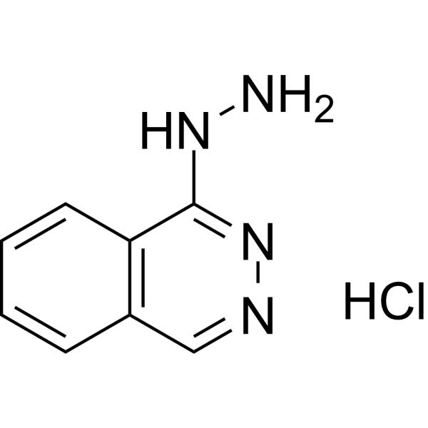
Hydralazine hydrochloride is an orally active, blood-brain barrier-permeable DNA methyltransferase inhibitor with vasodilatory, arterial smooth muscle relaxant and hypotensive activities. Hydralazine hydrochloride reactivates silenced tumor suppressor genes via mediating DNA demethylation, while exerting neuroprotective and anti-inflammatory properties. Hydralazine hydrochloride inhibits NOS-2 (iNOS) and COX-2, and reduces the production of NO and PGEE2; meanwhile, Hydralazine hydrochloride scavenges reactive oxygen species and inhibits macrophage activation. Hydralazine hydrochloride alleviates motor dysfunction, neuropathic inflammatory pain, and formalin-induced somatic and emotional pain responses. In addition, Hydralazine hydrochloride directly induces DNA strand breaks and sister chromatid exchange, exhibiting certain mutagenic characteristics. Hydralazine hydrochloride has been widely used in studies on hypertension, various cancers (such as cervical cancer, leukemia), spinal cord injury and the mechanisms of inflammatory pain.
For research use only. We do not sell to patients.
- Purity : 99.94%
- CAS No.: 304-20-1
- Formula: C8H9ClN4
- Molecular Weight:196.64
-
Storage:
4°C, sealed storage, away from moisture
* In solvent : -80°C, 6 months; -20°C, 1 month (sealed storage, away from moisture)

To place orders, for customer services and technical support, please contact: MedChemExpress USA
Tel: 609-228-6898 E-mail: [email protected] [email protected]


-
 Biological Activity
-
Chemical Information
-
Solvent & Solubility
- Protocols
- Purity & Documentation
- References
-
 Help & FAQs


-
HY-10586
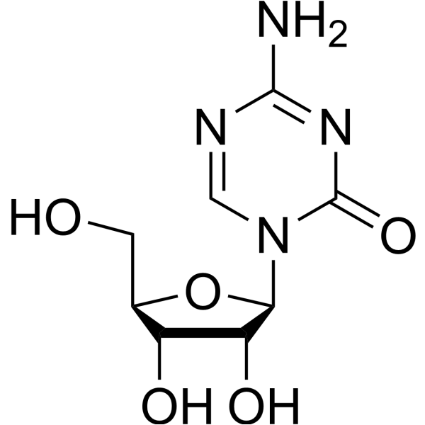
5-AzacytidineDNMT Inhibitor -
HY-14519
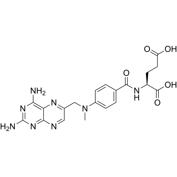
MethotrexateDihydrofolate Reductase Inhibitor -
HY-14596
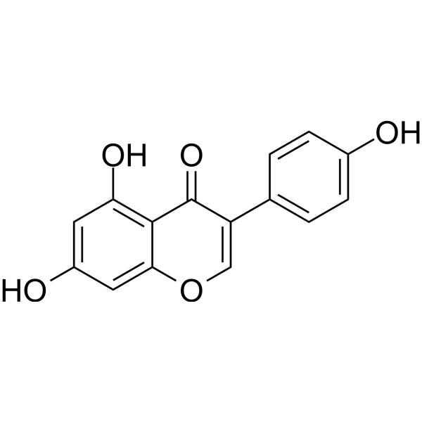
GenisteinAutophagy Inducer -
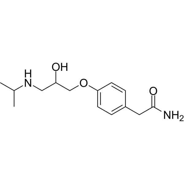
HY-17498
Atenololβ1-Adrenergic Receptor Blocker -
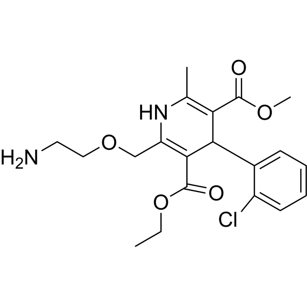
HY-B0317
AmlodipineCalcium Channel Blocker -
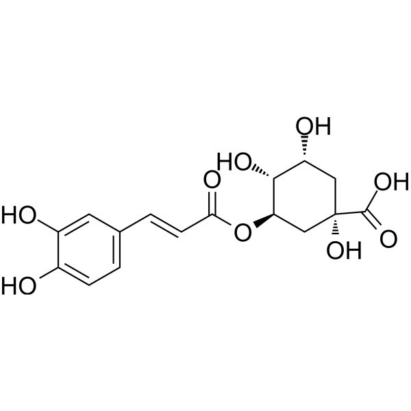
HY-N0055
Chlorogenic acidAntioxidant -

HY-B0464R
Hydralazine hydrochloride (Standard)Antihypertensive Agent -

HY-N0055R
Chlorogenic acid (Standard)Antioxidant -
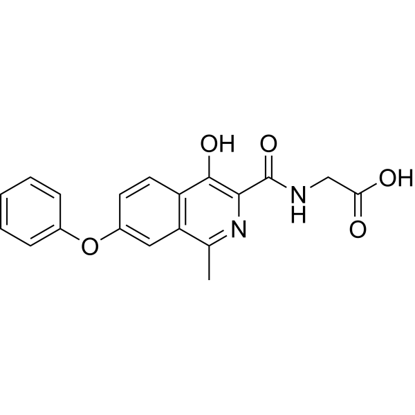
HY-13426
RoxadustatHIF-PH Inhibitor -
HY-N0293
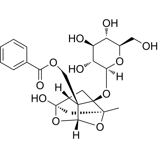
PaeoniflorinHSP Activator -
HY-125840
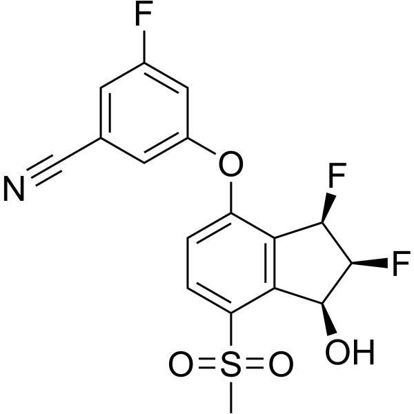
BelzutifanHIF-2α Inhibitor -
HY-13671
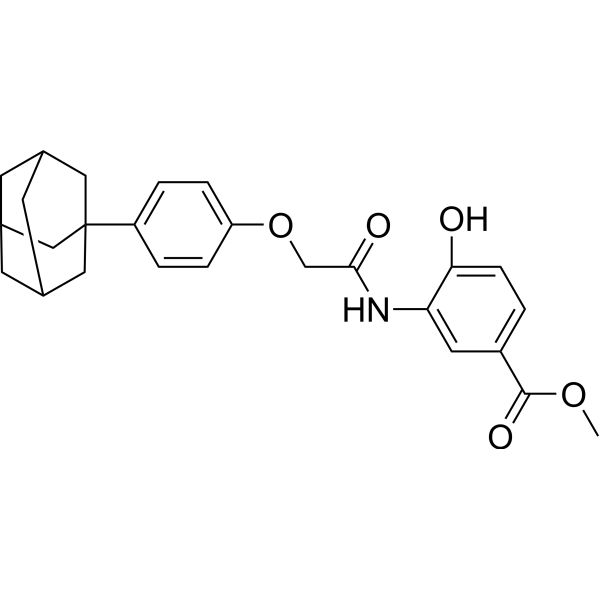
LW6Apoptosis Inducer
Publications Citing Use of MedChemExpress (MCE) Hydralazine hydrochloride
More Customer Validation & Images

-
WB
All DNA Methyltransferase Isoforms
More

Biological Activity
Description
IC50 & Target
|
Bcl-2 |
Bcl-xL |
Caspase-8 |
Caspase-9 |
Caspase 3 |
Cellular Effect
|
Cell Line

|
Type | Value | Description | References |
|---|---|---|---|---|
| HepG2 | EC50 |
0.45 μM
Compound: Hydralazine Hydrochloride
|
Activation of rat PXR expressed in human HepG2 cells after 24 hrs by luciferase reporter gene based luminescent analysis
Activation of rat PXR expressed in human HepG2 cells after 24 hrs by luciferase reporter gene based luminescent analysis
|
[PMID: 20966043] |
In Vitro
Hydralazine hydrochloride (10 μM; 5 days) demethylates and reactivates expression of silenced tumor suppressor genes (ER, RARβ, p16) in MDA-231, MCF-7, and T24 cancer cell lines, respectively[1].
Hydralazine hydrochloride (10 μM; 24 hours) reduces DNMT1 and DNMT3a mRNA expression in MCF-7 breast cancer cells[1].
Hydralazine hydrochloride (0.1-10.0 mM) does not impair phagocytic function or viability of thioglycollate-prestimulated rat peritoneal macrophages[2].
Hydralazine hydrochloride (0.1-10.0 mM) significantly inhibits nitrite production by LPS/IFN-γ-stimulated thioglycollate-prestimulated rat peritoneal macrophages, with an IC50 of 0.43 ± 0.03 mM[2].
Hydralazine hydrochloride (1.0-10.0 mM) inhibits both NOS-2 and COX-2 protein synthesis in LPS/IFNγ-stimulated thioglycollate-prestimulated rat peritoneal macrophages[2].
Hydralazine hydrochloride (3.1-50 nmol, plate incorporation test without S-9 mix; 1 mg per plate, spot test with/without S-9 mix) is a direct-acting, low-potency mutagen that induces mixed genetic mutation mechanisms in Salmonella typhimurium strains TA1535, TA100, TA1537, TA97, and TA98, with highest potency in strain TA97[3].
Hydralazine hydrochloride (5 mg per spot, spot test; serial twofold dilutions, micromethod assay with/without S-9 mix; 16 h incubation at 37°C) is a direct-acting genotoxin that is preferentially lethal to repair-deficient Escherichia coli strains WP67 and CM871, indicating involvement of recA recombination repair, lexA post-replication repair, and polA repair mechanisms, with no dependence on excision repair systems[3].
Hydralazine hydrochloride (≤25 μM) significantly mitigates acrolein-mediated cell death in PC12 cells[4].
MedChemExpress (MCE) has not independently confirmed the accuracy of these methods. They are for reference only. Further protocols information, click here.
-
Cell Line:Human peritoneal mesothelial cell (HPMC)
-
Concentration:3μg/mL, 6 μg/mL, 12 μg/mL, 25 μg/mL, 50 μg/mL, 100 μg/mL
-
Incubation Time:5 days
-
Result:Caused a dose-dependent inhibition of HPMC growth.
-
Cell Line:Jurkat, CEM-6, MOLT-4
-
Concentration:40 μM, 80 μM, 200 μM, 400 μM, 600 μM
-
Incubation Time:16 h, 24 h, 48 h
-
Result:Observed significant apoptosis in Jurkat and MOLT-4 cells at concentrations of 200 μM.
Induced a slight decrease in DNMT1 protein expression at 24 hr and nearly a complete loss after 48 hr.
Observed proteolytic processing of the initiator caspases-8 and -9, as well as cleavage of the effector caspase-3.
Resulted in generation of ROS and disruption of ΔΨm in Jurkat cells.
Observed that overexpression of Bcl-2 and Bcl-xL proteins prevented cell death.
In Vivo
Hydralazine hydrochloride (used in combination with valproic acid (HY-10585)) eliminates tumor recurrence of HT1080 fibrosarcoma xenografts in nude mice[1].
Hydralazine (83 mg/kg; i.p.; once daily; 5 days) hydrochloride induces DNA fragmentation in the liver, kidney and spleen (but not the lung) of mice at 6 hours after the last administration, and the damage is completely repaired at 12 hours post-administration; meanwhile, it moderately increases the sister chromatid exchange rate of mouse bone marrow cells by 33%[3].
Hydralazine (5 mg/kg; i.p.; twice; 14 d) hydrochloride reduces acrolein levels by 50-70% in the injured spinal cord of rats, decreases post-injury cyst formation by 70%, improves motor function recovery, and alleviates mechanical hyperalgesia in rat models of spinal cord injury[4].
Hydralazine (0.1-10 mg/kg; i.p.; single dose) hydrochloride dose-dependently alleviates formalin-induced somatic and emotional inflammatory pain in male C57BL/6 mice, with a ED50 of 0.239-1.0160 mg/kg, while inhibiting excessive acrolein production and neuronal activation in the spinal cord[5].
Hydralazine (10 mg/kg; i.p.; single dose) hydrochloride does not impair central nervous system function in male C57BL/6 mice[5].
MedChemExpress (MCE) has not independently confirmed the accuracy of these methods. They are for reference only.
-
Animal Model:Spinal cord injury mice[4]
-
Dosage:5 mg/kg
-
Administration:Intraperitoneal injection (i.p.)
-
Result:Improved not only motor function and hypersensitivity but also spontaneous pain and emotion response in SCI mice.
Inhibited both of BMDM recruitment and acrolein accumulation in the lesion epicenter of spinal cord in mice.
-
Animal Model:Spontaneously hypertensive rats (SHR)[5]
-
Dosage:14 mg/kg
-
Administration:Supplemented in drinking water
-
Result:Reduced systolic blood pressure values and the number of adherent and migrating leukocytes in SHR.
Reduce the number of adherent and migrating leukocytes in SHR.
Decreased the levels of ICAM-1 mRNA.


Chemical Information
-
CAS No. 304-20-1
-
Appearance Solid
-
Molecular Weight 196.64
-
Formula C8H9ClN4
-
Color White to off-white
-
SMILES
NNC1=NN=CC2=C1C=CC=C2.Cl
-
Shipping
Room temperature in continental US; may vary elsewhere.
-
Storage
4°C, sealed storage, away from moisture
* In solvent : -80°C, 6 months; -20°C, 1 month (sealed storage, away from moisture)


Publications (4)
-
Journal Impact Factor
-
Most Recent
-
J Pharm Anal
Hydralazine represses Fpn ubiquitination to rescue injured neurons via competitive binding to UBA52. [Abstract]2024 Jan;14(1):86-99. PMID: 38352945 -
Neurosci Bull
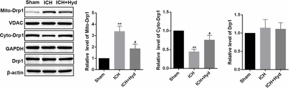
Acrolein Aggravates Secondary Brain Injury After Intracerebral Hemorrhage Through Drp1-Mediated Mitochondrial Oxidative Damage in Mice. [Abstract]2020 Oct;36(10):1158-1170. PMID: 32436179
Hydralazine hydrochloride purchased from MedChemExpress. Usage Cited in: Neurosci Bull. 2020 Oct;36(10):1158-1170. [Abstract]
Western blots (left) and analysis (right) of cyto-Drp1, mito-Drp1, and T-Drp1 in different groups of mice. Hydralazine (acrolein scavenger) suppresses the translocation of Drp1 and alleviates the morphological disruption of mitochondria after ICH in mice.
-
-


Solvent & Solubility
In Vitro:
H2O : 25 mg/mL (127.14 mM; Need ultrasonic)
Please refer to the solubility information to select the appropriate solvent. Once prepared, please aliquot and store the solution to prevent product inactivation from repeated freeze-thaw cycles.
Storage method and period of stock solution: -80°C, 6 months; -20°C, 1 month (sealed storage, away from moisture). When stored at -80°C, please use it within 6 months. When stored at -20°C, please use it within 1 month.
* Note: If you choose water as the stock solution, please dilute it to the working solution, then filter and sterilize it with a 0.22 μm filter before use.
Please refer to the solubility information to select the appropriate solvent. Once prepared, please aliquot and store the solution to prevent product inactivation from repeated freeze-thaw cycles.
Storage method and period of stock solution: -80°C, 6 months; -20°C, 1 month (sealed storage, away from moisture). When stored at -80°C, please use it within 6 months. When stored at -20°C, please use it within 1 month.
* Note: If you choose water as the stock solution, please dilute it to the working solution, then filter and sterilize it with a 0.22 μm filter before use.
Concentration (start) × Volume (start) = Concentration (final) × Volume (final)

Protocols
-
ROS/oxidative-stress fluorescent staining
ROS/oxidative-stress fluorescent staining uses cell-permeant fluorogenic probes that become fluorescent after oxidation inside cells or tissues; commonly used examples include DCFH-DA/DCFDA for broad cellular oxidant detection, DHE for superoxide-related signal detection, MitoSOX for mitochondrial superoxide-related signal detection, and CellROX probes for oxidative-stress-associated fluorescence readouts. The assay detects probe oxidation rather than a single ROS species unless the probe and analysis method have been chemically validated for that species. DCFH-DA enters cells, is deacetylated by intracellular esterases to DCFH, and produces fluorescent DCF after oxidation, so the readout is used as an operational measure of total cellular oxidative stress rather than a species-specific ROS measurement. DHE and MitoSOX can report superoxide-related oxidation, but red fluorescence alone can include non-specific ethidium-like oxidation products; HPLC or optimized spectral approaches are
-
Formalin-Induced Paw Inflammation/Nociceptive Inflammation
The formalin-induced paw inflammation/nociceptive test is a chemical persistent pain model in rodents in which subcutaneous injection of formalin into the hind paw produces spontaneous nocifensive behaviors such as flinching and licking. The response is classically biphasic, consisting of an early acute phase (Phase I) reflecting direct activation of peripheral nociceptors (particularly C-fiber afferents), followed by a later prolonged phase (Phase II) associated with central sensitization in the spinal dorsal horn driven by sustained afferent input and inflammatory signaling. This model is widely used to evaluate analgesic and anti-inflammatory interventions because it captures both peripheral nociception and central sensitization processes within a single assay system.
-
Research Protocol for Cardiovascular Diseases
Cardiovascular disease can be modeled as maladaptive cardiac remodeling, where ischemic injury or pressure overload activates inflammatory signaling, fibroblast activation, extracellular-matrix deposition, cardiomyocyte hypertrophy, vascular remodeling, and progressive ventricular dysfunction. The TGF-β/SMAD axis is a central profibrotic pathway after myocardial injury and pressure overload, while innate immune and cytokine pathways regulate leukocyte recruitment, scar formation, and adverse remodeling. Key unresolved questions include which inflammatory signals are reparative versus harmful, when fibrosis is protective versus maladaptive, and whether pathway inhibition improves function without weakening necessary infarct healing or compensatory remodeling.
-
Research Protocol for Inflammation-related Diseases
The NLRP3 inflammasome is a cytosolic innate immune signaling platform that integrates priming signals and danger-signal activation to promote caspase-1 activation, maturation of IL-1β and IL-18, and gasdermin D-mediated pyroptotic cell death. The core experimental logic is to determine whether inflammatory disease phenotypes are driven by increased NLRP3 expression, ASC-containing inflammasome assembly, caspase-1 cleavage, GSDMD cleavage, and extracellular release of IL-1β/IL-18 rather than by nonspecific cell injury alone. The pathway is strongly linked to inflammation-related disease phenotypes because monosodium urate crystals activate NALP3/NLRP3 inflammasome signaling in gout-like crystal inflammation, cholesterol crystals activate NLRP3 inflammasomes in atherogenesis models, and DSS-induced intestinal inflammation has been reported to involve NLRP3 inflammasome activity. However, experimental colitis studies also show context-dependent protective effects of NLRP3 inflammasome co
-
Genotoxicity/Mutagenicity Study
The bacterial reverse mutation assay detects point mutations that restore amino-acid prototrophy in auxotrophic Salmonella typhimurium or Escherichia coli tester strains; after exposure to a test article, mutagenic activity is read out as an increased number of revertant colonies on minimal agar compared with the vehicle control. The assay uses tester strains with different mutation targets so that base-substitution and frameshift mutagens can be detected, and testing is performed with and without exogenous mammalian metabolic activation because some chemicals require biotransformation to become mutagenic.


Purity & Documentation
-
Data Sheet (286 KB)
-
SDS (421 KB)
- English - EN (421 KB)
- Français - FR (421 KB)
- Deutsch - DE (421 KB)
- Norwegian - NO (421 KB)
- Español - ES (421 KB)
- Swedish - SV (421 KB)
- Italian - IT (421 KB)
- Korean - KR (421 KB)
- Portuguese - PT (421 KB)
-
Handling Instructions (2659 KB)
References
[1]. Arce C, et al. Hydralazine target: from blood vessels to the epigenome. J Transl Med. 2006;4:10. Published 2006 Feb 28. [Content Brief]
[2]. Leiro JM, et al. Antioxidant activity and inhibitory effects of hydralazine on inducible NOS/COX-2 gene and protein expression in rat peritoneal macrophages. Int Immunopharmacol. 2004;4(2):163-177. [Content Brief]
[3]. de Flora S, et al. In vivo and in vitro genotoxicity of three antihypertensive hydrazine derivatives (hydralazine, dihydralazine, and endralazine). Environ Mutagen. 1982;4(5):605-619. [Content Brief]
[4]. Park J, et al. Neuroprotective role of hydralazine in rat spinal cord injury-attenuation of acrolein-mediated damage. J Neurochem. 2014;129(2):339-349. [Content Brief]
[5]. Bai L, et al. Attenuation of mouse somatic and emotional inflammatory pain by hydralazine through scavenging acrolein and inhibiting neuronal activation. Pain Physician. 2012;15(4):311-326. [Content Brief]


Complete Stock Solution Preparation Table
Please refer to the solubility information to select the appropriate solvent. Once prepared, please aliquot and store the solution to prevent product inactivation from repeated freeze-thaw cycles.
Storage method and period of stock solution: -80°C, 6 months; -20°C, 1 month (sealed storage, away from moisture). When stored at -80°C, please use it within 6 months. When stored at -20°C, please use it within 1 month.
| Optional Solvent | Concentration Solvent Mass | 1 mg | 5 mg | 10 mg | 25 mg |
|---|---|---|---|---|---|
| H2O | 1 mM | 5.0854 mL | 25.4272 mL | 50.8544 mL | 127.1359 mL |
| 5 mM | 1.0171 mL | 5.0854 mL | 10.1709 mL | 25.4272 mL | |
| 10 mM | 0.5085 mL | 2.5427 mL | 5.0854 mL | 12.7136 mL | |
| 15 mM | 0.3390 mL | 1.6951 mL | 3.3903 mL | 8.4757 mL | |
| 20 mM | 0.2543 mL | 1.2714 mL | 2.5427 mL | 6.3568 mL | |
| 25 mM | 0.2034 mL | 1.0171 mL | 2.0342 mL | 5.0854 mL | |
| 30 mM | 0.1695 mL | 0.8476 mL | 1.6951 mL | 4.2379 mL | |
| 40 mM | 0.1271 mL | 0.6357 mL | 1.2714 mL | 3.1784 mL | |
| 50 mM | 0.1017 mL | 0.5085 mL | 1.0171 mL | 2.5427 mL | |
| 60 mM | 0.0848 mL | 0.4238 mL | 0.8476 mL | 2.1189 mL | |
| 80 mM | 0.0636 mL | 0.3178 mL | 0.6357 mL | 1.5892 mL | |
| 100 mM | 0.0509 mL | 0.2543 mL | 0.5085 mL | 1.2714 mL |
* Note: If you choose water as the stock solution, please dilute it to the working solution, then filter and sterilize it with a 0.22 μm filter before use.


Hydralazine hydrochloride Related Classifications
HY-B0464 Related Classifications
 Powered by Bioz