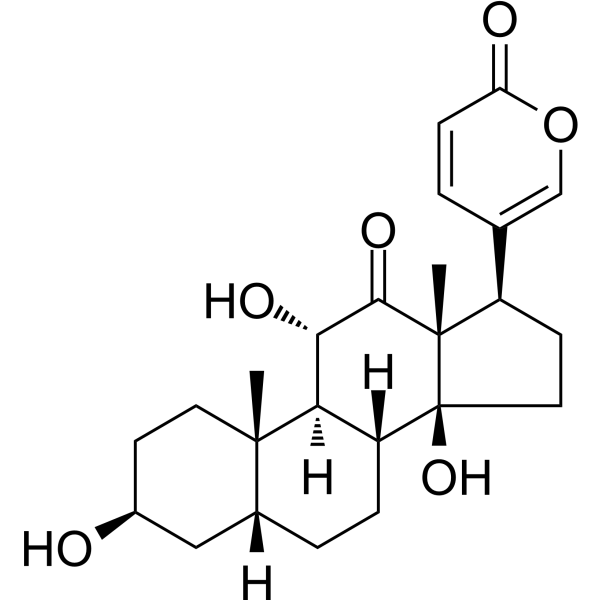
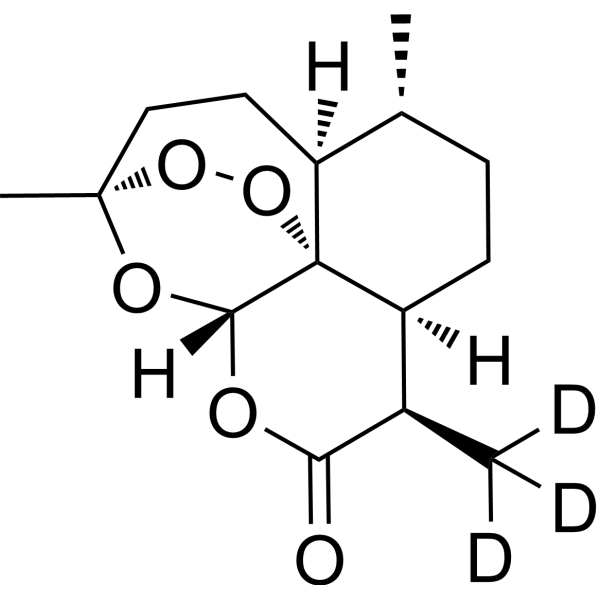
Alborixin
Alborixin is a polycyclic polyether ionophore Antibiotic. Alborixin is isolated from cultures of Streptomyces albus. Alborixin induces Autophagy via PTEN-mediated inhibition of the AKT pathway, thereby clearing Amyloid-β. Alborixin exhibits activity against Gram-positive bacteria and fungi. Alborixin can be used in the research of Alzheimer's disease.
For research use only. We do not sell to patients.
- CAS No.: 57760-36-8
- Formula: C48H84O14
- Molecular Weight:885.17
-
Storage:
Please store the product under the recommended conditions in the Certificate of Analysis.
All Antibiotic Isoforms
More
Biological Activity
Description
In Vitro
Alborixin (30-125 nM; 3-24 h) induces autophagy in mouse microglial N9 cells, with the maximal effect observed after treatment with 125 nM for 24 h; its mechanism of action involves upregulating LC3B-II levels, downregulating SQSTM1 levels, promoting GFP-LC3 puncta formation and increasing the number of autophagosomes[1].
Alborixin (125 nM) enhances soluble and fibrillar Aβ-induced autophagy in mouse microglial N9 cells, as evidenced by increased LC3B-II levels and decreased SQSTM1 levels[1].
Alborixin (125-250 nM; 12-24 h) eliminates soluble Aβ and fibrillary Aβ in mouse microglial N9 cells and human microglial HMC3 cells, and clears soluble Aβ in primary mouse neurons at 250 nM for 24 h. Its mechanism of action relies on the PTEN-AKT-dependent autophagy pathway, which requires functional ATG5 and BECN1[1].
Alborixin exhibits activity against Gram-positive bacteria and fungi[1].
MedChemExpress (MCE) has not independently confirmed the accuracy of these methods. They are for reference only. Further protocols information, click here.
-
Cell Line:mouse microglial N9 cells
-
Concentration:30 nM; 125 nM
-
Incubation Time:12 h; 3 h, 6 h, 12 h, 24 h
-
Result:Induced autophagy in a concentration-dependent manner, with increased LC3B-II levels and decreased SQSTM1 levels detectable at 30 nM for 12 h.
Increased LC3B-II:ACTB ratio in a time-dependent manner at 125 nM for 3 h, 6 h, 12 h, 24 h, while decreasing SQSTM1:ACTB ratio.
Confirmed autophagy flux via further increases in LC3B-II levels when combined with bafilomycin A1.
Increased GFP-LC3 puncta per cell.
Increased average autophagosomes per cell.
-
Cell Line:mouse microglial N9 cells
-
Concentration:125 nM
-
Incubation Time:3 h, 6 h, 9 h, 12 h, 24 h
-
Result:Upregulated autophagy-related proteins BECN1, ATG7, ATG5, and ATG12 in a time-dependent manner, with peak expression at 24 h.
Increased PTEN:ACTB ratio in a time-dependent manner.
Reduced the ratios of p-AKT (S473):AKT, p-AKT (T308):AKT, p-MTOR (S2448):MTOR, and RPTOR:ACTB over 24 h.
Chemical Information
-
CAS No. 57760-36-8
-
Molecular Weight 885.17
-
Formula C48H84O14
-
SMILES
O[C@@]1([C@]2(O[C@](CC2)([H])[C@H]([C@@]3(O[C@@H]([C@H](C[C@H]3C)C)C[C@]4(O[C@@](CC[C@@H]4C)([H])[C@@H](C)[C@H](O)C[C@@H]5O[C@]([C@H](C[C@H]5C)C)([H])[C@@H](C)C(O)=O)O)O)O)C)O[C@@](C[C@H]1C)([C@]6([H])O[C@H]([C@](O)(CC6)C)CC)C
-
Structure Classification
-
Initial Source
streptomyces scabrisporus
-
Shipping
Room temperature in continental US; may vary elsewhere.
-
Storage
Please store the product under the recommended conditions in the Certificate of Analysis.
Protocols
-
Autophagy
Autophagy is a process in which eukaryotic cells use lysosomes to degrade their own cytoplasmic proteins and damaged organelles under the regulation of autophagy related gene (Atg). Microtubule-associated proteins light chain 3 (LC3) is recognized as autophagy marker, which transfers from cytoplasmic LC3 (LC3-I) to membrane type (LC3-II). LC3-II/I ratio could be detected by Western Blot and fluorescence microscopy.
-
Lysosome and acidic-vesicle live-cell staining
Lysosome and acidic-vesicle live-cell staining detects acidic intracellular compartments by using membrane-permeant acidotropic probes that accumulate in low-pH vesicles, including lysosomes, late endosomes, autolysosomes, and acidic phagosomes. LysoTracker staining is commonly used as an intensity-based readout of acidic lysosomal compartment abundance or enlargement, while acridine orange produces green fluorescence in less concentrated compartments and red fluorescence after concentration-dependent accumulation in acidic vesicular organelles. Loss or reduction of acridine-orange red signal can be used as a readout of lysosomal membrane permeabilization or reduced acidic-vesicle integrity. This protocol is designed for live cultured cells and can be adapted for fluorescence microscopy, high-content imaging, plate-reader readout, or flow cytometry when the selected literature supports the readout. Because these dyes report acidotropic accumulation rather than lysosome identity alone,
-
Gram Staining of Tissue Sections
Gram staining of tissue sections is a histochemical technique used to differentiate Gram-positive and Gram-negative bacteria within histological specimens based on differences in bacterial cell wall structure and dye retention, adapted from classical bacteriological Gram staining into tissue-compatible “histological Gram stain” variants. In tissue applications, modifications of the Brown-Hopps and Brown-Brenn methods are commonly used to improve differentiation of microorganisms embedded within host connective tissue and to reduce overstaining or loss of Gram-negative signal, which are known limitations of earlier approaches. The principle relies on crystal violet-iodine complex retention in Gram-positive organisms and subsequent decolorization and counterstaining steps that allow contrast visualization of Gram-negative organisms against tissue background.
-
Macroautophagy Solutions
Macroautophagy is a conserved lysosome-dependent degradation pathway in which cytoplasmic material is sequestered into double-membrane autophagosomes and delivered to lysosomes for degradation and recycling. The pathway supports cellular homeostasis during nutrient limitation, organelle stress, protein-aggregate accumulation, infection, differentiation, and tissue remodeling by coupling cargo sequestration, autophagosome maturation, lysosomal fusion, and degradation of cargo-derived macromolecules. The core molecular sequence includes initiation by nutrient- and stress-regulated autophagy machinery, autophagosome nucleation, LC3/ATG8-family conjugation to autophagosomal membranes, cargo selection through receptors such as SQSTM1/p62, autophagosome-lysosome fusion, and lysosomal degradation. LC3 was identified as a mammalian homolog of yeast Atg8 that localizes to autophagosomal membranes after processing, and p62/SQSTM1 was shown to connect ubiquitinated cargo with autophagic degradati
-
Amyloid: Congo Red Amyloid Staining
Congo red amyloid staining is a histochemical method used to detect extracellular amyloid deposits in tissue sections based on the affinity of Congo red dye for β-pleated sheet-rich protein aggregates. When bound to amyloid, Congo red produces characteristic apple-green birefringence under polarized light microscopy, which is widely regarded as a diagnostic feature of amyloid deposition in histopathology. The diagnostic principle relies on the combination of dye binding (congophilia) and optical anisotropy under polarized illumination, which distinguishes amyloid from most non-amyloid eosinophilic extracellular deposits in routine histological evaluation. Amyloid identification by Congo red staining remains a cornerstone in diagnostic pathology despite the availability of adjunct methods such as immunohistochemistry and mass spectrometry, particularly because of its ability to localize deposits directly within tissue architecture. The specificity of Congo red-positive deposits is incre
-
Alzheimer’s Disease Modeling
Alzheimer’s Disease (AD) is a neurodegenerative disorder characterized by a progressive decline in cognitive functions and loss of specific types of neurons and synapses. Alzheimer's symptoms can be simulated in mice by injecting drugs (such as Aβ) or genetically modified.
Purity & Documentation
References
[1]. Wani A, et al. Alborixin clears amyloid-β by inducing autophagy through PTEN-mediated inhibition of the AKT pathway. Autophagy. 2019;15(10):1810-1828. [Content Brief]
[2]. Gachon P, et al. Alborixin, a new antibiotic ionophore: isolation, structure, physical and chemical properties. J Antibiot (Tokyo). 1976;29(6):603-610. [Content Brief]
Calculators
Concentration (start) × Volume (start) = Concentration (final) × Volume (final)