



Anti-Mouse TIGIT Antibody (1G9)

Based on 1 Customer Validation
Anti-Mouse TIGIT Antibody (1G9) is a mouse-derived agonistic TIGIT IgG1 κ type antibody. Anti-Mouse TIGIT Antibody (1G9) blocks CD155 binding to TIGIT. Anti-Mouse TIGIT Antibody (1G9) shows potent anti-infection and anti-immune effects in septic and experimental autoimmune encephalomyelitis.
For research use only. We do not sell to patients.
- Purity: 99.71%
- Molecular Weight:150 kDa
-
Storage:
Please store the product under the recommended conditions in the Certificate of Analysis.

To place orders, for customer services and technical support, please contact: MedChemExpress USA
Tel: 609-228-6898 E-mail: [email protected] [email protected]


-
 Biological Activity
-
Chemical Information
- Purity & Documentation
- References
-
 Help & FAQs




Biological Activity
Description
Isotype
Mouse IgG1 kappa
Recommend Isotype Controls
Species Reactivity
Mouse
IC50 & Target
TIGIT
In Vitro
Anti-Mouse TIGIT Antibody (1G9) (30 mins) blocks CD155 binding to TIGIT in P815 cells[3].
MedChemExpress (MCE) has not independently confirmed the accuracy of these methods. They are for reference only.
In Vivo
Anti-Mouse TIGIT Antibody (1G9) (400 μg/mouse, s.c., 1 h after cecal ligation and puncture) mitigates lymphocyte depletion and sepsis mortality in cancer septic mice [2].
Anti-Mouse TIGIT Antibody (1G9) (100 μg, i.p., at days 0, 2, and 4, 10 and 17) reduces severity in experimental autoimmune encephalomyelitis mice models[3].
MedChemExpress (MCE) has not independently confirmed the accuracy of these methods. They are for reference only.
-
Animal Model:C57BL/6 mice were infected with lymphocytic choriomeningitis clone 13[1]
-
Dosage:100 μg
-
Administration:Intraperitoneally injection, at days 0, 2, 4, 10, 17, and 24
-
Result:Showed significantly higher expression of IL-10-Thy1.1.
Reduced production of IFN-γ, activation markers and pro-inflammatory cytokine.
Had no weight loss.
Decreased the scores of portal vein inflammation and central vein infiltration.
Showed a markedly increased frequency of IL-10-producing T cells in the liver.
Shifted the ratio of IL-10+ to IFN-γ+ cells toward an anti-inflammatory cytokine profile.
-
Animal Model:Cancer mice inoculated with lung carcinoma cells were made septic by cecal ligation and puncture[2]
-
Dosage:400 μg/mouse
-
Administration:Subcutaneously injection, 1 h after cecal ligation and puncture
-
Result:Improved survival of mice.
Increased the CD4+ and CD8+ T cell and B cell compartments.
Reduced the frequency of PD-1+ cells within the CD4+, CD8+, and Foxp3+ Treg compartments.
Inhibited lymphocyte apoptosis.
-
Animal Model:Experimental autoimmune encephalomyelitis (EAE) mice models[3]
-
Dosage:100 μg
-
Administration:Intraperitoneally injection, at days 0, 2, and 4, 10 and 17
-
Result:Reduced in the secretion of IFN-γ and IL-17.
Reduced EAE severity.
Gene ID
Accession
NP_001139797
Conjugated
Unconjugated
Reconsititution
The product can be reconstituted/diluted with sterile PBS or saline.
Format
-
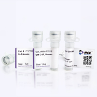
Product Image
Application
in vivo TIGIT stimulation; Flow cytometry


Chemical Information
-
Appearance Liquid
-
Molecular Weight 150 kDa
-
Color Colorless to light yellow
-
SMILES
[Anti-Mouse TIGIT Antibody (1G9)]
-
Formulation
Please refer to the lot-specific COA for specific buffer information.
-
Storage
Please store the product under the recommended conditions in the Certificate of Analysis.


Purity & Documentation
-
Data Sheet (262 KB)
-
SDS (251 KB)
- English - EN (251 KB)
- Français - FR (251 KB)
- Deutsch - DE (251 KB)
- Norwegian - NO (251 KB)
- Español - ES (251 KB)
- Swedish - SV (251 KB)
- Italian - IT (251 KB)
- Korean - KR (251 KB)
- Portuguese - PT (251 KB)
-
Inhibitory Antibodies User Guide (603 KB)
References
[1]. Schorer M, et al. TIGIT limits immune pathology during viral infections. Nat Commun. 2020 Mar 9;11(1):1288. [Content Brief]
[2]. Zhang W, et al. TIGIT modulates sepsis-induced immune dysregulation in mice with preexisting malignancy. JCI Insight. 2021 Jun 8;6(11):e139823. [Content Brief]
[3]. Dixon KO, et al. Functional Anti-TIGIT Antibodies Regulate Development of Autoimmunity and Antitumor Immunity. J Immunol. 2018 Apr 15;200(8):3000-3007. [Content Brief]

Calculators
Concentration (start) × Volume (start) = Concentration (final) × Volume (final)


Anti-Mouse TIGIT Antibody (1G9) Related Classifications
HY-P990155 Related Classifications
 Powered by Bioz