



Aurora A Antibody (YA859)
(Synonyms: Aurora A)Based on 1 publication(s) in Google Scholar
Aurora A Antibody (YA859) is a Rabbit-derived and non-conjugated monoclonal antibody, targeting to Aurora A.
-
Host:
Rabbit
-
Application:
WB, ICC/IF, IHC-P, FC
-
Reactivity :
Human, Mouse, Rat
-
Formulation:
Supplied in 1*TBS (pH7.4), 0.05% BSA, 40% Glycerol. Preservative: 0.05% Sodium Azide.
-
Conjugation:
Non-conjugated


To place orders, for customer services and technical support, please contact: MedChemExpress USA
Tel: 609-228-6898 E-mail: [email protected] [email protected]



Publications Citing Use of MedChemExpress (MCE) Aurora A Antibody (YA859)
More

Applications
| Application |
WB

WB: Western Blot
|
ICC/IF
ICC/IF: Immunocytochemistry/
Immunofluorescence |
IHC-P
IHC-P: Immunohistochemistry-Paraffin
|
FC
FC: Flow Cytometry
|
|---|---|---|---|---|
| Dilution Ratio | 1:500-1:2000 | 1:50-1:100 | 1:100-1:500 | 1:50-1:100 |


Product Details
Aurora A Antibody (YA859) is a Rabbit-derived and non-conjugated monoclonal antibody, targeting to Aurora A.
-
Host Rabbit

-
Clonality Recombinant,Monoclonal

-
Species ReactivityHuman, Mouse, Rat
-
Observed Molecular WeightObserved band size: 46 kDa
Note: Due to possible protein modifications or aggregation, the molecular weight should be confirmed by actual measurement, and the predicted value is for reference only.

-
Calculated Molecular Weight Predicted band size: 46 kDa

Endogenous
Protein A affinity purified.
Non-conjugated
Unmodified


Product Properties
-
Appearance
Solution
-
Formulation
Supplied in 1*TBS (pH7.4), 0.05% BSA, 40% Glycerol. Preservative: 0.05% Sodium Azide.
-
Concentration
Batch-dependent, Please check the COA for the concentration of each lot. Check Lot Concentration
-
Storage & Stability
Stored at -20°C for 1 year. Avoid repeated freeze / thaw cycles.
-
Shipping
Shipping with blue ice.


Publications (1)
-
Journal Impact Factor
-
Most Recent
-
Cell Death Discov
AURKA/PHB2 signaling drives acquired resistance to KRAS G12C inhibitors in KRAS G12C-mutant NSCLC. [Abstract]2026 Apr 25. PMID: 42031711


Verification Images
-
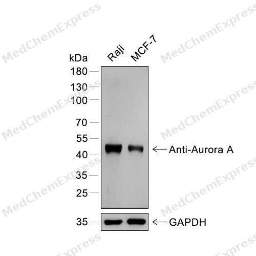 Western blot analysis of extracts from Raji (lane 1) and MCF-7 (lane 2) using Aurora A antibody. Proteins were transferred to a PVDF membrane and blocked with 5% nonfat powdered milk in PBST for 2 hour at room temperature. The primary antibody (1/1000) and loading control antibody (GAPDH, 1/3000) was diluted with 5% nonfat powdered milk in PBST at 4°C overnight. Goat Anti-Rabbit IgG-HRP Secondary Antibody (1/8,000) was incubated for 45min at room temperature.
 -
 Flow cytometric analysis of 1X10⁶ HeLa cells labeling Aurora A Antibody (HY-P80971, red). Cells were fixed with 4% paraformaldehyde and permeabilised with 90% methanol. Then stained with the primary antibody at 1/100 dilution for an hour at 4℃. AF488-conjugated Goat Anti-Rabbit IgG H&L (HY-P8002) was used as the secondary antibody at 1/1,000 dilution for 30 minutes at 4℃. Rabbit IgG Isotype Control (HY-P80879, blue) was used as the isotype control, cells without incubation with primary antibody were used as the unlabeled control (black).
 -
 Immunocytochemistry analysis of Hela cells labeling Aurora A with Aurora A Antibody (HY-P80971) at 1/50 dilution. Cells were fixed in 4% paraformaldehyde for 15 minutes at room temperature, permeabilized with 0.1% Triton X-100 in PBS for 15 minutes at room temperature, then blocked with quick block buffer for 10 minutes at room temperature. Cells were then incubated with Aurora A Antibody (HY-P80971) at 1/50 dilution in quick block buffer overnight at 4 ℃. AF488-conjugated Goat Anti-Rabbit IgG H&L(HY-P8002, Green) was used as the secondary antibody at 1/1,000 dilution. PBS instead of the primary antibody was used as the secondary antibody only control. The Nuclear counterstain was DAPI (Blue).
 -
 Immunocytochemistry analysis of HepG2 cells labeling Aurora A with Aurora A Antibody (HY-P80971) at 1/50 dilution. Cells were fixed in 4% paraformaldehyde for 15 minutes at room temperature, permeabilized with 0.1% Triton X-100 in PBS for 15 minutes at room temperature, then blocked with quick block buffer for 10 minutes at room temperature. Cells were then incubated with Aurora A Antibody (HY-P80971) at 1/50 dilution in quick block buffer overnight at 4 ℃. AF488-conjugated Goat Anti-Rabbit IgG H&L(HY-P8002, Green) was used as the secondary antibody at 1/1,000 dilution. PBS instead of the primary antibody was used as the secondary antibody only control. The Nuclear counterstain was DAPI (Blue).


Background
-
Function
Aurora A kinase (AURKA) is a conserved serine/threonine kinase that primarily regulates centrosome maturation, centrosome separation, and bipolar spindle assembly during mitosis, thereby supporting accurate chromosome segregation and cell-cycle progression[1][2]. Mechanistically, AURKA functions within mitotic spindle regulatory networks and is activated through interactions with spindle-associated cofactors such as TPX2, which promotes spatial control of kinase activity during mitosis[3][4]. Through its role in microtubule organization and spindle formation, AURKA contributes to the maintenance of genomic stability, whereas dysregulated AURKA activity is associated with abnormal mitotic progression and chromosomal instability in cancer models[1][5]. In disease-relevant experimental systems, aberrant Aurora kinase signaling has been linked to tumor development and progression, supporting the use of AURKA as a molecular target in studies of mitotic control and oncogenic transformation[5][6]. Compared with related isoforms, Aurora B predominantly regulates chromosome-microtubule attachment, spindle checkpoint signaling, chromosome alignment, and cytokinesis as a core component of the chromosomal passenger complex, whereas AURKA mainly functions at centrosomes and spindle poles during early mitosis[1][2][3]. This spatial and functional distinction provides an important framework for isoform-selective experimental design and interpretation of mitotic phenotypes[1][3]. For research applications, Aurora kinase inhibitors are widely used to investigate mitotic signaling pathways and chromosome segregation mechanisms, and selective targeting strategies continue to be explored in anticancer research models[6].
-
Subcellular Localization
Cytoplasm, cytoskeleton, microtubule organizing center, centrosome; Cytoplasm, cytoskeleton, spindle pole; Cytoplasm, cytoskeleton, microtubule organizing center, centrosome, centriole; Cell projection, neuron projection; Cell projection, cilium; Cytoplasm, cytoskeleton, cilium basal body; Basolateral cell membrane
-
Expression
Tissue_specificity:Highly expressed in testis and weakly in skeletal muscle, thymus and spleen. Also highly expressed in colon, ovarian, prostate, neuroblastoma, breast and cervical cancer cell lines
Induction:Expression is cell-cycle regulated, low in G1/S, accumulates during G2/M, and decreases rapidly after -
Subunit
Part of a complex composed of NEDD9, AURKA and CTTN; within the complex NEDD9 acts as a scaffold protein and is required for complex formation (PubMed:24574519).
-
SwissProt ID
-
Synonyms
Aurora A
Documentation
-
Data Sheet (262 KB)
-
SDS (251 KB)
- English - EN (251 KB)
- Français - FR (251 KB)
- Deutsch - DE (251 KB)
- Norwegian - NO (251 KB)
- Español - ES (251 KB)
- Swedish - SV (251 KB)
- Italian - IT (251 KB)
- Korean - KR (251 KB)
- Portuguese - PT (251 KB)
-
User Guide for Antibodies (1077 KB)
References
[1]. Willems E, et al. The functional diversity of Aurora kinases: a comprehensive review. Cell Div. 2018 Sep 19;13:7. [Content Brief]
[3]. Carmena M, et al. Making the Auroras glow: regulation of Aurora A and B kinase function by interacting proteins. Curr Opin Cell Biol. 2009 Dec;21(6):796-805. [Content Brief]
[4]. Wikipedia.
[6]. Kitzen JJ, et al. Aurora kinase inhibitors. Crit Rev Oncol Hematol. 2010 Feb;73(2):99-110. [Content Brief]

 Powered by Bioz
