



ATP13A2 Antibody (YA6939)
(Synonyms: AT132_HUMAN antibody)
Based on 1 Customer Validation
ATP13A2 Antibody (YA6939) is a Rabbit-derived and non-conjugated IgG monoclonal antibody, targeting to ATP13A2.
-
Host:
Rabbit
-
Isotype:
IgG
-
Application:
WB, IHC-P, IHC-F
-
Reactivity :
Human, Mouse, Rat
-
Formulation:
Supplied in PBS (pH7.4), 0.1% BSA, 40% Glycerol. Preservative: 0.05% Sodium Azide.
-
Conjugation:
Non-conjugated


To place orders, for customer services and technical support, please contact: MedChemExpress USA
Tel: 609-228-6898 E-mail: [email protected] [email protected]





Applications
| Application |
WB

WB: Western Blot
|
IHC-P
IHC-P: Immunohistochemistry-Paraffin
|
IHC-F
IHC-F: Immunohistochemistry-Frozen
|
|---|---|---|---|
| Dilution Ratio | 1:1000 | 1:1000 | 1:500 |


Product Details
ATP13A2 Antibody (YA6939) is a Rabbit-derived and non-conjugated IgG monoclonal antibody, targeting to ATP13A2.
-
Host Rabbit

-
Clonality Recombinant,Monoclonal

-
Species ReactivityHuman, Mouse, Rat Predicted Reactivity: Cynomolgus monkey,Pig
Note: The predicted reactivity is for reference only and should not be considered a guarantee of product performance.
-
Observed Molecular WeightObserved band size: 160 kDa
Note: Due to possible protein modifications or aggregation, the molecular weight should be confirmed by actual measurement, and the predicted value is for reference only.

-
Calculated Molecular Weight Predicted band size:129 kDa

Recombinant protein within human ATP13A2 aa 485-900.
affinity purified.
Non-conjugated
Unmodified
IgG


Product Properties
-
Appearance
Liquid
-
Formulation
Supplied in PBS (pH7.4), 0.1% BSA, 40% Glycerol. Preservative: 0.05% Sodium Azide.
-
Concentration
Batch-dependent, Please check the COA for the concentration of each lot. Check Lot Concentration
-
Storage & Stability
Stored at -20°C for 1 year. Avoid repeated freeze / thaw cycles.
-
Shipping
Shipping with blue ice.


Verification Images
-
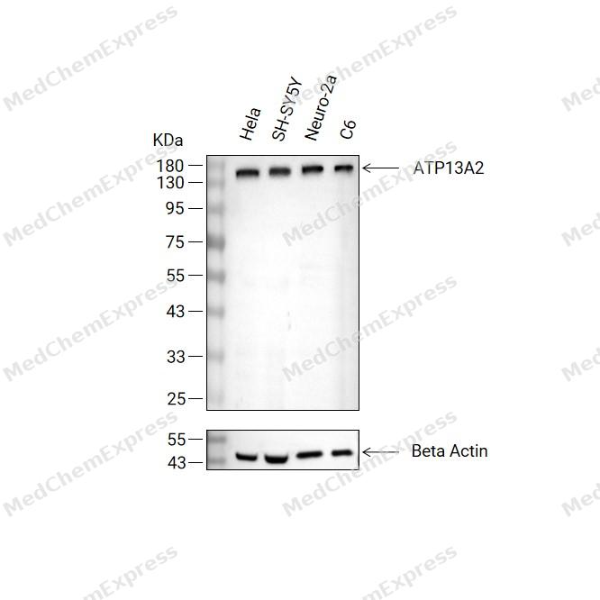 Western blot analysis of extracts from Hela(lane 2(20μg) , SH-SY5Y(lane 3(20μg) ,Neuro-2a(lane 4(20μg)and C6( lane 5(20μg) using ATP13A2 Antibody (HY-P87251). Proteins were transferred to a PVDF membrane and blocked with 5% non-fat milk in TBST for 2 hour at room temperature. The primary antibody (1/1000) and Loading control antibody (Beta Actin, HY-P80993,1/10000) was used in 5% non-fat milk in TBST at 4°C overnight. Goat Anti-Rabbit IgG-HRP Secondary Antibody (HY-P8001,1/10,000) was used for 1 hour at room temperature.
 -
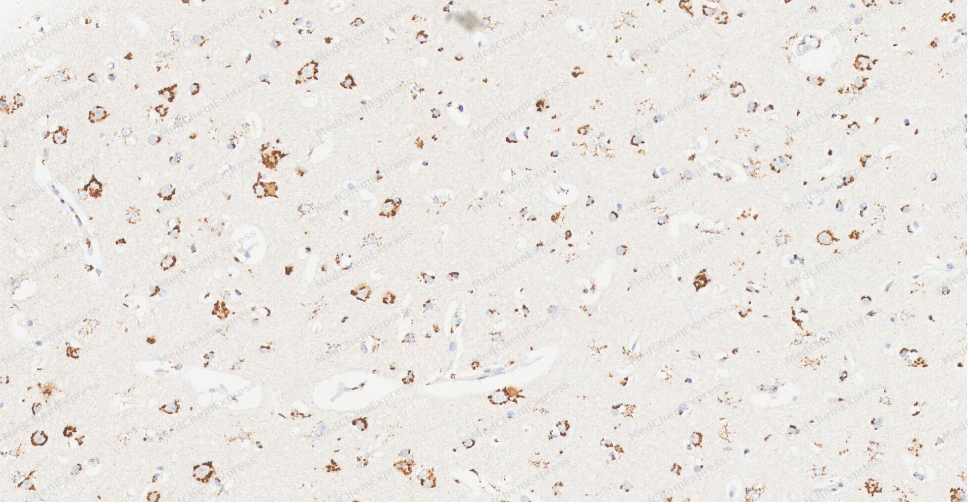 Immunohistochemical analysis of paraffin-embedded human Brain tissue using ATP13A2 antibody was performed. The section was pretreated using high-temperature and high-pressure mediated EDTA antigen retrieval buffer (pH 9.0), for 5 minutes. The tissues were incubated with primary antibody (HY-P87251, 1:1000 dilution) at room temperature for 60 minutes. Detection was performed using an HRP conjugated compact polymer system. DAB was used as the chromogen. The tissues were counterstained with hematoxylin and mounted with neutral balsam mounting medium.
 -
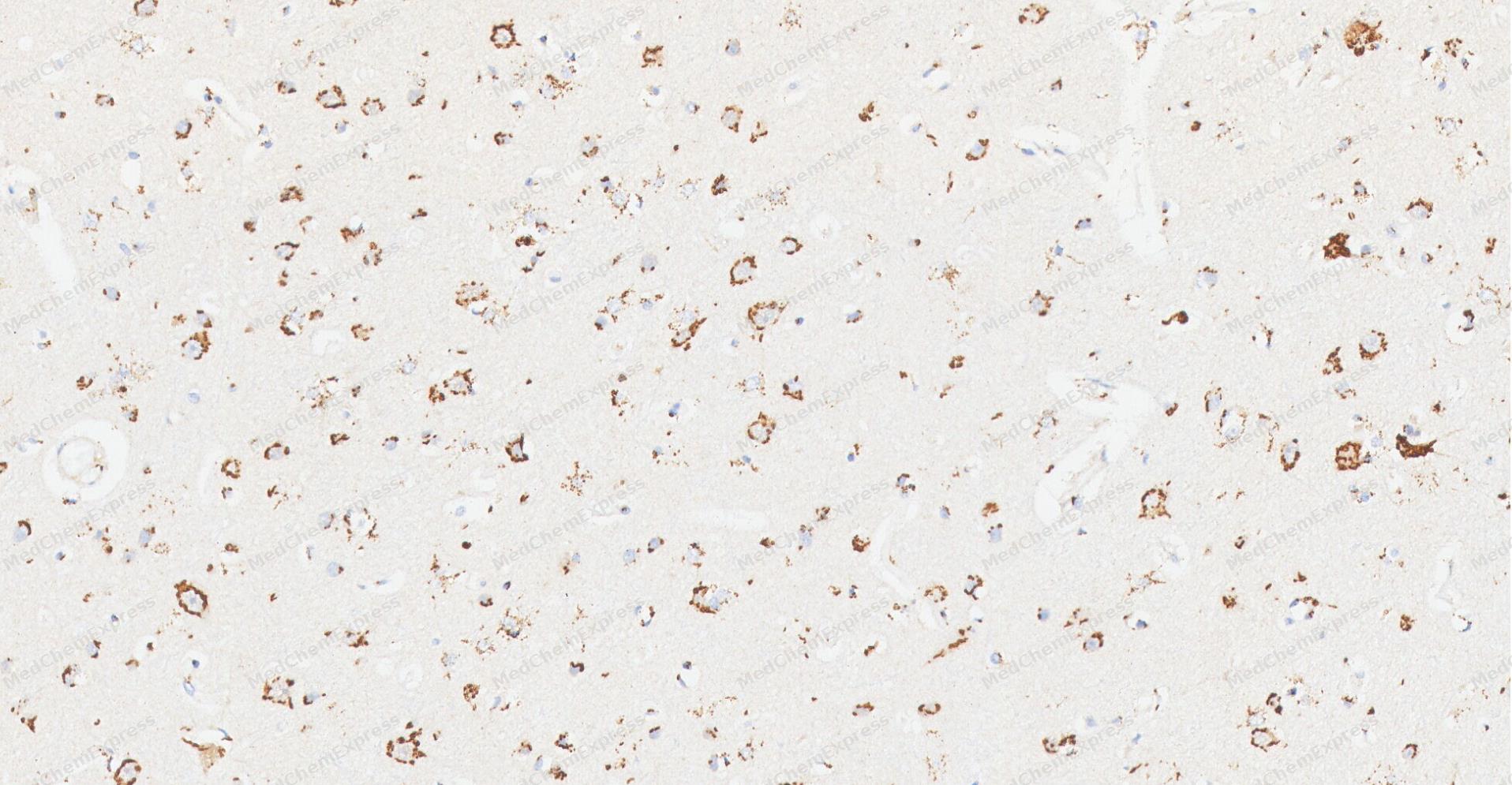 Immunohistochemical analysis of paraffin-embedded human Brain tissue using ATP13A2 antibody was performed. The section was pretreated using high-temperature and high-pressure mediated EDTA antigen retrieval buffer (pH 9.0), for 5 minutes. The tissues were incubated with primary antibody (HY-P87251, 1:1000 dilution) at room temperature for 60 minutes. Detection was performed using an HRP conjugated compact polymer system. DAB was used as the chromogen. The tissues were counterstained with hematoxylin and mounted with neutral balsam mounting medium.
 -
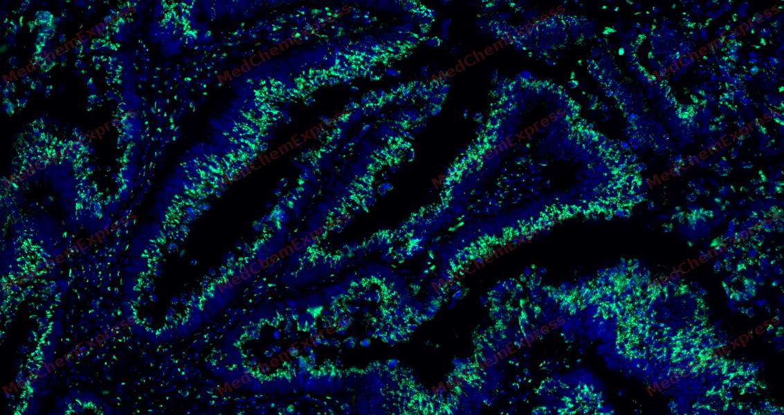 Tyramide signaling amplification based immunofluorescence was performed on paraffin-embedded human Colon cancer tissue using ATP13A2 antibody. Antigen retrieval was performed in EDTA buffer pH 9.0 (95 °C, 20 min) followed by cooling to RT. Then incubated with primary antibody (HY-P87251, 1:1500 dilution) at room temperature for 60 minutes and HRP conjugated secondary antibody for 10 minutes. Fluorescence was then developed with Vari Fluor 532 TSA (200×)(HY-D1832). The tissues were counterstained with DAPI and mounted with Anti-fade mounting medium.
 -
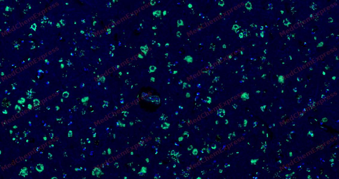 Tyramide signaling amplification based immunofluorescence was performed on paraffin-embedded human Brain tissue using ATP13A2 antibody. Antigen retrieval was performed in EDTA buffer pH 9.0 (95 °C, 20 min) followed by cooling to RT. Then incubated with primary antibody (HY-P87251, 1:1500 dilution) at room temperature for 60 minutes and HRP conjugated secondary antibody for 10 minutes. Fluorescence was then developed with Vari Fluor 532 TSA (200×)(HY-D1832). The tissues were counterstained with DAPI and mounted with Anti-fade mounting medium.
 -
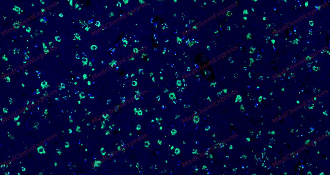 Tyramide signaling amplification based immunofluorescence was performed on paraffin-embedded human Brain tissue using ATP13A2 antibody. Antigen retrieval was performed in EDTA buffer pH 9.0 (95 °C, 20 min) followed by cooling to RT. Then incubated with primary antibody (HY-P87251, 1:1500 dilution) at room temperature for 60 minutes and HRP conjugated secondary antibody for 10 minutes. Fluorescence was then developed with Vari Fluor 532 TSA (200×)(HY-D1832). The tissues were counterstained with DAPI and mounted with Anti-fade mounting medium.


Background
-
Function
ATP13A2 is an ATPase which acts as a lysosomal polyamine exporter with high affinity for spermine. Also stimulates cellular uptake of polyamines and protects against polyamine toxicity. Plays a role in intracellular cation homeostasis and the maintenance of neuronal integrity. Contributes to cellular zinc homeostasis. Confers cellular protection against Mn(2+) and Zn(2+) toxicity and mitochondrial stress. Required for proper lysosomal and mitochondrial maintenance. Regulates the autophagy-lysosome pathway through the control of SYT11 expression at both transcriptional and post-translational levels. Facilitates recruitment of deacetylase HDAC6 to lysosomes to deacetylate CTTN, leading to actin polymerization, promotion of autophagosome-lysosome fusion and completion of autophagy. Promotes secretion of exosomes as well as secretion of SCNA via exosomes. Plays a role in lipid homeostasis[1][2][3][4][5][6][7][8][9][10].
-
Subcellular Localization
Lysosome membrane,Late endosome membrane,Endosome, multivesicular body membrane,Cytoplasmic vesicle, autophagosome membrane
-
Expression
Tissue_Specificity: Expressed in brain; protein levels are markedly increased in brain from subjects with Parkinson disease and subjects with dementia with Lewy bodies. Detected in pyramidal neurons located throughout the cingulate cortex (at protein level). In the substantia nigra, it is found in neuromelanin-positive dopaminergic neurons (at protein level) -
Isoforms & Post-Translational Modification
Q9NQ11 has three isomers: Q9NQ11-1: 128794 Da (predicted); Q9NQ11-2: 125977 Da (predicted); Q9NQ11-3: 128323 Da (predicted).
Autophosphorylated (PubMed:26134396, PubMed:28137957) -
Subunit
Interacts with MYCBP2; the interaction inhibits the ubiquitination of TSC2 by MYCBP2 (PubMed:27278822)
-
SwissProt ID
-
Synonyms
AT132_HUMAN antibody


Documentation
-
Data Sheet (231 KB)
-
SDS (313 KB)
- English - EN (313 KB)
- Français - FR (313 KB)
- Deutsch - DE (313 KB)
- Norwegian - NO (313 KB)
- Español - ES (313 KB)
- Swedish - SV (313 KB)
- Italian - IT (313 KB)
- Korean - KR (313 KB)
- Portuguese - PT (313 KB)
-
User Guide for Antibodies (1077 KB)

 Powered by Bioz
