



Bcl-2 Antibody (YA824)
(Synonyms: BCL2; Apoptosis regulator Bcl-2)Based on 1 publication(s) in Google Scholar
Bcl-2 Antibody (YA824) is a Mouse-derived and non-conjugated IgG1 monoclonal antibody, targeting to Bcl-2.
-
Host:
Mouse
-
Isotype:
IgG
-
Application:
WB
-
Reactivity :
Human, Rat, Mouse
-
Formulation:
Supplied in 1*PBS (pH 7.3), 50% glycerol and 0.5% BSA. Preservative: 0.02% sodium azide.
-
Conjugation:
Non-conjugated


To place orders, for customer services and technical support, please contact: MedChemExpress USA
Tel: 609-228-6898 E-mail: [email protected] [email protected]




Publications Citing Use of MedChemExpress (MCE) Bcl-2 Antibody (YA824)
More

Applications
| Application |
WB

WB: Western Blot
|
|---|---|
| Dilution Ratio | 1:500-1:1000 |


Product Details
Bcl-2 Antibody (YA824) is a Mouse-derived and non-conjugated IgG1 monoclonal antibody, targeting to Bcl-2.
-
Host Mouse

-
Clonality Monoclonal

-
Species ReactivityHuman, Rat, Mouse
-
Observed Molecular WeightObserved band size: 26 kDa
Note: Due to possible protein modifications or aggregation, the molecular weight should be confirmed by actual measurement, and the predicted value is for reference only.

-
Calculated Molecular Weight Predicted band size: 26 kDa

Entrez Gene: 596 Human ; 12043 Mouse ; 24224 Rat
SwissProt: P10415 Human ; P10417 Mouse ; P49950 Rat
OMIM: 151430 Human
Synthetic peptide corresponding to Human Bcl-2 conjugated to KLH.The exact sequence is proprietary to MCE.
Endogenous
affinity purified
Non-conjugated
Unmodified
IgG


Product Properties
-
Appearance
Solution
-
Formulation
Supplied in 1*PBS (pH 7.3), 50% glycerol and 0.5% BSA. Preservative: 0.02% sodium azide.
-
Concentration
Batch-dependent, Please check the COA for the concentration of each lot. Check Lot Concentration
-
Storage & Stability
Stored at -20°C for 1 year. Avoid repeated freeze / thaw cycles.
-
Shipping
Shipping with blue ice.


Publications (1)
-
Journal Impact Factor
-
Most Recent


Verification Images
-
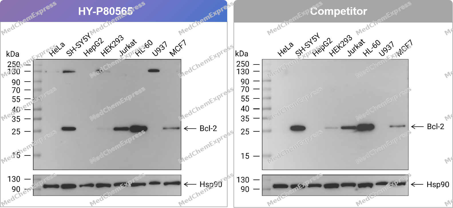 Western blot analysis of extracts from HeLa (lane 1(20μg)) 、SH-SY5Y (lane 2(20μg)) 、HepG2 (lane 3(20μg)) 、HEK293 (lane 4(20μg)) 、Jurkat (lane 5(20μg)) 、HL-60 (lane 6(20μg)) 、U937 (lane 7(20μg)) and MCF7 (lane 8(20μg)) using Bcl-2 Antibody. Proteins were transferred to a PVDF membrane and blocked with 5% nonfat dry milk in TBST for 1.5 hour at room temperature. The primary antibody (HY-P80565, 1/1000) , competitor's antibody (1/1000) and Loading control antibody (Hsp90, 1/10000) was used in 5% nonfat dry milk in TBST at 4℃ overnight. Goat Anti-Rabbit IgG-HRP Secondary Antibody (1/10,000) was used for 1 hour at room temperature.
 -
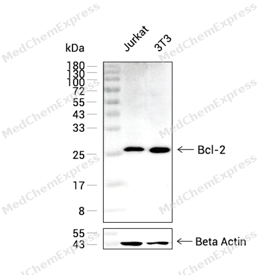 Western blot analysis was performed on extracts from Jurkat (lane 1, 20 μg), 3T3(lane 2, 20 μg) using AP2M1 Rabbit mAb.Proteins were transferred to a PVDF membrane and blocked with 5% non - fat milk in TBST at 4°C overnight.The primary antibody (1:1000 dilution) and the loading control antibody (beta-Actin, HY-P83730, 1:5000 dilution) were incubated in 5% non-fat milk in TBST for 1 hour at 37°C.Goat Anti - Mouse/Rabbit IgG - HRP Secondary Antibody (1:20000 dilution) was then applied for 40 minutes at 37°C.
 -
 Western blot analysis of extracts from HeLa (lane 1(20μg)) 、SH-SY5Y (lane 2(20μg)) 、HepG2 (lane 3(20μg)) 、HEK293 (lane 4(20μg)) 、Jurkat (lane 5(20μg)) 、HL-60 (lane 6(20μg)) 、U937 (lane 7(20μg)) and MCF7 (lane 8(20μg)) using Bcl-2 Antibody (HY-P80565) . Proteins were transferred to a PVDF membrane and blocked with 5% nonfat dry milk in TBST for 1.5 hour at room temperature. The primary antibody (1/1000) and Loading control antibody (Hsp90, 1/10000) was used in 5% nonfat dry milk in TBST at 4℃ overnight. Goat Anti-Rabbit IgG-HRP Secondary Antibody (1/10,000) was used for 1 hour at room temperature.
 -
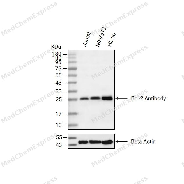 Western blot analysis of extracts from Jurkat(lane 2(20ug) , NIH/3T3(lane 3(20ug) and HL-60(lane 4(20ug) using Bcl-2 Antibody (HY-P80565) Mouse mAb. Proteins were transferred to a PVDF membrane and blocked with 5% non-fat milk in TBST for 2 hour at room temperature. The primary antibody (1/1000) and Loading control antibody (Beta Actin, HY-P83730, 1/10000) was used in 5% non-fat milk in TBST at 4°C overnight. Goat Anti-Mouse/Rabbit IgG-HRP Secondary Antibody (1/10000) was used for 1 hour at room temperature.


Background
-
Function
Bcl-2 (B-cell lymphoma 2) is a central anti-apoptotic regulator that preserves mitochondrial integrity by preventing mitochondrial outer membrane permeabilization (MOMP), a critical checkpoint controlling cytochrome c release and caspase activation during intrinsic apoptosis[1][2]. Mechanistically, Bcl-2 functions through direct interactions with pro-apoptotic BCL-2 family proteins, including Bax and Bak, thereby suppressing their oligomerization and limiting mitochondrial membrane disruption[3][4]. The protein also integrates signals transmitted by BH3-only proteins, which regulate the balance between cell survival and programmed cell death under physiological and pathological stress conditions[3][5]. Dysregulated Bcl-2 expression contributes to apoptosis resistance in multiple malignancies and supports tumor cell survival by maintaining mitochondrial homeostasis despite cellular stress[6][7]. In disease models, the anti-apoptotic activity of Bcl-2 is closely linked to mitochondrial apoptosis pathways that represent major therapeutic targets in hematologic and solid cancers[6][8]. Compared with related anti-apoptotic isoforms such as Mcl-1 and Bcl-B, Bcl-2 displays broader interactions with both Bax and Bak, whereas Mcl-1 and Bcl-B show preferential regulation of Bak- and Bax-dependent apoptotic pathways, respectively[4]. For experimental applications, BH3-mimetic inhibitors disrupt the interaction between Bcl-2 and pro-apoptotic partners, thereby restoring mitochondrial apoptosis and providing a widely used strategy for mechanistic studies and targeted cancer research[7][8].
-
Subcellular Localization
Mitochondrion outer membrane; Single-pass membrane protein; Nucleus membrane; Single-pass membrane protein; Endoplasmic reticulum membrane; Single-pass membrane protein; Cytoplasm
-
Expression
Tissue_specificity:Expression in multiple organizations -
Isoforms & Post-Translational Modification
P10415 has 2 isomers: P10415-1: 26266 Da (predicted); P10415-2: 22337 Da (predicted).
Phosphorylation/dephosphorylation on Ser-70 regulates anti-apoptotic activity (PubMed:11368354). Growth factor-stimulated phosphorylation on Ser-70 by PKC is required for the anti-apoptosis activity and occurs during the G2/M phase of the cell cycle (PubMed:11368354). In the absence of growth factors, BCL2 appears to be phosphorylated by other protein kinases such as ERKs and stress-activated kinases (PubMed:11368354). Phosphorylated by MAPK8/JNK1 at Thr-69, Ser-70 and Ser-87, which stimulates starvation-induced autophagy (PubMed:10567572, PubMed:18570871). Dephosphorylated by protein phosphatase 2A (PP2A) (By similarity);Proteolytically cleaved by caspases during apoptosis. The cleaved protein, lacking the BH4 motif, has pro-apoptotic activity, causes the release of cytochrome c into the cytosol promoting further caspase activity;Monoubiquitinated by PRKN, leading to an increase in its stability (PubMed:20889974). Ubiquitinated by SCF(FBXO10), leading to its degradation by the proteasome (PubMed:23431138). Ubiquitinated by XIAP, leading to its degradation by the proteasome (PubMed:29020630) -
Subunit
Forms homodimers, and heterodimers with BAX, BAD, BAK and Bcl-X(L). Heterodimerization with BAX requires intact BH1 and BH2 motifs, and is necessary for anti-apoptotic activity (PubMed:25609812, PubMed:8183370).
-
SwissProt ID
-
Synonyms
BCL2; Apoptosis regulator Bcl-2
-
Research Field
Cell Biology
Documentation
-
Data Sheet (261 KB)
-
SDS (251 KB)
- English - EN (251 KB)
- Français - FR (251 KB)
- Deutsch - DE (251 KB)
- Norwegian - NO (251 KB)
- Español - ES (251 KB)
- Swedish - SV (251 KB)
- Italian - IT (251 KB)
- Korean - KR (251 KB)
- Portuguese - PT (251 KB)
-
User Guide for Antibodies (1077 KB)
References
[4]. Zhai D, et al. Differential regulation of Bax and Bak by anti-apoptotic Bcl-2 family proteins Bcl-B and Mcl-1. J Biol Chem. 2008 Apr 11;283(15):9580-6. [Content Brief]
[5]. Lomonosova E, et al. BH3-only proteins in apoptosis and beyond: an overview. Oncogene. 2008 Dec;27 Suppl 1(Suppl 1):S2-19. [Content Brief]
[8]. Kang MH, et al. Bcl-2 inhibitors: targeting mitochondrial apoptotic pathways in cancer therapy. Clin Cancer Res. 2009 Feb 15;15(4):1126-32. [Content Brief]

 Powered by Bioz
