



Bax Antibody (YA825)
(Synonyms: Apoptosis regulator BAX; BAX; Bcl-2-like protein 4; BCL2-associated X protein; Bcl2-L-4; BCL2L4)Based on 4 publication(s) in Google Scholar
Bax Antibody (YA825) is a Mouse-derived and non-conjugated IgG2b monoclonal antibody, targeting to Bax.
-
Host:
Mouse
-
Isotype:
IgG
-
Application:
WB, IHC-P
-
Reactivity :
Human, Mouse, Rat
-
Formulation:
Supplied in 1*PBS (pH7.4), 0.2% BSA and 40% Glycerol. Preservative: 0.05% Sodium Azide.
-
Conjugation:
Non-conjugated


To place orders, for customer services and technical support, please contact: MedChemExpress USA
Tel: 609-228-6898 E-mail: [email protected] [email protected]



Publications Citing Use of MedChemExpress (MCE) Bax Antibody (YA825)
More

Applications
| Application |
WB

WB: Western Blot
|
IHC-P
IHC-P: Immunohistochemistry-Paraffin
|
|---|---|---|
| Dilution Ratio | 1:500-1:1000 | 1:50-1:100 |


Product Details
Bax Antibody (YA825) is a Mouse-derived and non-conjugated IgG2b monoclonal antibody, targeting to Bax.
-
Host Mouse

-
Clonality Monoclonal

-
Species ReactivityHuman, Mouse, Rat
-
Observed Molecular WeightObserved band size: 21 kDa
Note: Due to possible protein modifications or aggregation, the molecular weight should be confirmed by actual measurement, and the predicted value is for reference only.

-
Calculated Molecular Weight Predicted band size: 21 kDa

Entrez Gene: 581 Human ; 12028 Mouse ; 24887 Rat
SwissProt: Q07812 Human ; Q07813 Mouse ; Q63690 Rat
OMIM: 600040 Human
Synthetic peptide corresponding to Human BAX.The exact sequence is proprietary to MCE.
Endogenous
Protein A affinity purified.
Non-conjugated
Unmodified
IgG


Product Properties
-
Appearance
Solution
-
Formulation
Supplied in 1*PBS (pH7.4), 0.2% BSA and 40% Glycerol. Preservative: 0.05% Sodium Azide.
-
Concentration
Batch-dependent, Please check the COA for the concentration of each lot. Check Lot Concentration
-
Storage & Stability
Stored at -20°C for 1 year. Avoid repeated freeze / thaw cycles.
-
Shipping
Shipping with blue ice.


Publications (4)
-
Journal Impact Factor
-
Most Recent
-
Ecotoxicol Environ Saf
A network toxicology and machine learning approach to investigate the mechanism of kidney injury from melamine and cyanuric acid co-exposure. [Abstract]2025 Mar 15:293:118029. PMID: 40088607 -
Neurotoxicol Teratol
Sevoflurane induced hippocampal neuron cell injury via the miR-132-5p/BDNF axis and further caused cognitive impairment in rats. [Abstract]2025 Oct 26:112:107567. PMID: 41151746 -
Cryobiology
Effect of temperature on acute kidney injury in a rat model of renal ischemia‒reperfusion. [Abstract]2025 Sep:120:105276. PMID: 40516228


Verification Images
-
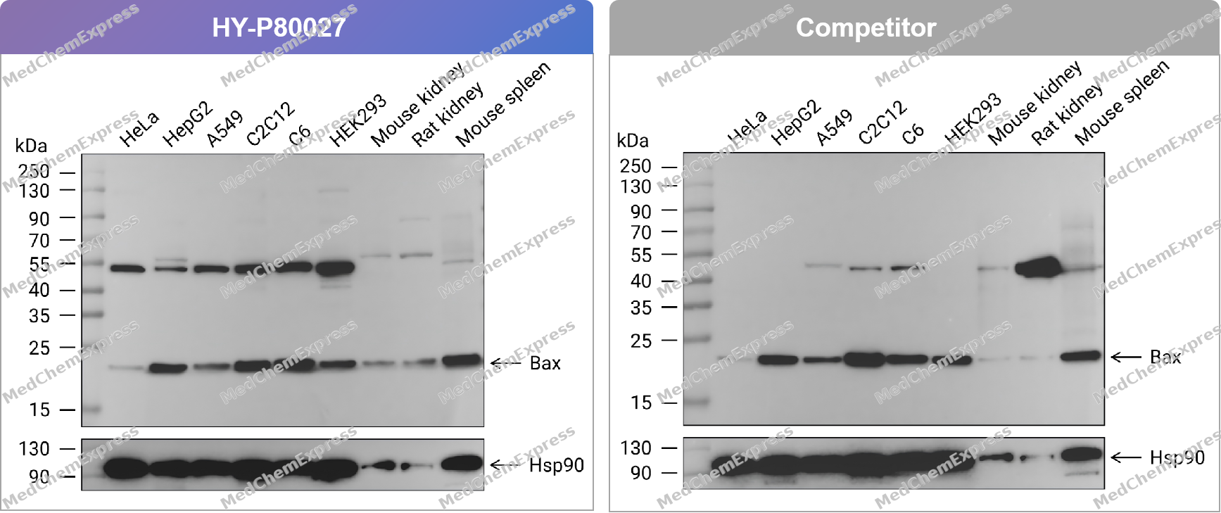 Western blot analysis of extracts from HeLa (lane 1(20μg)) 、HepG2 (lane 2(20μg)) 、A549 (lane 3(20μg)) 、C2C12 (lane 4(20μg)) 、C6 (lane 5(20μg)) 、HEK293 (lane 6(20μg)) 、Mouse kidney (lane 7(20μg)) 、Rat kidney (lane 8(20μg)) and Mouse spleen (lane 9(20μg)) using Bax Antibody. Proteins were transferred to a PVDF membrane and blocked with 5% nonfat dry milk in TBST for 1.5 hour at room temperature. The primary antibody (HY-P80027, 1/1000) , competitor's antibody (1/1000) and Loading control antibody (Hsp90, 1/10000) was used in 5% nonfat dry milk in TBST at 4℃ overnight. Goat Anti-Mouse IgG-HRP Secondary Antibody (1/10,000) was used for 1 hour at room temperature.
 -
 Western blot analysis of extracts from HeLa (lane 1(20μg)) 、HepG2 (lane 2(20μg)) 、A549 (lane 3(20μg)) 、C2C12 (lane 4(20μg)) 、C6 (lane 5(20μg)) 、HEK293 (lane 6(20μg)) 、Mouse kidney (lane 7(20μg)) 、Rat kidney (lane 8(20μg)) and Mouse spleen (lane 9(20μg)) using Bax Antibody (HY-P80027) . Proteins were transferred to a PVDF membrane and blocked with 5% nonfat dry milk in TBST for 1.5 hour at room temperature. The primary antibody (1/1000) and Loading control antibody (Hsp90, 1/10000) was used in 5% nonfat dry milk in TBST at 4℃ overnight. Goat Anti-Mouse IgG-HRP Secondary Antibody (1/10,000) was used for 1 hour at room temperature.
 -
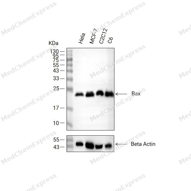 Western blot analysis of extracts from Hela (lane2(20μg), MCF-7 (lane3(20μg), C2C12 (lane4(20μg) and C6 (lane5(20μg) using Bax Antibody (HY-P80027). Proteins were transferred to a PVDF membrane and blocked with 5% non-fat milk in TBST at 4°C overnight. The primary antibody (1/1000) and Loading control antibody (Beta Actin, HY-P80993, 1/10,000) was used in 5% non-fat milk in TBST for 2 hour at room temperature. Goat Anti-Mouse IgG-HRP Secondary Antibody (HY-P8004, 1/10,000) was used for 1 hour at room temperature.
 -
 Immunohistochemical analysis of paraffin-embedded endometrial cancer tissue using Bax Antibody. The section was pre-treated using heat mediated antigen retrieval with Tris/EDTA buffer (pH 9.0) for 20 minutes. The tissues were probed with the primary antibody (HY-P80027, 1/100) , competitor's antibody (1/100) overnight at 4℃. The detection was performed using an HRP conjugated compact polymer system. DAB was used as the chromogen. Tissues were counterstained with hematoxylin and mounted with neutral balsam.
 -
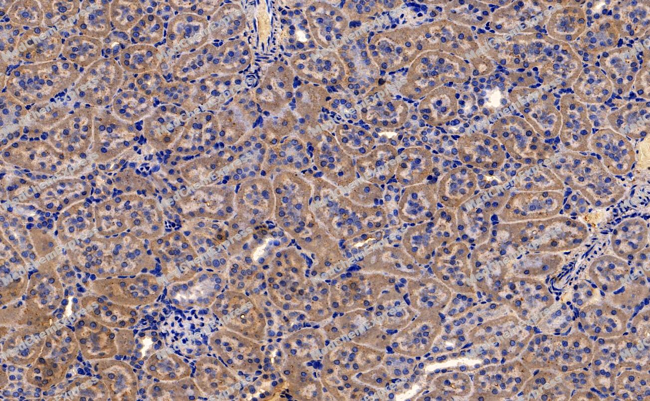 Immunohistochemical analysis of paraffin-embedded Rat kidney tissue using BAX Antibody (YA825). The section was pre-treated using heat mediated antigen retrieval with Tris-EDTA buffer (pH 9.0) for 8 minutes. The tissues were blocked in QuickBlock for 20 minutes at room temperature, washed with ddH2O and PBS, and then probed with the primary antibody (HY-P80027, 1/100) in 4℃ overnight. The detection was performed using an HRP conjugated compact polymer system. DAB was used as the chromogen. Tissues were counterstained with hematoxylin and mounted with DPX.
 -
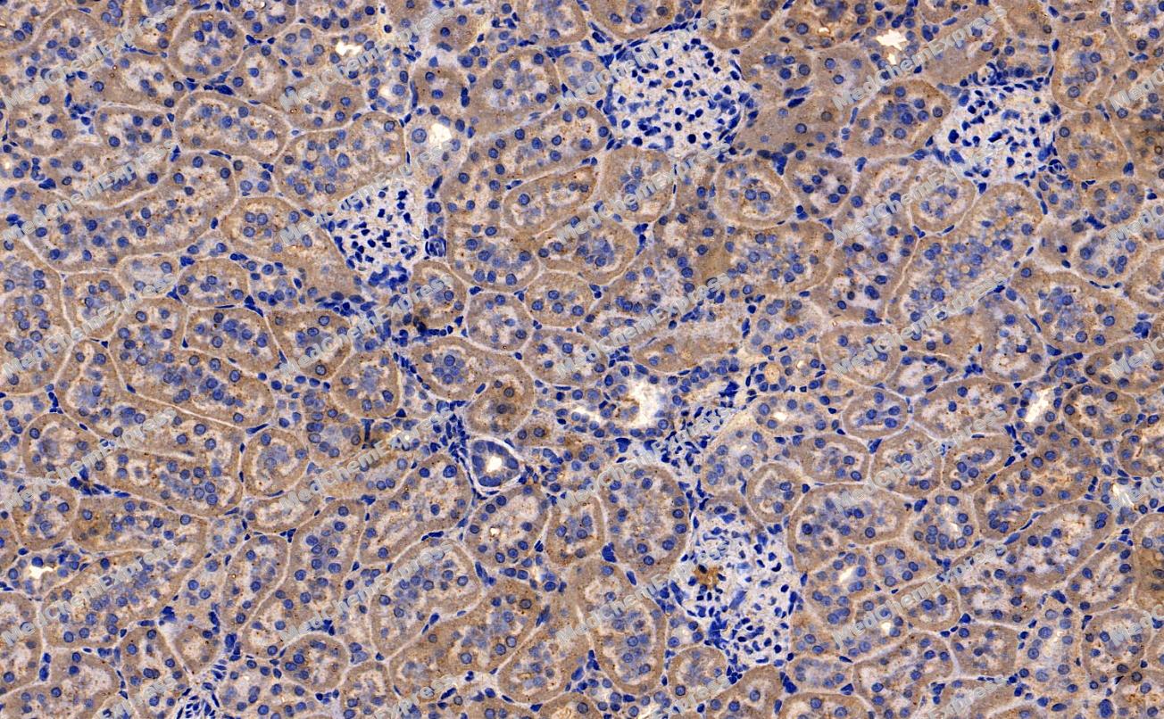 Immunohistochemical analysis of paraffin-embedded Rat kidney tissue using BAX Antibody (YA825). The section was pre-treated using heat mediated antigen retrieval with Tris-EDTA buffer (pH 9.0) for 8 minutes. The tissues were blocked in QuickBlock for 20 minutes at room temperature, washed with ddH2O and PBS, and then probed with the primary antibody (HY-P80027, 1/100) in 4℃ overnight. The detection was performed using an HRP conjugated compact polymer system. DAB was used as the chromogen. Tissues were counterstained with hematoxylin and mounted with DPX.


Background
-
Function
BAX (BCL2-associated X protein) is a pro-apoptotic member of the BCL-2 family that regulates programmed cell death through mitochondrial apoptosis and functions as a critical determinant of cellular survival following apoptotic stimuli[1][2]. BAX forms heterodimers with anti-apoptotic BCL-2 proteins, and the balance between BAX and BCL-2 influences mitochondrial outer membrane permeabilization (MOMP), cytochrome c release, and activation of the intrinsic apoptotic pathway[2][3][4]. Mechanistically, BAX belongs to the pore-forming executioner subgroup of the BCL-2 family and undergoes activation in response to cellular stress signals, including p53-dependent apoptotic signaling, thereby promoting mitochondrial membrane permeabilization and irreversible cell death commitment[1][3][5]. In disease models, dysregulated BAX activity contributes to the pathophysiology of cancer, neurodegenerative disorders, and other apoptosis-associated conditions, making mitochondrial apoptosis a major target for therapeutic intervention[3][4][6]. Compared with related BCL-2 family isoforms, BAX is distinguished by its direct executioner function in apoptosis, whereas many alternative BCL-2 family isoforms display variable anti-apoptotic or context-dependent activities that remain incompletely characterized[3]. A notable BAX isoform, BaxΔ2, retains pro-apoptotic activity despite N-terminal alterations, highlighting functional diversity within the BAX locus and providing experimental models for studying non-canonical apoptotic mechanisms[7]. For experimental applications, BAX-dependent signaling is widely used to evaluate mitochondrial apoptosis, while therapeutic strategies targeting BCL-2 family interactions, including BH3 mimetics, are actively investigated to modulate BAX-mediated cell death in cancer research[3][6].
-
Subcellular Localization
Mitochondrion outer membrane; Single-pass membrane protein; Cytoplasm; Nucleus; Cytoplasm; Cytoplasm; Cytoplasm
-
Expression
Tissue_specificity:This gene is expressed in a variety of tissues. The Psi isoform is present in gliomas. The α isoform is expressed in the spleen, breast, ovary, testis, colon, and brain, and at low levels in the skin and lungs. The Sigma isoform is expressed in the spleen, breast, ovary, testis, lung, colon, and brain, and at low levels in the skin. Both the α and Sigma isoforms are expressed in promyelocytic leukemia, histiocytic lymphoma, Burkitt lymphoma, T-cell lymphoma, lymphoblastic leukemia, breast adenocarcinoma, ovarian adenocarcinoma, prostate cancer, prostate adenocarcinoma, lung cancer, epidermoid carcinoma, small cell lung cancer, and colon adenocarcinoma cell lines. -
Subunit
Homodimer. Forms higher oligomers under stress conditions. Forms heterooligomers with BAK (PubMed:29531808). Interacts with BCL2L11. Interaction with BCL2L11 promotes BAX oligomerization and association with mitochondrial membranes, with subsequent release of cytochrome c. Forms heterodimers with BCL2, BCL2L1 isoform Bcl-X(L), BCL2L2, MCL1 and A1 (PubMed:25609812). Interacts with SH3GLB1. Interacts with humanin; forms fibers with humanin which results in BAX conformational changes and sequestering of BAX into the fibers, preventing BAX activation (PubMed:12732850, PubMed:31690630). Interacts with SFN and YWHAZ; the interaction occurs in the cytoplasm. Under stress conditions, JNK-mediated phosphorylation of SFN and YWHAZ, releases BAX to mitochondria. Interacts with RNF144B, which regulates the ubiquitin-dependent stability of BAX. Interacts with CLU under stress conditions that cause a conformation change leading to BAX oligomerization and association with mitochondria. Does not interact with CLU in unstressed cells. Interacts with FAIM2/LFG2. Interacts with RTL10/BOP. Interacts (via a C-terminal 33 residues) with NOL3 (via CARD domain); inhibits BAX activation and translocation and consequently cytochrome c release from mitochondria. Interacts with GIMAP3/IAN4 and GIMAP5/IAN5; this interaction is increased, when cells initiate apoptosis upon IL2 withdrawal (PubMed:16509771). Interacts with IRF3; the interaction is direct, increases upon Sendai virus infection and mediates the formation of the apoptosis complex TOMM70:HSP90AA1:IRF3:BAX (PubMed:25609812). Interacts with MOAP1, facilitating BAX-dependent mitochondrial outer membrane permeabilization and apoptosis (PubMed:11060313, PubMed:16199525). Interacts with BCL2L10/BCL-B (PubMed:23235460). Interacts with non-acetylated XRCC6/Ku70; this interaction leads to BAX sequestration in the cytosol, away from the mitochondria, preventing BAX-mediated apoptosis (PubMed:15023334)
-
SwissProt ID
-
Synonyms
Apoptosis regulator BAX; BAX; Bcl-2-like protein 4; BCL2-associated X protein; Bcl2-L-4; BCL2L4
-
Research Field
Cell Biology
Documentation
-
Data Sheet (262 KB)
-
SDS (252 KB)
- English - EN (252 KB)
- Français - FR (252 KB)
- Deutsch - DE (252 KB)
- Norwegian - NO (252 KB)
- Español - ES (252 KB)
- Swedish - SV (252 KB)
- Italian - IT (252 KB)
- Korean - KR (252 KB)
- Portuguese - PT (252 KB)
-
User Guide for Antibodies (1077 KB)
References
[1]. Brady HJ, et al. Bax. The pro-apoptotic Bcl-2 family member, Bax. Int J Biochem Cell Biol. 1998 Jun;30(6):647-50. [Content Brief]
[2]. Bax gene information from NCBI.
[3]. Warren CFA, et al. BCL-2 family isoforms in apoptosis and cancer. Cell Death Dis. 2019 Feb 21;10(3):177. [Content Brief]
[4]. Vogler M, et al. The BCL2 family: from apoptosis mechanisms to new advances in targeted therapy. Signal Transduct Target Ther. 2025 Mar 21;10(1):91. [Content Brief]
[5]. Maes ME, et al. BAX to basics: How the BCL2 gene family controls the death of retinal ganglion cells. Prog Retin Eye Res. 2017 Mar;57:1-25. [Content Brief]
[6]. Huang Y, et al. The adjuvant treatment role of ω-3 fatty acids by regulating gut microbiota positively in the acne vulgaris. J Dermatolog Treat. 2024 Dec;35(1):2299107. [Content Brief]
[7]. Rabezanahary H, et al. Live virus neutralizing antibodies against pre and post Omicron strains in food and retail workers in Québec, Canada. Heliyon. 2024 May 21;10(10):e31026. [Content Brief]

 Powered by Bioz
