



Caspase-12 Antibody (YA3489)
(Synonyms: CASP 12 antibody; CASP-12 antibody; Casp12 antibody; CASP12P1 antibody; caspase 12 (gene/pseudogene) antibody; caspase 12 pseudogene 1 antibody; CASPC_HUMAN antibody; Inactive caspase-12 antibody; OTTHUMP00000207032 antibody; )
Based on 1 Customer Validation
Caspase-12 Antibody (YA3489) is a Rabbit-derived and non-conjugated IgG monoclonal antibody, targeting to Caspase-12.
-
Host:
Rabbit
-
Isotype:
IgG
-
Application:
WB, IHC-P
-
Reactivity :
Human, Mouse, Rat
-
Formulation:
Supplied in PBS (pH7.4), 0.1% BSA, 40% Glycerol. Preservative: 0.05% Sodium Azide.
-
Conjugation:
Non-conjugated


To place orders, for customer services and technical support, please contact: MedChemExpress USA
Tel: 609-228-6898 E-mail: [email protected] [email protected]





Applications
| Application |
WB

WB: Western Blot
|
IHC-P
IHC-P: Immunohistochemistry-Paraffin
|
|---|---|---|
| Dilution Ratio | 1:1000 | 1:200-1:1000 |


Product Details
Caspase-12 Antibody (YA3489) is a Rabbit-derived and non-conjugated IgG monoclonal antibody, targeting to Caspase-12.
-
Host Rabbit

-
Clonality Monoclonal,Recombinant

-
Species ReactivityHuman, Mouse, Rat
-
Observed Molecular WeightObserved band size: 53 kDa
Note: Due to possible protein modifications or aggregation, the molecular weight should be confirmed by actual measurement, and the predicted value is for reference only.

-
Calculated Molecular Weight Predicted band size: 48 kDa

Entrez Gene: 100506742 Human ; 12364 Mouse ; 156117 Rat
SwissProt: Q6UXS9 Human ; O08736 Mouse ; Q920D5 Rat
OMIM: 608633 Human
Recombinant protein within mouse Caspase-12 aa 101-419.
Endogenous
affinity purified
Non-conjugated
Unmodified
IgG


Product Properties
-
Appearance
Solution
-
Formulation
Supplied in PBS (pH7.4), 0.1% BSA, 40% Glycerol. Preservative: 0.05% Sodium Azide.
-
Concentration
Batch-dependent, Please check the COA for the concentration of each lot. Check Lot Concentration
-
Storage & Stability
Stored at -20°C for 1 year. Avoid repeated freeze / thaw cycles.
-
Shipping
Shipping with blue ice.


Verification Images
-
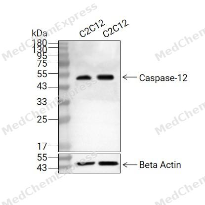 Western blot analysis of extracts from C2C12 (lane 2(20μg) and C2C12 (lane 3(40μg) using Caspase-12 (HY-P81095) Rabbit mAb. Proteins were transferred to a PVDF membrane and blocked with 5% non-fat milk in TBST for 2 hour at room temperature. The primary antibody (1/1000) and Loading control antibody (Beta Actin, HY-P80438, 1/10000) was used in 5% non-fat milk in TBST at 4°C overnight. Goat Anti-Mouse/Rabbit IgG-HRP Secondary Antibody (1/10000) was used for 1 hour at room temperature.
 -
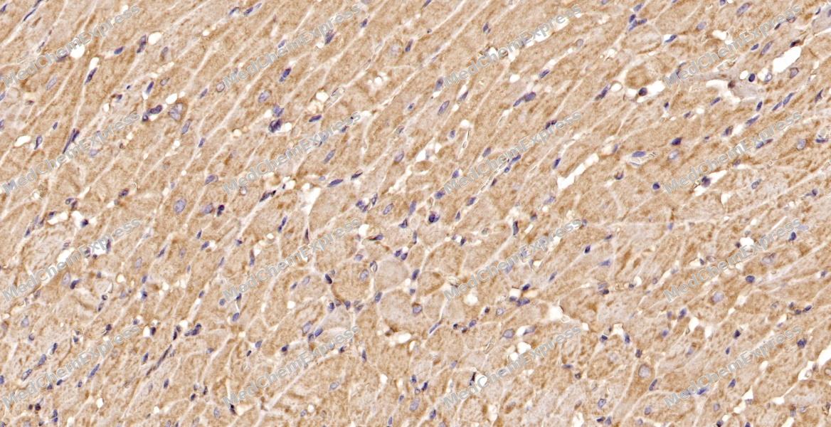 Immunohistochemical analysis of paraffin-embedded rat heart tissue using Caspase-12 Antibody. The section was pre-treated using heat mediated antigen retrieval with sodium citrate buffer (pH 6.0) for 8 minutes. The tissues were blocked in QuickBlock for 20 minutes at room temperature, washed with ddH2O and PBS, and then probed with the primary antibody at 1/100 dilution in 4℃ overnight. The detection was performed using an HRP conjugated compact polymer system. DAB was used as the chromogen. Tissues were counterstained with hematoxylin and mounted with DPX.
 -
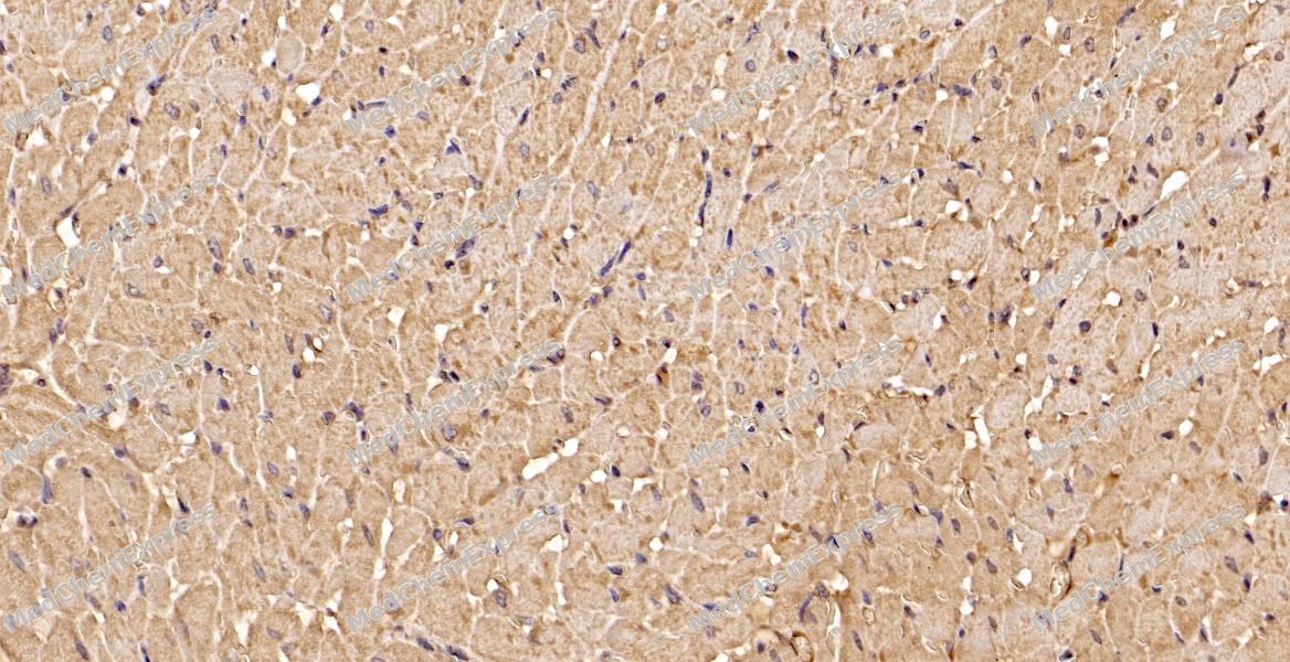 Immunohistochemical analysis of paraffin-embedded rat heart tissue using Caspase-12 Antibody. The section was pre-treated using heat mediated antigen retrieval with sodium citrate buffer (pH 6.0) for 8 minutes. The tissues were blocked in QuickBlock for 20 minutes at room temperature, washed with ddH2O and PBS, and then probed with the primary antibody at 1/100 dilution in 4℃ overnight. The detection was performed using an HRP conjugated compact polymer system. DAB was used as the chromogen. Tissues were counterstained with hematoxylin and mounted with DPX.


Background
-
Function
Caspase-12 belongs to the inflammatory caspase group, but its biology differs from caspase-1, -4, -5, and -11 because it can attenuate endotoxin-induced cytokine production rather than directly promote cytokine maturation[1][2]. Mechanistically, human full-length Caspase-12 reduced lipopolysaccharide-stimulated cytokine production in ex vivo whole blood and was linked to severe sepsis risk in African American individuals[1]. In viral immunity, mouse Caspase-12 controlled West Nile virus infection by regulating TRIM25-mediated RIG-I ubiquitination and type I interferon production[3]. ER-stress studies connect Caspase-12 with apoptosis, because overexpressed full-length caspase-12 underwent processing and ER-stress models showed caspase-12-associated caspase-3 activation[4][5]. In intestinal inflammation, IKKα loss increased unfolded protein response and caspase-12 activation, reducing protective IL-18 secretion during acute colitis[6]. Compared with caspase-1 and caspase-11, however, triple-knockout mouse data found no additional caspase-12 contribution to LPS response, apoptosis, necroptosis, or tunicamycin-induced ER stress in that model[7]. For experimental applications, caspase-12 inhibition with Z-ATAD-FMK reduced oxygen-glucose-deprivation injury, apoptosis, NLRP3, caspase-1, IL-1β, and cleaved caspase-3 in primary astrocytes[8].
-
Expression
Tissue_specificity:Mainly expressed in skeletal muscle and lung
Positive sample: C2C12 cell lysate, J774A.1 cell lysate, human heart tissue, mouse heart tissue, rat heart tissue. -
Subunit
Heterotetramer that consists of two anti-parallel arranged heterodimers, each one formed by two subunits (Potential). Interacts with TRAF2 under resting conditions; this interaction is reduced in ER stress conditions
-
SwissProt ID
-
Synonyms
CASP 12 antibody; CASP-12 antibody; Casp12 antibody; CASP12P1 antibody; caspase 12 (gene/pseudogene) antibody; caspase 12 pseudogene 1 antibody; CASPC_HUMAN antibody; Inactive caspase-12 antibody; OTTHUMP00000207032 antibody;


Documentation
-
Data Sheet (261 KB)
-
SDS (252 KB)
- English - EN (252 KB)
- Français - FR (252 KB)
- Deutsch - DE (252 KB)
- Norwegian - NO (252 KB)
- Español - ES (252 KB)
- Swedish - SV (252 KB)
- Italian - IT (252 KB)
- Korean - KR (252 KB)
- Portuguese - PT (252 KB)
-
User Guide for Antibodies (1077 KB)
References
[1]. Saleh M, et al. Differential modulation of endotoxin responsiveness by human caspase-12 polymorphisms. Nature. 2004 May 6;429(6987):75-9. [Content Brief]
[2]. Martinon F, et al. Inflammatory caspases and inflammasomes: master switches of inflammation. Cell Death Differ. 2007 Jan;14(1):10-22. [Content Brief]
[3]. Wang P, et al. Caspase-12 controls West Nile virus infection via the viral RNA receptor RIG-I. Nat Immunol. 2010 Oct;11(10):912-9. [Content Brief]
[4]. Kalai M, et al. Regulation of the expression and processing of caspase-12. J Cell Biol. 2003 Aug 4;162(3):457-67. [Content Brief]
[5]. Hitomi J, et al. Apoptosis induced by endoplasmic reticulum stress depends on activation of caspase-3 via caspase-12. Neurosci Lett. 2004 Mar 4;357(2):127-30. [Content Brief]
[6]. Diamanti MA, et al. IKKα controls ATG16L1 degradation to prevent ER stress during inflammation. J Exp Med. 2017 Feb;214(2):423-437. [Content Brief]
[7]. Salvamoser R, et al. Characterisation of mice lacking the inflammatory caspases-1/11/12 reveals no contribution of caspase-12 to cell death and sepsis. Cell Death Differ. 2019 Jun;26(6):1124-1137. [Content Brief]
[8]. Liu L, et al. Inhibiting Caspase-12 Mediated Inflammasome Activation protects against Oxygen-Glucose Deprivation Injury in Primary Astrocytes. Int J Med Sci. 2020 Jul 19;17(13):1936-1945. [Content Brief]

 Powered by Bioz
