



Fos B Antibody (YA431)
(Synonyms: G0S3, FOSB, Protein FosB, G0/G1 switch regulatory protein 3, Transcription factor AP-1 subunit FosB)
Based on 1 Customer Validation
Fos B Antibody (YA431) is a Rabbit-derived and non-conjugated IgG monoclonal antibody, targeting to Fos B.
-
Host:
Rabbit
-
Isotype:
IgG
-
Application:
WB, IHC-P, IP
-
Reactivity :
Human, Mouse
-
Formulation:
Supplied in 1*TBS (pH7.4), 0.05% BSA and 40% Glycerol. Preservative: 0.05% Sodium Azide.
-
Conjugation:
Non-conjugated


To place orders, for customer services and technical support, please contact: MedChemExpress USA
Tel: 609-228-6898 E-mail: [email protected] [email protected]





Applications
| Application |
WB

WB: Western Blot
|
IHC-P
IHC-P: Immunohistochemistry-Paraffin
|
IP
IP: Immunoprecipitation
|
|---|---|---|---|
| Dilution Ratio | 1:500-1:1000 | 1:50-1:2000 | Use at an assay dependent concentration. |


Product Details
Fos B Antibody (YA431) is a Rabbit-derived and non-conjugated IgG monoclonal antibody, targeting to Fos B.
-
Host Rabbit

-
Clonality Recombinant,Monoclonal

-
Species ReactivityHuman, Mouse
-
Observed Molecular WeightObserved band size: 40 kDa
Note: Due to possible protein modifications or aggregation, the molecular weight should be confirmed by actual measurement, and the predicted value is for reference only.

-
Calculated Molecular Weight Predicted band size: 36 kDa

Synthetic peptide corresponding to Human FosB.AA range:6-189.
Endogenous
Protein A affinity purified.
Non-conjugated
Unmodified
IgG


Product Properties
-
Appearance
Solution
-
Formulation
Supplied in 1*TBS (pH7.4), 0.05% BSA and 40% Glycerol. Preservative: 0.05% Sodium Azide.
-
Concentration
Batch-dependent, Please check the COA for the concentration of each lot. Check Lot Concentration
-
Storage & Stability
Stored at -20°C for 1 year. Avoid repeated freeze / thaw cycles.
-
Shipping
Shipping with blue ice.


Verification Images
-
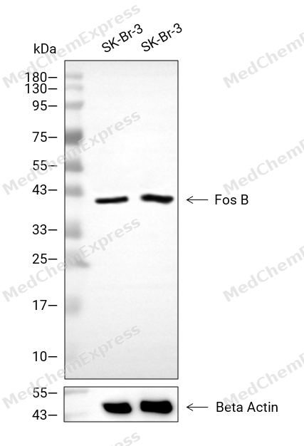 Western blot analysis of extracts from SK-Br-3 (lane 2(20μg), SK-Br-3 (lane 3(40μg), using Fos B Antibody. Proteins were transferred to a PVDF membrane and blocked with 5% BSA in TBST for 2 hour at room temperature. The primary antibody and Loading control antibody (Beta Actin, HY-P80438, 1/3000) was used in 5% BSA in TBST at 4°C overnight. Goat Anti-Mouse/Rabbit IgG-HRP Secondary Antibody (HY-P8004/HY-P8001, 1/10,000) was used for 1 hour at room temperature.
 -
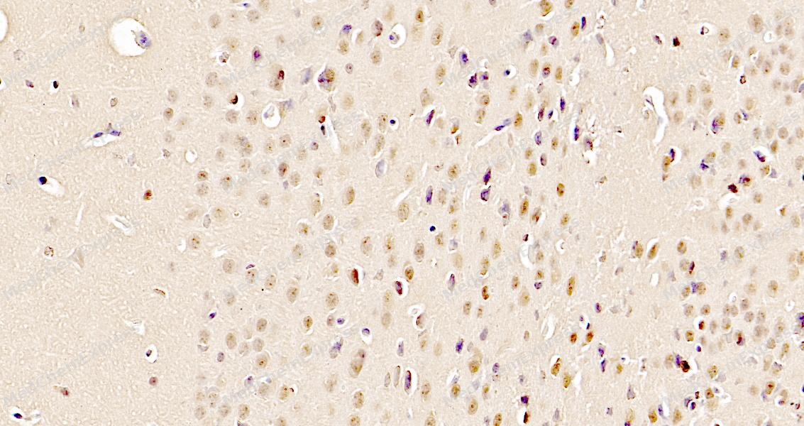 Immunohistochemical analysis of paraffin-embedded mouse brain tissue using Fos B Antibody. The section was pre-treated using heat mediated antigen retrieval with sodium citrate buffer (pH 6.0) for 8 minutes. The tissues were blocked in QuickBlock for 20 minutes at room temperature, washed with ddH2O and PBS, and then probed with the primary antibody at 1/100 dilution in 4℃ overnight. The detection was performed using an HRP conjugated compact polymer system. DAB was used as the chromogen. Tissues were counterstained with hematoxylin and mounted with DPX.
 -
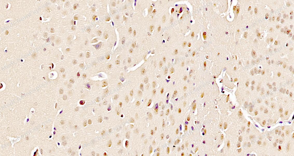 Immunohistochemical analysis of paraffin-embedded mouse brain tissue using Fos B Antibody. The section was pre-treated using heat mediated antigen retrieval with sodium citrate buffer (pH 6.0) for 8 minutes. The tissues were blocked in QuickBlock for 20 minutes at room temperature, washed with ddH2O and PBS, and then probed with the primary antibody at 1/100 dilution in 4℃ overnight. The detection was performed using an HRP conjugated compact polymer system. DAB was used as the chromogen. Tissues were counterstained with hematoxylin and mounted with DPX.


Background
-
Function
FosB is a member of the Activator Protein-1 (AP-1) transcription factor family that regulates gene expression in response to extracellular stimuli[1][2]. Mechanistically, FosB integrates signals from pathways including P2RX7 nucleotide receptor activation, TGF-β signaling, and IL-1-mediated inflammatory pathways, modulating cellular proliferation, differentiation, migration, and osteogenic or immune responses[1][3]. In disease models, FosB isoforms such as ΔFosB exhibit region-specific effects in the brain, influencing reward circuitry, stress response, and cognitive function in addiction, depression, and Parkinson’s disease[4][5][6]. Compared with related Fos isoforms, ΔFosB demonstrates enhanced nuclear stability and prolonged transcriptional activity, which allows sustained regulation of target genes including GluA2, parkin, and COX-2[4][5][1]. FosB also forms heterodimeric complexes with other transcription factors such as NFATc3 to regulate tissue factor expression and monocyte trafficking in inflammatory models[7]. Functionally, pharmacological or genetic manipulation of FosB or ΔFosB alters migration, invasion, and fibrosis-related pathways, highlighting their utility as targets in neurodegeneration and cancer research[8][3]. Isoform-specific transcriptional activity and inducibility make FosB a critical node in linking extracellular signals to downstream gene networks, offering mechanistic insight for experimental designs involving transcriptional regulation, stress response, and tissue-specific pathology[1][2].
-
Subcellular Localization
Nucleus
-
Expression
Tissue_specificity:Expression (protein level) in the nucleus accumbens of the striatum. -
Subunit
Heterodimer; binds to DNA as heterodimer (PubMed:28981703). Component of an AP-1 transcription factor complex; composed of FOS-JUN heterodimers (By similarity). As part of the AP-1 transcription factor complex, forms heterodimers with JUN, JUNB or JUND, thereby binding to the AP-1 consensus sequence and stimulating transcription (PubMed:28981703). Interacts with the BAF multiprotein chromatin-remodeling complex subunits SMARCB1 and SMARCD1 (By similarity). Interacts with ARID1A and JUN (By similarity)
-
SwissProt ID
-
Synonyms
G0S3, FOSB, Protein FosB, G0/G1 switch regulatory protein 3, Transcription factor AP-1 subunit FosB
-
Research Field
Epigenetics and Nuclear Signaling


Documentation
-
Data Sheet (264 KB)
-
SDS (251 KB)
- English - EN (251 KB)
- Français - FR (251 KB)
- Deutsch - DE (251 KB)
- Norwegian - NO (251 KB)
- Español - ES (251 KB)
- Swedish - SV (251 KB)
- Italian - IT (251 KB)
- Korean - KR (251 KB)
- Portuguese - PT (251 KB)
-
User Guide for Antibodies (1077 KB)
References
[1]. Gavala ML, et al. Activation of the transcription factor FosB/activating protein-1 (AP-1) is a prominent downstream signal of the extracellular nucleotide receptor P2RX7 in monocytic and osteoblastic cells. J Biol Chem. 2010 Oct 29;285(44):34288-98. [Content Brief]
[2]. Gajewski PA, et al. Differential Expression of FosB Proteins and Potential Target Genes in Select Brain Regions of Addiction and Depression Patients. PLoS One. 2016 Aug 5;11(8):e0160355. [Content Brief]
[3]. Patterson JR, et al. FosB and ΔFosB expression in brain regions containing differentially susceptible dopamine neurons following acute neurotoxicant exposure. Brain Res. 2016 Oct 15;1649(Pt A):53-66. [Content Brief]
[4]. Saaoud F, et al. Organelle stresses and energetic metabolisms promote endothelial-to-mesenchymal transition and fibrosis via upregulating FOSB and MEOX1 in Alzheimer's disease. Front Mol Neurosci. 2025 Aug 22;18:1605012. [Content Brief]
[5]. Kotla S, et al. Heterodimers of the transcriptional factors NFATc3 and FosB mediate tissue factor expression for 15(S)-hydroxyeicosatetraenoic acid-induced monocyte trafficking. J Biol Chem. 2017 Sep 8;292(36):14885-14901. [Content Brief]
[6]. Wagner EF, et al. Signalling in osteoclasts and the role of Fos/AP1 proteins. Ann Rheum Dis. 2003 Nov;62 Suppl 2(Suppl 2):ii83-5. [Content Brief]
[7]. Barrett CS, et al. TGF-β Effects on Prostate Cancer Cell Migration and Invasion Require FosB. Prostate. 2017 Jan;77(1):72-81. [Content Brief]
[8]. Grueter BA, et al. ∆FosB differentially modulates nucleus accumbens direct and indirect pathway function. Proc Natl Acad Sci U S A. 2013 Jan 29;110(5):1923-8. [Content Brief]

 Powered by Bioz
