



HDAC10 Antibody (YA394)
(Synonyms: Polyamine deacetylase HDAC10, Histone deacetylase 10, HD10, HDAC10)
Based on 1 Customer Validation
HDAC10 Antibody (YA394) is a Rabbit-derived and non-conjugated IgG monoclonal antibody, targeting to HDAC10.
-
Host:
Rabbit
-
Isotype:
IgG
-
Application:
WB, ICC/IF, IP
-
Reactivity :
Human
-
Formulation:
Supplied in 1*TBS (pH7.4), 0.05% BSA and 40% Glycerol. Preservative: 0.05% Sodium Azide.
-
Conjugation:
Non-conjugated


To place orders, for customer services and technical support, please contact: MedChemExpress USA
Tel: 609-228-6898 E-mail: [email protected] [email protected]





Applications
| Application |
WB

WB: Western Blot
|
ICC/IF
ICC/IF: Immunocytochemistry/
Immunofluorescence |
IHC
|
|---|---|---|---|
| Dilution Ratio | 1:1000 | 1:50-1:200 | 1:50-1:200 |


Product Details
HDAC10 Antibody (YA394) is a Rabbit-derived and non-conjugated IgG monoclonal antibody, targeting to HDAC10.
-
Host Rabbit

-
Clonality Recombinant,Monoclonal

-
Species ReactivityHuman
-
Observed Molecular WeightObserved band size: 75 kDa
Note: Due to possible protein modifications or aggregation, the molecular weight should be confirmed by actual measurement, and the predicted value is for reference only.

-
Calculated Molecular Weight Predicted band size: 71 kDa

Synthetic peptide corresponding to Human HDAC10.AA range:339-388.
Endogenous
Protein A affinity purified.
Non-conjugated
Unmodified
IgG


Product Properties
-
Appearance
Solution
-
Formulation
Supplied in 1*TBS (pH7.4), 0.05% BSA and 40% Glycerol. Preservative: 0.05% Sodium Azide.
-
Concentration
Batch-dependent, Please check the COA for the concentration of each lot. Check Lot Concentration
-
Storage & Stability
Stored at -20°C for 1 year. Avoid repeated freeze / thaw cycles.
-
Shipping
Shipping with blue ice.


Verification Images
-
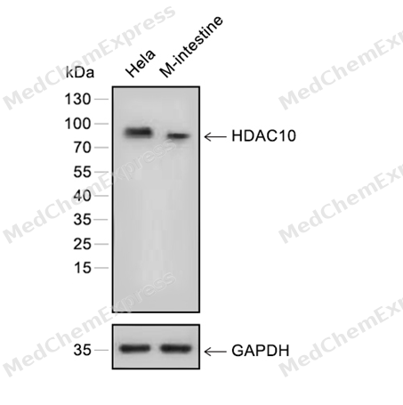 Western blot analysis of extracts from Hela (lane 1) and M-intestine (lane 2) using HDAC10 antibody. Proteins were transferred to a PVDF membrane and blocked with 5% nonfat powdered milk in PBST for 2 hour at room temperature. The primary antibody (1/1000) and loading control antibody (GAPDH, 1/3000) was diluted with 5% nonfat powdered milk in PBST at 4°C overnight. Goat Anti-Rabbit IgG-HRP Secondary Antibody (1/8,000) was incubated for 45min at room temperature.
 -
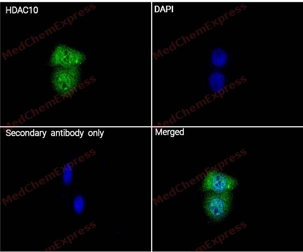 Immunocytochemistry analysis of A431 cells labeling HDAC10 with HDAC10 Antibody (HY-P80150) at 1/100 dilution . Cells were fixed in 4% paraformaldehyde for 15 minutes at room temperature, permeabilized with 0.1% Triton X-100 in PBS for 15 minutes at room temperature, then blocked with quick block buffer for 10 minutes at room temperature. Cells were then incubated with HDAC10 Antibody (HY-P80150) at 1/100 dilution in quick block buffer overnight at 4 ℃. AF488-conjugated Goat Anti-Rabbit IgG H&L(HY-P8002, Green) was used as the secondary antibody at 1/1,000 dilution. PBS instead of the primary antibody was used as the secondary antibody only control. The Nuclear counterstain was DAPI (Blue).
 -
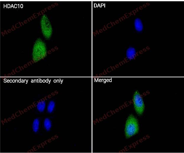 Immunocytochemistry analysis of Hela cells labeling HDAC10 with HDAC10 Antibody (HY-P80150) at 1/100 dilution . Cells were fixed in 4% paraformaldehyde for 15 minutes at room temperature, permeabilized with 0.1% Triton X-100 in PBS for 15 minutes at room temperature, then blocked with quick block buffer for 10 minutes at room temperature. Cells were then incubated with HDAC10 Antibody (HY-P80150) at 1/100 dilution in quick block buffer overnight at 4 ℃. AF488-conjugated Goat Anti-Rabbit IgG H&L(HY-P8002, Green) was used as the secondary antibody at 1/1,000 dilution. PBS instead of the primary antibody was used as the secondary antibody only control. The Nuclear counterstain was DAPI (Blue).


Background
-
Function
HDAC10 (histone deacetylase 10) is a zinc-dependent class IIb histone deacetylase that functions primarily as a polyamine deacetylase, representing a distinct enzymatic activity within the HDAC family[1]. Mechanistically, HDAC10 regulates polyamine metabolism and contributes to cellular homeostasis through pathways linked to autophagy and lysosomal function, making it an important epigenetic regulator beyond canonical histone deacetylation[1][2]. In disease-related studies, HDAC10 has attracted considerable interest in cancer biology, where altered HDAC10 activity has been associated with tumor-relevant cellular processes and therapeutic response mechanisms[2][3]. Experimental evidence further indicates that pharmacological or genetic inhibition of HDAC10 can promote autolysosome accumulation in neuroblastoma models, supporting a functional connection between HDAC10 and autophagy regulation[3][4]. Compared with its closest class IIb homolog HDAC6, HDAC10 exhibits unique substrate selectivity and is currently recognized as the only identified HDAC family member with established polyamine deacetylase activity, highlighting a major functional distinction between these related isoforms[1][2]. Structural studies have identified characteristic features, including the acidic gatekeeper residue Glu274, that contribute to substrate recognition and isoform selectivity[4]. For experimental applications, the development of selective HDAC10 inhibitors has enabled more precise interrogation of HDAC10-dependent biology, and recent compounds have demonstrated high selectivity over HDAC6 while serving as valuable chemical probes for investigating autophagy, cancer-associated pathways, and polyamine-dependent cellular processes[1][4].
-
Subcellular Localization
Cytoplasm; Nucleus
-
Expression
Tissue_specificity:Widely expressed with high levels in liver and kidney -
Isoforms & Post-Translational Modification
Q969S8 has 4 isomers: Q969S8-1: 71445 Da (predicted); Q969S8-2: 69385 Da (predicted); Q969S8-4: 70033 Da (predicted); Q969S8-5: 43016 Da (predicted).
-
Subunit
Interacts with HDAC3 (PubMed:11861901). Interacts with HDAC2 and NCOR2/SMRT (PubMed:11739383). Interacts with HSPA8/HSC70 (PubMed:23801752). Interacts with MSH2 (PubMed:26221039)
-
SwissProt ID
-
Synonyms
Polyamine deacetylase HDAC10, Histone deacetylase 10, HD10, HDAC10
-
Research Field
Epigenetics and Nuclear Signaling


Documentation
-
Data Sheet (261 KB)
-
SDS (251 KB)
- English - EN (251 KB)
- Français - FR (251 KB)
- Deutsch - DE (251 KB)
- Norwegian - NO (251 KB)
- Español - ES (251 KB)
- Swedish - SV (251 KB)
- Italian - IT (251 KB)
- Korean - KR (251 KB)
- Portuguese - PT (251 KB)
-
User Guide for Antibodies (1077 KB)
References
[1]. Lambona C, et al. Histone deacetylase 10: A polyamine deacetylase from the crystal structure to the first inhibitors. Curr Opin Struct Biol. 2023 Oct;82:102668. [Content Brief]
[2]. Delcuve GP, et al. Roles of histone deacetylases in epigenetic regulation: emerging paradigms from studies with inhibitors. Clin Epigenetics. 2012 Mar 12;4(1):5. [Content Brief]
[4]. Zeyen P, et al. Identification of histone deacetylase 10 (HDAC10) inhibitors that modulate autophagy in transformed cells. Eur J Med Chem. 2022 Apr 15;234:114272. doi: 10.1016/j.ejmech.2022.114272. Epub 2022 Mar 11. PMID: 35306288; PMCID: PMC9007901. [Content Brief]

 Powered by Bioz
