



HLA-C Antibody (YA3019)
(Synonyms: MHC; HLAC; HLC-C; D6S204; PSORS1; HLA-JY3)
Based on 1 Customer Validation
HLA-C Antibody (YA3019) is a Rabbit-derived and non-conjugated IgG monoclonal antibody, targeting to HLA-C.
-
Host:
Rabbit
-
Isotype:
IgG
-
Application:
WB, IP
-
Reactivity :
Human, Mouse
-
Formulation:
Supplied in 50mM Tris-Glycine(pH 7.4), 0.15M NaCl, 40% Glycerol, 0.01% Sodium azide and 0.05% BSA
-
Conjugation:
Non-conjugated


To place orders, for customer services and technical support, please contact: MedChemExpress USA
Tel: 609-228-6898 E-mail: [email protected] [email protected]





Applications
| Application |
WB

WB: Western Blot
|
IP
IP: Immunoprecipitation
|
|---|---|---|
| Dilution Ratio | 1:500-1:1000 | 1:20 |


Product Details
HLA-C Antibody (YA3019) is a Rabbit-derived and non-conjugated IgG monoclonal antibody, targeting to HLA-C.
-
Host Rabbit

-
Clonality Recombinant, Monoclonal

-
Species ReactivityHuman, Mouse
-
Observed Molecular WeightObserved band size: 41 kDa
Note: Due to possible protein modifications or aggregation, the molecular weight should be confirmed by actual measurement, and the predicted value is for reference only.

-
Calculated Molecular Weight Predicted band size: 41 kDa

Entrez Gene: 3107 Human ;
SwissProt: P04222 Human ; P10321 Human ; Q07000 Human ;
OMIM: 177900 Human
A synthetic peptide of human HLA-C
Endogenous
Affinity Purified
Non-conjugated
Unmodified
IgG


Product Properties
-
Appearance
Liquid
-
Formulation
Supplied in 50mM Tris-Glycine(pH 7.4), 0.15M NaCl, 40% Glycerol, 0.01% Sodium azide and 0.05% BSA
-
Concentration
Batch-dependent, Please check the COA for the concentration of each lot. Check Lot Concentration
-
Storage & Stability
Stored at -20°C for 1 year. Avoid repeated freeze / thaw cycles.
-
Shipping
Shipping with blue ice.


Verification Images
-
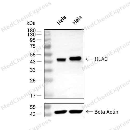 Western blot analysis of extracts from Hela (lane 2(20μg) and Hela (lane 3(40μg) using HLA C (HY-P83274) Rabbit mAb. Proteins were transferred to a PVDF membrane and blocked with 5% non-fat milk in TBST for 2 hour at room temperature. The primary antibody (1/1000) and Loading control antibody (Beta Actin, HY-P80438, 1/10000) was used in 5% non-fat milk in TBST at 4°C overnight. Goat Anti-Mouse IgG-HRP Secondary Antibody (1/10000) was used for 1 hour at room temperature.


Background
-
Function
HLA-C is an Antigen-presenting major histocompatibility complex class I (MHCI) molecule with an important role in reproduction and antiviral immunity. In complex with B2M/beta 2 microglobulin displays a restricted repertoire of self and viral peptides and acts as a dominant ligand for inhibitory and activating killer immunoglobulin receptors (KIRs) expressed on NK cells. In an allogeneic setting, such as during pregnancy, mediates interaction of extravillous trophoblasts with KIR on uterine NK cells and regulate trophoblast invasion necessary for placentation and overall fetal growth. During viral infection, may present viral peptides with low affinity for KIRs, impeding KIR-mediated inhibition through peptide antagonism and favoring lysis of infected cells. Presents a restricted repertoire of viral peptides on antigen-presenting cells for recognition by alpha-beta T cell receptor (TCR) on HLA-C-restricted CD8-positive T cells, guiding antigen-specific T cell immune response to eliminate infected cells, particularly in chronic viral infection settings such as HIV-1 or CMV infection. Both the peptide and the MHC molecule are recognized by TCR, the peptide is responsible for the fine specificity of antigen recognition and MHC residues account for the MHC restriction of T cells. Typically presents intracellular peptide antigens of 9 amino acids that arise from cytosolic proteolysis via proteasome. Can bind different peptides containing allele-specific binding motifs, which are mainly defined by anchor residues at position 2 and 9. Preferentially displays peptides having a restricted repertoire of hydrophobic or aromatic amino acids (Phe, Ile, Leu, Met, Val and Tyr) at the C-terminal anchor; ALLELE C*01:02: The peptide-bound form interacts with KIR2DL2 and KIR2DL3 inhibitory receptors on NK cells. The low affinity peptides compete with the high affinity peptides impeding KIR-mediated inhibition and favoring lysis of infected cells. Presents to CD8-positive T cells a CMV epitope derived from UL83/pp65 (RCPEMISVL), an immediate-early antigen necessary for initiating viral replication; ALLELE C*04:01: Presents a conserved HIV-1 epitope derived from env (SFNCGGEFF) to memory CD8-positive T cells, eliciting very strong IFNG responses. Presents CMV epitope derived from UL83/pp65 (QYDPVAALF) to CD8-positive T cells, triggering T cell cytotoxic response; ALLELE C*05:01: Presents HIV-1 epitope derived from rev (SAEPVPLQL) to CD8-positive T cells, triggering T cell cytotoxic response; ALLELE C*06:02: In trophoblasts, interacts with KIR2DS2 on uterine NK cells and triggers NK cell activation, including secretion of cytokines such as GMCSF that enhances trophoblast migration; ALLELE C*07:02: Plays an important role in the control of chronic CMV infection. Presents immunodominant CMV epitopes derived from IE1 (LSEFCRVL and CRVLCCYVL) and UL28 (FRCPRRFCF), both antigens synthesized during immediate-early period of viral replication. Elicits a strong anti-viral CD8-positive T cell immune response that increases markedly with age; ALLELE C*08:01: Presents viral epitopes derived from CMV UL83 (VVCAHELVC) and IAV M1 (GILGFVFTL), triggering CD8-positive T cell cytotoxic response; ALLELE C*12:02: Presents CMV epitope derived from UL83 (VAFTSHEHF) to CD8-positive T cells; ALLELE C*15:02: Presents CMV epitope derived from UL83 CC (VVCAHELVC) to CD8-positive T cells, triggering T cell cytotoxic response[1][2][3][4][5][6][7][8][9][10][11].
-
Subcellular Localization
Cell membrane; Single-pass type I membrane protein; Endoplasmic reticulum membrane; Single-pass membrane protein
-
Expression
Tissue_specificity:Ubiquitous. Highly expressed in fetal extravillous trophoblasts in the decidua basalis (at protein level) -
Isoforms & Post-Translational Modification
P10321 has 2 isomers: P10321-1: 40649 Da (predicted); P10321-2: 36608 Da (predicted).
N-linked glycosylation at Asn-110 is required for efficient interaction with CANX and CALR chaperones and appropriate HLA-C-B2M folded conformers prior to peptide loading -
Subunit
Heterotrimer that consists of an alpha chain HLA-C, a beta chain B2M and a peptide (peptide-HLA-C-B2M) (PubMed:10850706, PubMed:24990997, PubMed:28649982).
-
SwissProt ID
-
Synonyms
MHC; HLAC; HLC-C; D6S204; PSORS1; HLA-JY3
-
Research Field
Immunology


Documentation
-
Data Sheet (235 KB)
-
SDS (251 KB)
- English - EN (251 KB)
- Français - FR (251 KB)
- Deutsch - DE (251 KB)
- Norwegian - NO (251 KB)
- Español - ES (251 KB)
- Swedish - SV (251 KB)
- Italian - IT (251 KB)
- Korean - KR (251 KB)
- Portuguese - PT (251 KB)
-
User Guide for Antibodies (1077 KB)

 Powered by Bioz
