



Integrin alpha 5 Antibody (YA339)
(Synonyms: CD49e, FNRA, ITGA5, Integrin alpha-5, CD49 antigen-like family member E, Fibronectin receptor subunit alpha, Integrin alpha-F, VLA-5)Based on 3 publication(s) in Google Scholar
Integrin alpha 5 Antibody (YA339) is a Rabbit-derived and non-conjugated IgG monoclonal antibody, targeting to Integrin alpha 5.
-
Host:
Rabbit
-
Isotype:
IgG
-
Application:
WB, ICC/IF, IHC-P, IP, FC
-
Reactivity :
Human, Mouse
-
Formulation:
Supplied in 1*TBS (pH7.4), 0.05% BSA and 40% Glycerol. Preservative: 0.05% Sodium Azide.
-
Conjugation:
Non-conjugated


To place orders, for customer services and technical support, please contact: MedChemExpress USA
Tel: 609-228-6898 E-mail: [email protected] [email protected]



Publications Citing Use of MedChemExpress (MCE) Integrin alpha 5 Antibody (YA339)
More

Applications
| Application |
WB

WB: Western Blot
|
ICC/IF
ICC/IF: Immunocytochemistry/
Immunofluorescence |
IHC-P
IHC-P: Immunohistochemistry-Paraffin
|
FC
FC: Flow Cytometry
|
IP
IP: Immunoprecipitation
|
|---|---|---|---|---|---|
| Dilution Ratio | 1:1000-1:2000 | 1:50-1:200 | 1:50-1:200 | 1:50-1:100 | Use at an assay dependent concentration. |


Product Details
Integrin alpha 5 Antibody (YA339) is a Rabbit-derived and non-conjugated IgG monoclonal antibody, targeting to Integrin alpha 5.
-
Host Rabbit

-
Clonality Recombinant,Monoclonal

-
Species ReactivityHuman, Mouse
-
Observed Molecular WeightObserved band size: 140/19 kDa
Note: Due to possible protein modifications or aggregation, the molecular weight should be confirmed by actual measurement, and the predicted value is for reference only.

-
Calculated Molecular Weight Predicted band size: 115 kDa

Entrez Gene: 3678 Human ; 16402 Mouse ; 315346 Rat
SwissProt: P08648 Human ; P11688 Mouse ;
OMIM: 135620 Human
Synthetic peptide within C-terminal human Integrin alpha 5.
Endogenous
Protein A affinity purified.
Non-conjugated
Unmodified
IgG


Product Properties
-
Appearance
Solution
-
Formulation
Supplied in 1*TBS (pH7.4), 0.05% BSA and 40% Glycerol. Preservative: 0.05% Sodium Azide.
-
Concentration
Batch-dependent, Please check the COA for the concentration of each lot. Check Lot Concentration
-
Storage & Stability
Stored at -20°C for 1 year. Avoid repeated freeze / thaw cycles.
-
Shipping
Shipping with blue ice.


Publications (3)
-
Journal Impact Factor
-
Most Recent
-
Cell Rep
NAT10-dependent N4-acetylcytidine reprograms R-loops and promotes cancer stem cell growth. [Abstract]2026 Jun 23;45(6):117408. PMID: 42228575


Verification Images
-
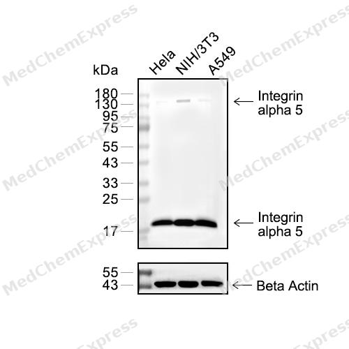 Western blot analysis of extracts from Hela (lane 2(20μg), NIH/3T3 (lane 3(20μg), A549 (lane 4(20μg) using Integrin alpha 5 Antibody (HY-P80416) Rabbit mAb. Proteins were transferred to a PVDF membrane and blocked with 5% BSA in TBST for 2 hour at room temperature. The primary antibody (HY-P80416, 1/1000) and Loading control antibody (Beta Actin, HY-P80438, 1/3000) was used in 5% BSA in TBST at 4°C overnight. Goat Anti-Mouse/Rabbit IgG-HRP Secondary Antibody (HY-P8004/HY-P8001, 1/10,000) was used for 1 hour at room temperature.
 -
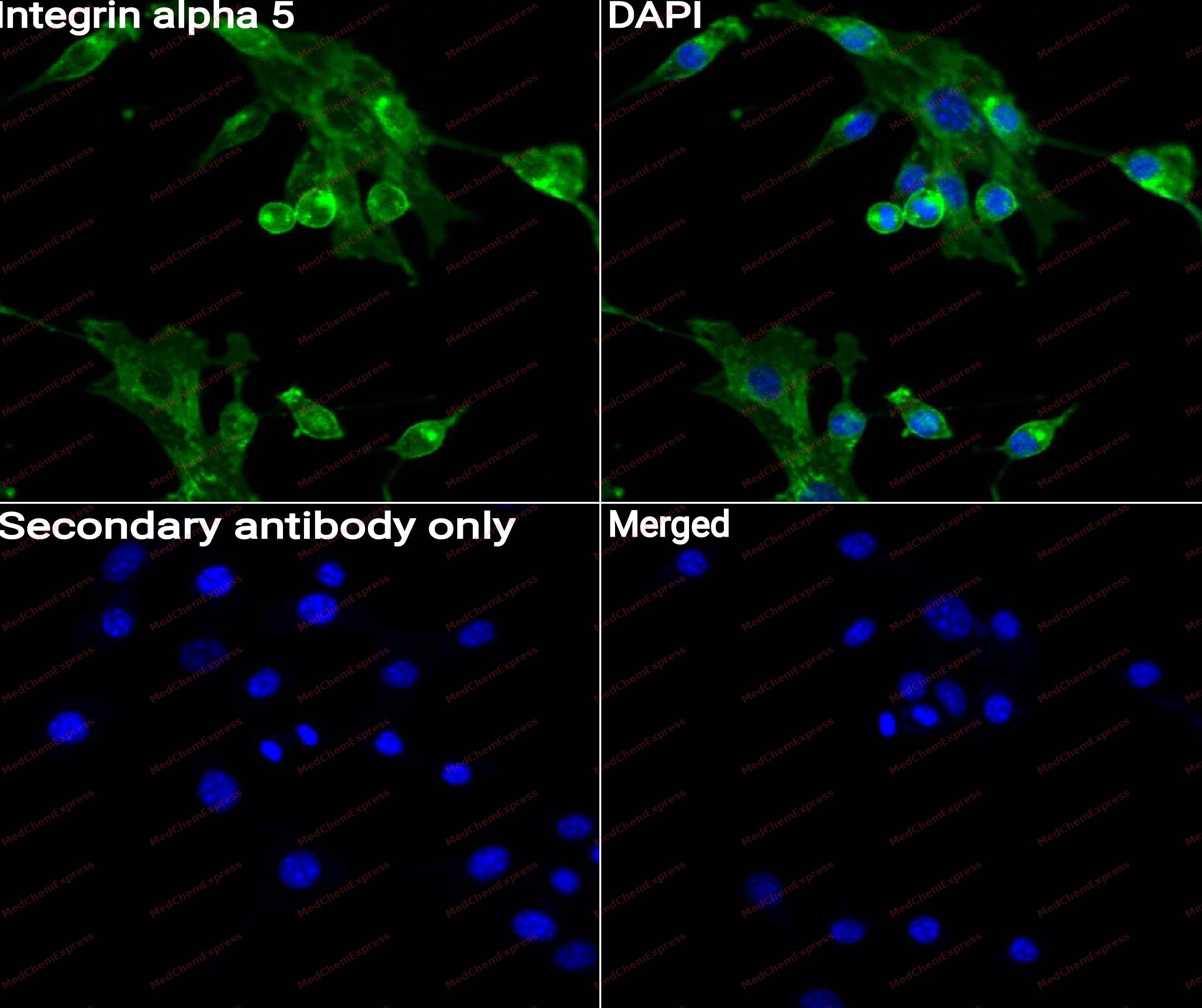 Immunocytochemistry analysis of NIH3T3 cells labeling Integrin alpha 5 with Integrin alpha 5 Antibody (HY-P80416) at 1/50 dilution. Cells were fixed in 4% paraformaldehyde for 15 minutes at room temperature, permeabilized with 0.1% Triton X-100 for 10 minutes at room temperature, then blocked with QuickBlock™ Blocking Buffer for Immunol Staining for 10 min at room temperature. Cells were then incubated with Integrin alpha 5 Antibody (HY-P80416)at 1/50 dilution in QuickBlock™ Blocking Buffer for Immunol Staining at 4 ℃. Alexa Fluor® 488-conjugated AffiniPure Goat Anti-Rabbit IgG H&L(HY-P8002, Green) was used as the secondary antibody at 1/1,000 dilution. PBS instead of the primary antibody was used as the secondary antibody only control. The Nuclear counterstain was DAPI (Blue).
 -
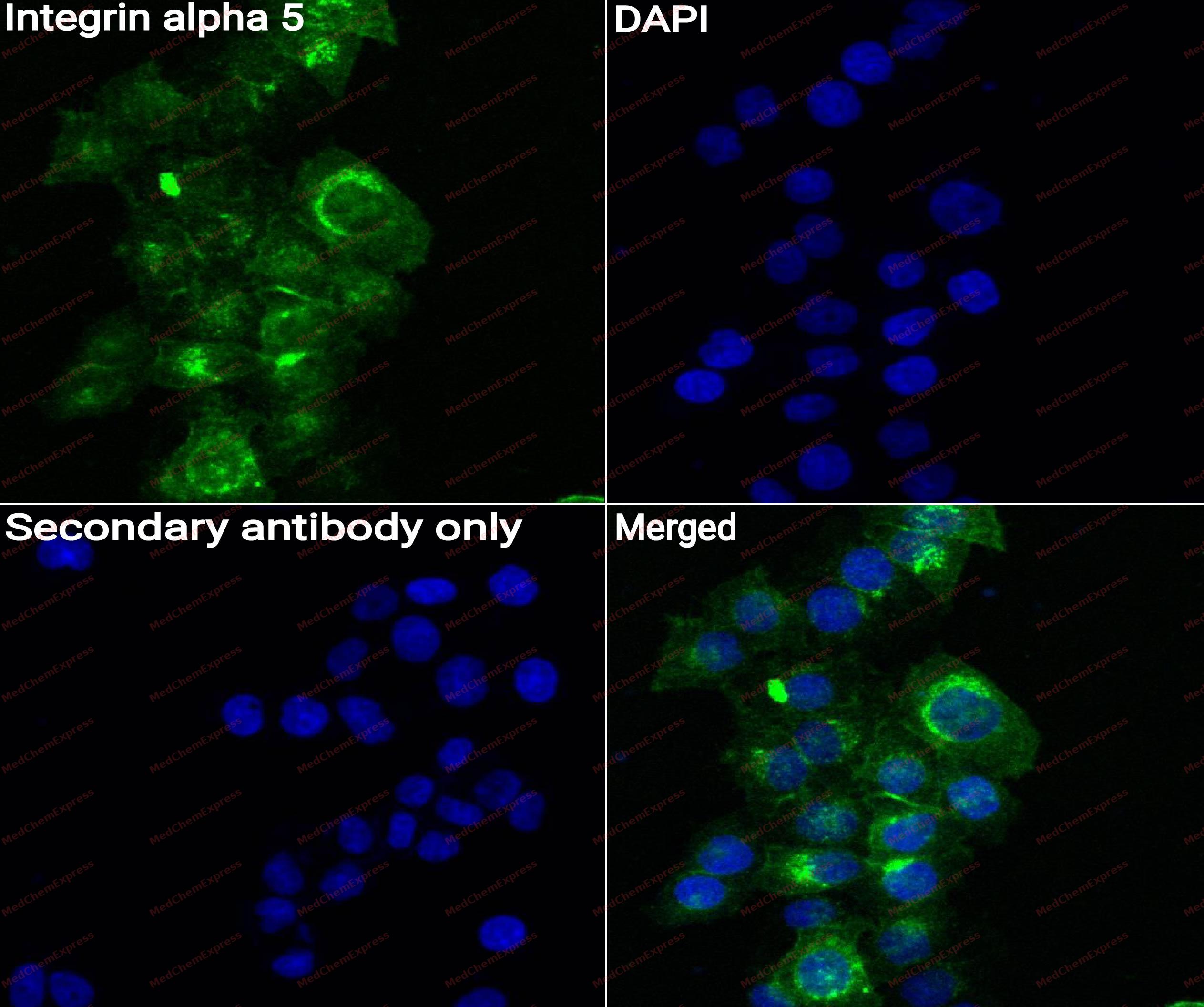 Immunocytochemistry analysis of Hela cells labeling Integrin alpha 5 with Integrin alpha 5 Antibody (HY-P80416)at 1/50 dilution. Cells were fixed in 4% paraformaldehyde for 15 minutes at room temperature, permeabilized with 0.1% Triton X-100 for 10 minutes at room temperature, then blocked with QuickBlock™ Blocking Buffer for Immunol Staining for 10 min at room temperature. Cells were then incubated with Integrin alpha 5 Antibody (HY-P80416) at 1/50 dilution in QuickBlock™ Blocking Buffer for Immunol Staining at 4 ℃. Alexa Fluor® 488-conjugated AffiniPure Goat Anti-Rabbit IgG H&L(HY-P8002, Green) was used as the secondary antibody at 1/1,000 dilution. PBS instead of the primary antibody was used as the secondary antibody only control. The Nuclear counterstain was DAPI (Blue).
 -
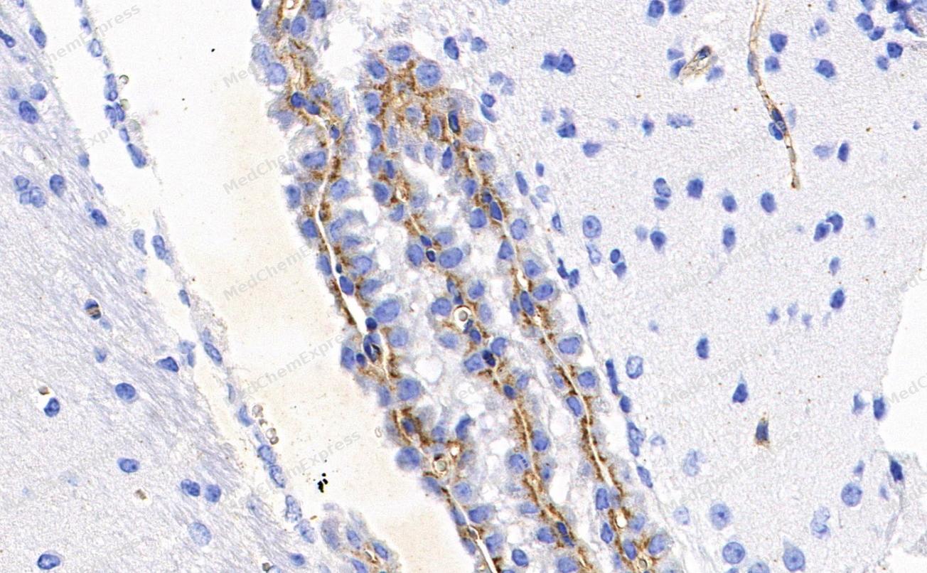 Immunohistochemical analysis of paraffin-embedded Rat cerebral cortex tissue using Integrin alpha 5 Antibody. The section was pre-treated using heat mediated antigen retrieval with Tris-EDTA buffer (pH 9.0) for 8 minutes. The tissues were blocked in QuickBlock for 20 minutes at room temperature, washed with ddH2O and PBS, and then probed with the primary antibody (HY-P80416, 1/400) in 4℃ overnight. The detection was performed using an HRP conjugated compact polymer system. DAB was used as the chromogen. Tissues were counterstained with hematoxylin and mounted with DPX.
 -
Immunohistochemical analysis of paraffin-embedded Rat cerebral cortex tissue using Integrin alpha 5 Antibody. The section was pre-treated using heat mediated antigen retrieval with Tris-EDTA buffer (pH 9.0) for 8 minutes. The tissues were blocked in QuickBlock for 20 minutes at room temperature, washed with ddH2O and PBS, and then probed with the primary antibody (HY-P80416, 1/400) in 4℃ overnight. The detection was performed using an HRP conjugated compact polymer system. DAB was used as the chromogen. Tissues were counterstained with hematoxylin and mounted with DPX.
-
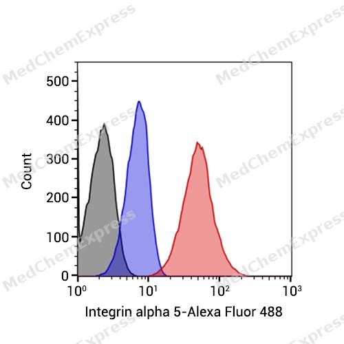 Flow cytometric analysis of 1X10^6 Jurkat cells labeling Integrin alpha 5 Antibody (HY-P80416, red). Cells were fixed with 4% paraformaldehyde and permeabilised with 90% methanol. Then stained with the primary antibody at 1/50 dilution for an hour at 4℃. Alexa Fluor® 488-conjugated AffiniPure Goat Anti-Rabbit IgG H&L (HY-P8002) was used as the secondary antibody at 1/1,000 dilution for 30 minutes at 4℃. Rabbit IgG Isotype Control (HY-P80879, blue) was used as the isotype control, cells without incubation with primary antibody were used as the unlabeled control (black).


Background
-
Function
Integrin alpha 5 is an Integrin alpha-5/beta-1 (ITGA5:ITGB1) is a receptor for fibronectin and fibrinogen. It recognizes the sequence R-G-D in its ligands. ITGA5:ITGB1 binds to PLA2G2A via a site (site 2) which is distinct from the classical ligand-binding site (site 1) and this induces integrin conformational changes and enhanced ligand binding to site 1. ITGA5:ITGB1 acts as a receptor for fibrillin-1 (FBN1) and mediates R-G-D-dependent cell adhesion to FBN1. ITGA5:ITGB1 acts as a receptor for fibronectin (FN1) and mediates R-G-D-dependent cell adhesion to FN1. ITGA5:ITGB1 is a receptor for IL1B and binding is essential for IL1B signaling. ITGA5:ITGB3 is a receptor for soluble CD40LG and is required for CD40/CD40LG signaling; (Microbial infection) Integrin ITGA5:ITGB1 acts as a receptor for Human metapneumovirus; (Microbial infection) Integrin ITGA2:ITGB1 acts as a receptor for Human parvovirus B19; (Microbial infection) In case of HIV-1 infection, the interaction with extracellular viral Tat protein seems to enhance angiogenesis in Kaposi's sarcoma lesions[1][2][3][4][5][6][7].
-
Subcellular Localization
Cell membrane; Single-pass type I membrane protein; Cell junction, focal adhesion
-
Expression
Tissue_specificity:Expression in the placenta (protein level) -
Subunit
Heterodimer of an alpha and a beta subunit. The alpha subunit is composed of a heavy and a light chain linked by a disulfide bond. ITGA5/Alpha-5 associates with ITGB1/beta-1 (PubMed:33962943). Interacts with NISCH (PubMed:11912194). Interacts with HPS5 (PubMed:10094488). Interacts with RAB21 and COMP. Interacts with CIB1. ITGA5:ITGB1 interacts with CCN3. ITGA5:ITGB1 interacts with FBN1 (PubMed:12807887, PubMed:17158881). ITGA5:ITGB1 interacts with IL1B (PubMed:29030430). ITGA5:ITGB1 interacts with ACE2 (PubMed:33102950). ITGA5:ITGB1 interacts with SELP (PubMed:37184585). Interacts with ANGPT2 (PubMed:32908006). ITGA5:ITGB1 interacts with IGFBP2 (PubMed:16569642, PubMed:26076738). ITGA5:ITGB1 interacts with IGFBP1 (PubMed:7504269)
-
SwissProt ID
-
Synonyms
CD49e, FNRA, ITGA5, Integrin alpha-5, CD49 antigen-like family member E, Fibronectin receptor subunit alpha, Integrin alpha-F, VLA-5
-
Research Field
Signal Transduction
Documentation
-
Data Sheet (263 KB)
-
SDS (252 KB)
- English - EN (252 KB)
- Français - FR (252 KB)
- Deutsch - DE (252 KB)
- Norwegian - NO (252 KB)
- Español - ES (252 KB)
- Swedish - SV (252 KB)
- Italian - IT (252 KB)
- Korean - KR (252 KB)
- Portuguese - PT (252 KB)
-
User Guide for Antibodies (1077 KB)
References
[1]. Saegusa J, et al. Pro-inflammatory secretory phospholipase A2 type IIA binds to integrins alphavbeta3 and alpha4beta1 and induces proliferation of monocytic cells in an integrin-dependent manner. J Biol Chem. 2008 Sep 19;283(38):26107-15. [Content Brief]
[2]. Fujita M, et al. Proinflammatory secreted phospholipase A2 type IIA (sPLA-IIA) induces integrin activation through direct binding to a newly identified binding site (site 2) in integrins αvβ3, α4β1, and α5β1. J Biol Chem. 2015 Jan 2;290(1):259-71. [Content Brief]
[3]. Bax DV, et al. Cell adhesion to fibrillin-1 molecules and microfibrils is mediated by alpha 5 beta 1 and alpha v beta 3 integrins. J Biol Chem. 2003 Sep 5;278(36):34605-16. [Content Brief]
[4]. Jovanovic J, et al. alphaVbeta6 is a novel receptor for human fibrillin-1. Comparative studies of molecular determinants underlying integrin-rgd affinity and specificity. J Biol Chem. 2007 Mar 2;282(9):6743-51. [Content Brief]
[5]. Schumacher S, et al. Structural insights into integrin α(5)β(1) opening by fibronectin ligand. Sci Adv. 2021 May;7(19):. [Content Brief]
[6]. Takada YK, et al. Direct binding to integrins and loss of disulfide linkage in interleukin-1β (IL-1β) are involved in the agonistic action of IL-1β. J Biol Chem. 2017 Dec 8;292(49):20067-20075. [Content Brief]
[7]. Takada YK, et al. Integrin Binding to the Trimeric Interface of CD40L Plays a Critical Role in CD40/CD40L Signaling. J Immunol. 2019 Sep 1;203(5):1383-1391. [Content Brief]

 Powered by Bioz
