



JNK3 Antibody (YA3150)
(Synonyms: Mitogen-activated protein kinase 10; MAP kinase 10; MAPK 10; MAP kinase p49 3F12; Stress-activated protein kinase 1b; SAPK1b; MAPK10; JNK3; JNK3A; PRKM10; SAPK1B)
Based on 1 Customer Validation
JNK3 Antibody (YA3150) is a Rabbit-derived and non-conjugated IgG monoclonal antibody, targeting to JNK3.
-
Host:
Rabbit
-
Isotype:
IgG
-
Application:
WB, ICC/IF, FC
-
Reactivity :
Human, Mouse, Rat
-
Formulation:
Supplied in rabbit IgG in 50 mM Tris-Glycine(pH 7.4), 0.15 M NaCl, 40% Glycerol, 0.01% sodium azide and 0.05% BSA
-
Conjugation:
Non-conjugated


To place orders, for customer services and technical support, please contact: MedChemExpress USA
Tel: 609-228-6898 E-mail: [email protected] [email protected]





Applications
| Application |
WB

WB: Western Blot
|
ICC/IF
ICC/IF: Immunocytochemistry/
Immunofluorescence |
FC
FC: Flow Cytometry
|
|---|---|---|---|
| Dilution Ratio | 1:500-1:1000 | 1:50-1:200 | 1:50-1:100 |


Product Details
JNK3 Antibody (YA3150) is a Rabbit-derived and non-conjugated IgG monoclonal antibody, targeting to JNK3.
-
Host Rabbit

-
Clonality Recombinant,Monoclonal

-
Species ReactivityHuman, Mouse, Rat
-
Observed Molecular WeightObserved band size: 53 kDa
Note: Due to possible protein modifications or aggregation, the molecular weight should be confirmed by actual measurement, and the predicted value is for reference only.

-
Calculated Molecular Weight Predicted band size: 53 kDa

A synthesized peptide derived from human JNK3 aa410-464.
Endogenous
Affinity Chromatography
Non-conjugated
Unmodified
IgG


Product Properties
-
Appearance
Solution
-
Formulation
Supplied in rabbit IgG in 50 mM Tris-Glycine(pH 7.4), 0.15 M NaCl, 40% Glycerol, 0.01% sodium azide and 0.05% BSA
-
Concentration
Batch-dependent, Please check the COA for the concentration of each lot. Check Lot Concentration
-
Storage & Stability
Stored at -20°C for 1 year. Avoid repeated freeze / thaw cycles.
-
Shipping
Shipping with blue ice.


Verification Images
-
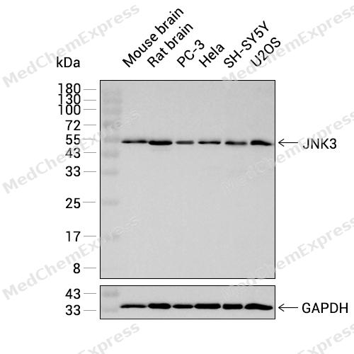
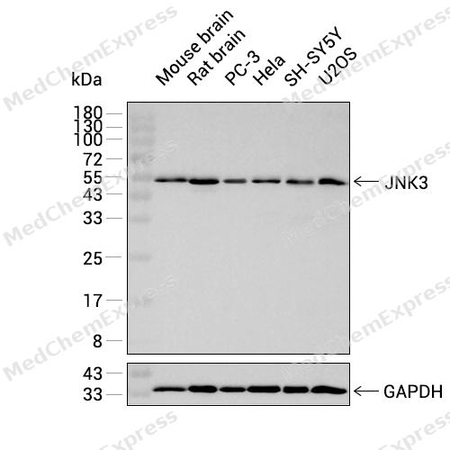 Western blot analysis was performed on extracts from Mouse brain (lane 1, 15 μg), Rat brain (lane 2, 15 μg), PC-3 (lane 3, 15 μg), Hela (lane 4, 15 μg), SH-SY5Y (lane 5, 15 μg), and U2OS (lane 6, 15 μg) using JNK3 Rabbit mAb.Proteins were transferred to a PVDF membrane and blocked with 5% non-fat milk in TBST at 4°C overnight.The primary antibody (1:1000 dilution) and the loading control antibody (GAPDH, HY-P80137, 1:80000 dilution) were incubated in 5% non-fat milk in TBST for 1 hour at 37°C.Goat Anti-Rabbit IgG-HRP Secondary Antibody (1:20000 dilution) was then applied for 40 minutes at 37°C.
 -
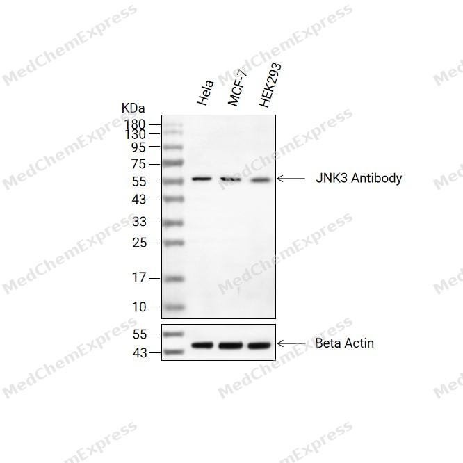 Western blot analysis of extracts from Hela(lane 2(20ug) , MCF-7(lane 3(20ug) and HEK293(lane 4(20ug) using JNK3 Antibody (HY-P83405) Rabbit mAb. Proteins were transferred to a PVDF membrane and blocked with 5% non-fat milk in TBST for 2 hour at room temperature. The primary antibody (1/1000) and Loading control antibody (Beta Actin, HY-P83730, 1/10000) was used in 5% non-fat milk in TBST at 4°C overnight. Goat Anti-Mouse/Rabbit IgG-HRP Secondary Antibody (1/10000) was used for 1 hour at room temperature.
 -
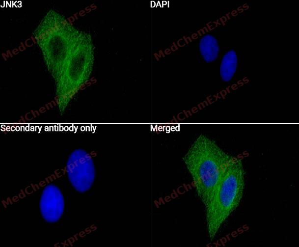 Immunocytochemistry analysis of HepG2 cells labeling JNK3 with JNK3 Antibody (HY-P83405) at 1/50 dilution. Cells were fixed in 4% paraformaldehyde for 15 minutes at room temperature, permeabilized with 0.1% Triton X-100 for 10 minutes at room temperature, then blocked with BSA for Immunol Staining for 10 min at room temperature. Cells were then incubated with JNK3 Antibody (HY-P83405) at 1/50 dilution in BSA for Immunol Staining the night at 4 ℃. AF488-conjugated AffiniPure Goat Anti-Rabbit IgG H&L(HY-P8002, Green) was used as the secondary antibody at 1/1,000 dilution. PBS instead of the primary antibody was used as the secondary antibody only control. The Nuclear counterstain was DAPI (Blue).
 -
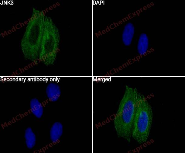 Immunocytochemistry analysis of PC-3 cells labeling JNK3 with JNK3 Antibody (HY-P83405) at 1/50 dilution. Cells were fixed in 4% paraformaldehyde for 15 minutes at room temperature, permeabilized with 0.1% Triton X-100 for 10 minutes at room temperature, then blocked with BSA for Immunol Staining for 10 min at room temperature. Cells were then incubated with JNK3 Antibody (HY-P83405) at 1/50 dilution in BSA for Immunol Staining the night at 4 ℃. AF488-conjugated AffiniPure Goat Anti-Rabbit IgG H&L(HY-P8002, Green) was used as the secondary antibody at 1/1,000 dilution. PBS instead of the primary antibody was used as the secondary antibody only control. The Nuclear counterstain was DAPI (Blue).


Background
-
Function
c-Jun N-terminal kinase 3 (JNK3) is a stress-activated protein kinase predominantly expressed in neurons, pancreatic β-cells, and cardiac tissue[1][2]. Mechanistically, JNK3 modulates transcriptional programs by phosphorylating c-Jun and regulating downstream factors such as Forkhead BoxO3A (FoxO3A) and Insulin Receptor Substrate 2 (IRS2), thereby maintaining cell survival under stress conditions[3][2]. In neuronal models, JNK3 contributes to axonal injury signaling and apoptosis, with combined JNK2/3 deficiency markedly reducing retinal ganglion cell death, highlighting its role in neurodegenerative processes[4][5]. Compared with ubiquitously expressed JNK1 and JNK2, JNK3 demonstrates nuclear localization in β-cells and isoform-specific protective effects against cytokine-induced apoptosis[2][3]. In the hypothalamus, JNK3 influences energy homeostasis by counteracting JNK1-mediated feeding and weight gain, illustrating its unique physiological role among isoforms[6][7]. JNK3 activation is scaffold-dependent, particularly via arrestin-3, enabling specific phosphorylation by upstream MKK4/7 kinases and contributing to selective signal propagation[8]. Structurally selective inhibitors targeting JNK2/3 demonstrate that hydrophobic pocket residues, including L144 in JNK3, determine isoform-specific binding, providing tools for experimental modulation of neuroprotection and β-cell survival[9][10]. Overall, JNK3 serves as a critical mediator of stress responses, with distinct subcellular localization and signaling effects compared with JNK1/2, making it a valuable target for studying neurodegeneration and metabolic regulation.
-
Subcellular Localization
Cytoplasm; Membrane; Lipid-anchor; Nucleus; Mitochondrion
-
Expression
Tissue_specificity:It is specifically expressed in specific neuronal subsets within the nervous system. It is present in the hippocampus and surrounding areas, cerebellum, striatum, and brainstem, with weaker expression in the spinal cord. Expression is extremely weak in the testes and kidneys. -
Isoforms & Post-Translational Modification
P53779 has 3 isomers: P53779-1: 52585 Da (predicted); P53779-2: 48554 Da (predicted); P53779-3: 48128 Da (predicted).
Dually phosphorylated on Thr-221 and Tyr-223 by MAP2K4 and MAP2K7, which activates the enzyme. MAP2K7 shows a strong preference for Thr-221 while MAP2K4 phosphorylates Tyr-223 preferentially. Weakly autophosphorylated on threonine and tyrosine residues in vitro;Palmitoylation regulates subcellular location and axonal development -
Subunit
Interacts with MAPKBP1 (By similarity). Interacts with MAPK8IP1/JIP-1 and MAPK8IP3/JIP-3/JSAP1 (By similarity). Interacts with SPAG9/MAPK8IP4/JIP4 (PubMed:15693750). Interacts with HDAC9 (PubMed:16611996). Interacts with ARRB2; the interaction enhances MAPK10 activation by MAP3K5 (PubMed:18435604). Interacts with SARM1 (By similarity). Interacts with JUND; interaction is inhibited in the presence of MEN1 (PubMed:22327296)
-
SwissProt ID
-
Synonyms
Mitogen-activated protein kinase 10; MAP kinase 10; MAPK 10; MAP kinase p49 3F12; Stress-activated protein kinase 1b; SAPK1b; MAPK10; JNK3; JNK3A; PRKM10; SAPK1B
-
Research Field
Neuroscience


Documentation
-
Data Sheet (262 KB)
-
SDS (252 KB)
- English - EN (252 KB)
- Français - FR (252 KB)
- Deutsch - DE (252 KB)
- Norwegian - NO (252 KB)
- Español - ES (252 KB)
- Swedish - SV (252 KB)
- Italian - IT (252 KB)
- Korean - KR (252 KB)
- Portuguese - PT (252 KB)
-
User Guide for Antibodies (1077 KB)
References
[1]. Nogueiras R, et al. Brain JNK and metabolic disease. Diabetologia. 2021 Feb;64(2):265-274. [Content Brief]
[2]. Abdelli S, et al. JNK3 is abundant in insulin-secreting cells and protects against cytokine-induced apoptosis. Diabetologia. 2009 Sep;52(9):1871-80. [Content Brief]
[3]. Nakano R, et al. Biological Properties of JNK3 and Its Function in Neurons, Astrocytes, Pancreatic β-Cells and Cardiovascular Cells. Cells. 2020 Jul 29;9(8):1802. [Content Brief]
[4]. Abdelli S, et al. JNK3 maintains expression of the insulin receptor substrate 2 (IRS2) in insulin-secreting cells: functional consequences for insulin signaling. PLoS One. 2012;7(5):e35997. [Content Brief]
[5]. Zhan X, et al. Arrestin-dependent activation of JNK family kinases. Handb Exp Pharmacol. 2014;219:259-80. [Content Brief]
[6]. Fernandes KA, et al. JNK2 and JNK3 are major regulators of axonal injury-induced retinal ganglion cell death. Neurobiol Dis. 2012 May;46(2):393-401. [Content Brief]
[7]. Ries V, et al. JNK2 and JNK3 combined are essential for apoptosis in dopamine neurons of the substantia nigra, but are not required for axon degeneration. J Neurochem. 2008 Dec;107(6):1578-88. [Content Brief]
[8]. Park H, et al. Structural basis and biological consequences for JNK2/3 isoform selective aminopyrazoles. Sci Rep. 2015 Jan 27;5:8047. [Content Brief]
[9]. Solinas G, et al. JNK at the crossroad of obesity, insulin resistance, and cell stress response. Mol Metab. 2016 Dec 8;6(2):174-184. [Content Brief]
[10]. Wydra VR, et al. A \"Ligand First\" Approach toward Selective, Covalent JNK2/3 Inhibitors. J Med Chem. 2025 Jun 12;68(11):12004-12028. [Content Brief]

 Powered by Bioz
