



MEK1 Antibody (YA300)
(Synonyms: MEK1, PRKMK1, MAP2K1, Dual specificity mitogen-activated protein kinase kinase 1, MAP kinase kinase 1, MAPKK 1, MKK1, ERK activator kinase 1, MAPK/ERK kinase 1, MEK 1)
Based on 1 Customer Validation
MEK1 Antibody (YA300) is a Rabbit-derived and non-conjugated IgG monoclonal antibody, targeting to MEK1.
-
Host:
Rabbit
-
Isotype:
IgG
-
Application:
WB, ICC/IF, IHC-P, IP, FC
-
Reactivity :
Human, Mouse
-
Formulation:
Supplied in 1*TBS (pH7.4), 0.05% BSA and 40% Glycerol. Preservative: 0.05% Sodium Azide.
-
Conjugation:
Non-conjugated


To place orders, for customer services and technical support, please contact: MedChemExpress USA
Tel: 609-228-6898 E-mail: [email protected] [email protected]





Applications
| Application |
WB

WB: Western Blot
|
ICC/IF
ICC/IF: Immunocytochemistry/
Immunofluorescence |
IHC-P
IHC-P: Immunohistochemistry-Paraffin
|
FC
FC: Flow Cytometry
|
IP
IP: Immunoprecipitation
|
|---|---|---|---|---|---|
| Dilution Ratio | 1:500-1:2000 | 1:50-1:200 | 1:50-1:200 | 1:50-1:100 | Use at an assay dependent concentration. |


Product Details
MEK1 Antibody (YA300) is a Rabbit-derived and non-conjugated IgG monoclonal antibody, targeting to MEK1.
-
Host Rabbit

-
Clonality Recombinant,Monoclonal

-
Species ReactivityHuman, Mouse
-
Observed Molecular WeightObserved band size: 45 kDa
Note: Due to possible protein modifications or aggregation, the molecular weight should be confirmed by actual measurement, and the predicted value is for reference only.

-
Calculated Molecular Weight Predicted band size: 43 kDa

Synthetic peptide within N-terminal human MEK1.
Endogenous
Protein A affinity purified.
Non-conjugated
Unmodified
IgG


Product Properties
-
Appearance
Solution
-
Formulation
Supplied in 1*TBS (pH7.4), 0.05% BSA and 40% Glycerol. Preservative: 0.05% Sodium Azide.
-
Concentration
Batch-dependent, Please check the COA for the concentration of each lot. Check Lot Concentration
-
Storage & Stability
Stored at -20°C for 1 year. Avoid repeated freeze / thaw cycles.
-
Shipping
Shipping with blue ice.


Verification Images
-
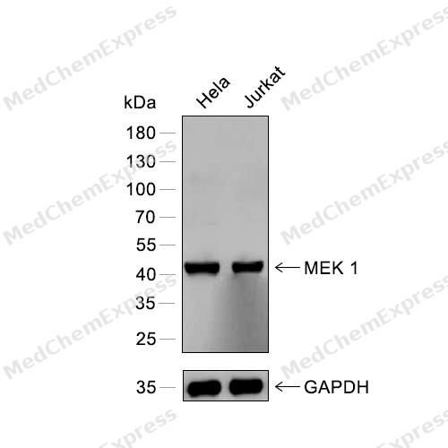 Western blot analysis of extracts from Hela (lane 1) and Jurkat (lane 2) using MEK1 antibody. Proteins were transferred to a PVDF membrane and blocked with 5% nonfat powdered milk in PBST for 2 hour at room temperature. The primary antibody (1/1000) and loading control antibody (GAPDH, 1/3000) was diluted with 5% nonfat powdered milk in PBST at 4°C overnight. Goat Anti-Rabbit IgG-HRP Secondary Antibody (1/8,000) was incubated for 45min at room temperature.
 -
 Immunohistochemical analysis of paraffin-embedded human colon cancer using MEK1 antibody. The section was pre-treated using heat mediated antigen retrieval with EDTA (pH 9.0) for 14 minutes. The tissues were blocked in 5% BSA for 30 minutes at room temperature, washed with TBST, and then probed with the primary antibody (HY-P80217, 1/200) for 1 hour at room temperature. The detection was performed using Polymer HRP-conjugated Goat Anti-Mouse/Rabbit lgG(H&L) secondary antibody (HY-P83652). DAB was used as the chromogen. Tissues were counterstained with hematoxylin and mounted with DPX.
 -
 Immunohistochemical analysis of paraffin-embedded human pancreas using MEK1 antibody. The section was pre-treated using heat mediated antigen retrieval with EDTA (pH 9.0) for 14 minutes. The tissues were blocked in 5% BSA for 30 minutes at room temperature, washed with TBST, and then probed with the primary antibody (HY-P80217, 1/200) for 1 hour at room temperature. The detection was performed using Polymer HRP-conjugated Goat Anti-Mouse/Rabbit lgG(H&L) secondary antibody (HY-P83652). DAB was used as the chromogen. Tissues were counterstained with hematoxylin and mounted with DPX.
 -
 Immunohistochemical analysis of paraffin-embedded human ovarian cancer using MEK1 antibody. The section was pre-treated using heat mediated antigen retrieval with EDTA (pH 9.0) for 14 minutes. The tissues were blocked in 5% BSA for 30 minutes at room temperature, washed with TBST, and then probed with the primary antibody (HY-P80217, 1/200) for 1 hour at room temperature. The detection was performed using Polymer HRP-conjugated Goat Anti-Mouse/Rabbit lgG(H&L) secondary antibody (HY-P83652). DAB was used as the chromogen. Tissues were counterstained with hematoxylin and mounted with DPX.
 -
 Immunohistochemical analysis of paraffin-embedded human liver cancer using MEK1 antibody. The section was pre-treated using heat mediated antigen retrieval with EDTA (pH 9.0) for 14 minutes. The tissues were blocked in 5% BSA for 30 minutes at room temperature, washed with TBST, and then probed with the primary antibody (HY-P80217, 1/200) for 1 hour at room temperature. The detection was performed using Polymer HRP-conjugated Goat Anti-Mouse/Rabbit lgG(H&L) secondary antibody (HY-P83652). DAB was used as the chromogen. Tissues were counterstained with hematoxylin and mounted with DPX.
 -
 Immunohistochemical analysis of paraffin-embedded human liver using MEK1 antibody. The section was pre-treated using heat mediated antigen retrieval with EDTA (pH 9.0) for 14 minutes. The tissues were blocked in 5% BSA for 30 minutes at room temperature, washed with TBST, and then probed with the primary antibody (HY-P80217, 1/200) for 1 hour at room temperature. The detection was performed using Polymer HRP-conjugated Goat Anti-Mouse/Rabbit lgG(H&L) secondary antibody (HY-P83652). DAB was used as the chromogen. Tissues were counterstained with hematoxylin and mounted with DPX.
 -
 Immunohistochemical analysis of paraffin-embedded human bladder cancer using MEK1 antibody. The section was pre-treated using heat mediated antigen retrieval with EDTA (pH 9.0) for 14 minutes. The tissues were blocked in 5% BSA for 30 minutes at room temperature, washed with TBST, and then probed with the primary antibody (HY-P80217, 1/200) for 1 hour at room temperature. The detection was performed using Polymer HRP-conjugated Goat Anti-Mouse/Rabbit lgG(H&L) secondary antibody (HY-P83652). DAB was used as the chromogen. Tissues were counterstained with hematoxylin and mounted with DPX.
 -
 Immunohistochemical analysis of paraffin-embedded human breast cancer using MEK1 antibody. The section was pre-treated using heat mediated antigen retrieval with EDTA (pH 9.0) for 14 minutes. The tissues were blocked in 5% BSA for 30 minutes at room temperature, washed with TBST, and then probed with the primary antibody (HY-P80217, 1/200) for 1 hour at room temperature. The detection was performed using Polymer HRP-conjugated Goat Anti-Mouse/Rabbit lgG(H&L) secondary antibody (HY-P83652). DAB was used as the chromogen. Tissues were counterstained with hematoxylin and mounted with DPX.
 -
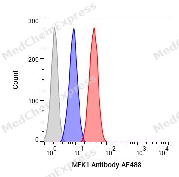 Flow cytometric analysis of 1X10⁶ HeLa cells labeling MEK1 Antibody(HY-P80217, red). Cells were fixed with 4% paraformaldehyde and permeabilised with 90% methanol. Then stained with the primary antibody at 1/100 dilution for an hour at 4℃. AF488-conjugated Goat Anti-Rabbit IgG H&L (HY-P8002) was used as the secondary antibody at 1/1,000 dilution for 30 minutes at 4℃. Rabbit IgG Isotype Control (HY-P80879, blue) was used as the isotype control, cells without incubation with primary antibody were used as the unlabeled control (black).


Background
-
Function
MEK1 is a MAP kinase or ERK kinase that phosphorylates the ERK gene product and acts as an immediate upstream activator of ERK signaling[1][2]. Mechanistically, MEK1 and MEK2 phosphorylate and activate ERK1/ERK2, positioning MEK proteins as pathway gatekeepers in the RAS-RAF-MEK-ERK cascade[3][4]. This cascade regulates cell proliferation, differentiation, survival, invasion, and other ERK-dependent cellular responses[3][5]. In disease models, ERK pathway activation is prominent in cancers driven by receptor tyrosine kinase, RAS, BRAF, CRAF, MEK1, or MEK2 alterations, which promote growth factor-independent ERK1/2 activation[5]. Compared with MEK2, MEK1 shares the same ERK substrates, but Mek1- embryos die from placental defects whereas Mek2- mice survive, indicating isoform-related developmental differences and functional redundancy controlled by total MEK protein level[4]. Structurally, MEK1 and MEK2 are closely related dual-specificity tyrosine/threonine kinases with a hydrophobic allosteric pocket adjacent to the ATP-binding site[6][5]. For experimental applications, PD098059 selectively inhibited MEK without significant inhibition of MAPK itself, and allosteric MEK blockade suppressed colon tumor growth in vivo[7][8].
-
Subcellular Localization
Cytoplasm, cytoskeleton, microtubule organizing center, centrosome; Cytoplasm, cytoskeleton, microtubule organizing center, spindle pole body; Cytoplasm; Nucleus; Membrane; Peripheral membrane protein
-
Expression
Tissue_specificity:It is widely expressed, but present in extremely low concentrations in the brain. -
Subunit
Found in a complex with at least BRAF, HRAS, MAP2K1, MAPK3/ERK1 and RGS14 (By similarity). Forms a heterodimer with MAP2K2/MEK2 (By similarity). Forms heterodimers with KSR2 which further dimerize to form tetramers (By similarity). Interacts with KSR1 or KSR2 and BRAF; the interaction with KSR1 or KSR2 mediates KSR1-BRAF or KSR2-BRAF dimerization (PubMed:10409742, PubMed:29433126). Interacts with ARBB2, LAMTOR3 and RAF1 (By similarity). Interacts with MAPK1/ERK2 (PubMed:32721402). Interacts with MORG1 (By similarity). Interacts with PPARG (PubMed:17101779). Interacts with isoform 1 of VRK2 (PubMed:20679487). Interacts with SGK1 (PubMed:19447520). Interacts with BIRC6/bruce (PubMed:18329369). Interacts with KAT7; the interaction promotes KAT7 phosphorylation (By similarity). Interacts with RAF1 and NEK10; the interaction is required for ERK1/2-signaling pathway activation in response to UV irradiation (PubMed:20956560). Interacts with TRAF3IP3 (PubMed:26195727). Interacts with MOS (PubMed:34779126, PubMed:35670744)
-
SwissProt ID
-
Synonyms
MEK1, PRKMK1, MAP2K1, Dual specificity mitogen-activated protein kinase kinase 1, MAP kinase kinase 1, MAPKK 1, MKK1, ERK activator kinase 1, MAPK/ERK kinase 1, MEK 1
-
Research Field
Signal Transduction


Documentation
-
Data Sheet (262 KB)
-
SDS (252 KB)
- English - EN (252 KB)
- Français - FR (252 KB)
- Deutsch - DE (252 KB)
- Norwegian - NO (252 KB)
- Español - ES (252 KB)
- Swedish - SV (252 KB)
- Italian - IT (252 KB)
- Korean - KR (252 KB)
- Portuguese - PT (252 KB)
-
User Guide for Antibodies (1077 KB)
References
[1]. Crews CM, et al. The primary structure of MEK, a protein kinase that phosphorylates the ERK gene product. Science. 1992 Oct 16;258(5081):478-80. [Content Brief]
[2]. Zheng CF, et al. Properties of MEKs, the kinases that phosphorylate and activate the extracellular signal-regulated kinases. J Biol Chem. 1993 Nov 15;268(32):23933-9. [Content Brief]
[3]. Shaul YD, et al. The MEK/ERK cascade: from signaling specificity to diverse functions. Biochim Biophys Acta. 2007 Aug;1773(8):1213-26. [Content Brief]
[4]. Aoidi R, et al. Functional redundancy of the kinases MEK1 and MEK2: Rescue of the Mek1 mutant phenotype by Mek2 knock-in reveals a protein threshold effect. Sci Signal. 2016 Jan 26;9(412):ra9. [Content Brief]
[5]. Caunt CJ, et al. MEK1 and MEK2 inhibitors and cancer therapy: the long and winding road. Nat Rev Cancer. 2015 Oct;15(10):577-92. [Content Brief]
[6]. Ohren JF, et al. Structures of human MAP kinase kinase 1 (MEK1) and MEK2 describe novel noncompetitive kinase inhibition. Nat Struct Mol Biol. 2004 Dec;11(12):1192-7. [Content Brief]
[7]. Dudley DT, et al. A synthetic inhibitor of the mitogen-activated protein kinase cascade. Proc Natl Acad Sci U S A. 1995 Aug 15;92(17):7686-9. [Content Brief]
[8]. Sebolt-Leopold JS, et al. Blockade of the MAP kinase pathway suppresses growth of colon tumors in vivo. Nat Med. 1999 Jul;5(7):810-6. [Content Brief]

 Powered by Bioz
