



Ras Antibody (YA106)
(Synonyms: NRAS; HRAS; KRAS)Based on 1 publication(s) in Google Scholar
Ras Antibody (YA106) is a Rabbit-derived and non-conjugated IgG monoclonal antibody, targeting to Ras.
-
Host:
Rabbit
-
Isotype:
IgG
-
Application:
WB, ICC/IF, IP, FC
-
Reactivity :
Human, Mouse, Rat, Zebrafish
-
Formulation:
Supplied in 1*TBS (pH7.4), 0.05% BSA and 40% Glycerol. Preservative: 0.05% Sodium Azide.
-
Conjugation:
Non-conjugated


To place orders, for customer services and technical support, please contact: MedChemExpress USA
Tel: 609-228-6898 E-mail: [email protected] [email protected]



Publications Citing Use of MedChemExpress (MCE) Ras Antibody (YA106)
More

Applications
| Application |
WB

WB: Western Blot
|
ICC/IF
ICC/IF: Immunocytochemistry/
Immunofluorescence |
FC
FC: Flow Cytometry
|
IP
IP: Immunoprecipitation
|
|---|---|---|---|---|
| Dilution Ratio | 1:1000-1:2000 | 1:100-1:500 | 1:1000 | Use at an assay dependent concentration. |


Product Details
Ras Antibody (YA106) is a Rabbit-derived and non-conjugated IgG monoclonal antibody, targeting to Ras.
-
Host Rabbit

-
Clonality Recombinant,Monoclonal

-
Species ReactivityHuman, Mouse, Rat, Zebrafish
-
Observed Molecular WeightObserved band size: 21 kDa
Note: Due to possible protein modifications or aggregation, the molecular weight should be confirmed by actual measurement, and the predicted value is for reference only.

-
Calculated Molecular Weight Predicted band size: 21 kDa

Entrez Gene: 3265 Human ; 3845 Human ; 4893 Human ; 15461 Mouse ; 16653 Mouse ; 18176 Mouse ;
SwissProt: P01111 Human ; P01112 Human ; P01116 Human ; P08556 Mouse ; P32883 Mouse ; Q61411 Mouse ;
OMIM: 607785 Human
Synthetic peptide corresponding to Human Ras.AA range:21-64.
Endogenous
Protein A affinity purified.
Non-conjugated
Unmodified
IgG


Product Properties
-
Appearance
Solution
-
Formulation
Supplied in 1*TBS (pH7.4), 0.05% BSA and 40% Glycerol. Preservative: 0.05% Sodium Azide.
-
Concentration
Batch-dependent, Please check the COA for the concentration of each lot. Check Lot Concentration
-
Storage & Stability
Stored at -20°C for 1 year. Avoid repeated freeze / thaw cycles.
-
Shipping
Shipping with blue ice.


Publications (1)
-
Journal Impact Factor
-
Most Recent
-
Biomed Pharmacother
Methyleugenol alleviates pulmonary vascular remodeling in rats with high-altitude pulmonary hypertension by improving pulmonary smooth muscle cell function. [Abstract]2025 Jun:187:118109. PMID: 40306181


Verification Images
-
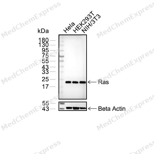 Western blot analysis of extracts from Hela (lane 2(20μg) , HEK293 (lane 3(20μg) , NIH/3T3 (lane 3(20μg),using Ras Antibody (HY-P80298). Proteins were transferred to a PVDF membrane and blocked with 5% BSA in TBST for 2 hour at room temperature. The primary antibody and Loading control antibody (Beta Actin, HY-P80438, 1/3000) was used in 5% BSA in TBST at 4°C overnight. Goat Anti-Mouse/Rabbit IgG-HRP Secondary Antibody (HY-P8004/HY-P8001, 1/10,000) was used for 1 hour at room temperature.
 -
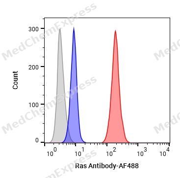 Flow cytometric analysis of 1X10⁶ Jurkat cells labeling Ras Antibody (HY-P80298, red). Cells were fixed with 4% paraformaldehyde and permeabilised with 90% methanol. Then stained with the primary antibody at 1/1000 dilution for an hour at 4℃. AF488-conjugated Goat Anti-Rabbit IgG H&L (HY-P8002) was used as the secondary antibody at 1/1,000 dilution for 30 minutes at 4℃. Rabbit IgG Isotype Control (HY-P80879, blue) was used as the isotype control, cells without incubation with primary antibody were used as the unlabeled control (black).
 -
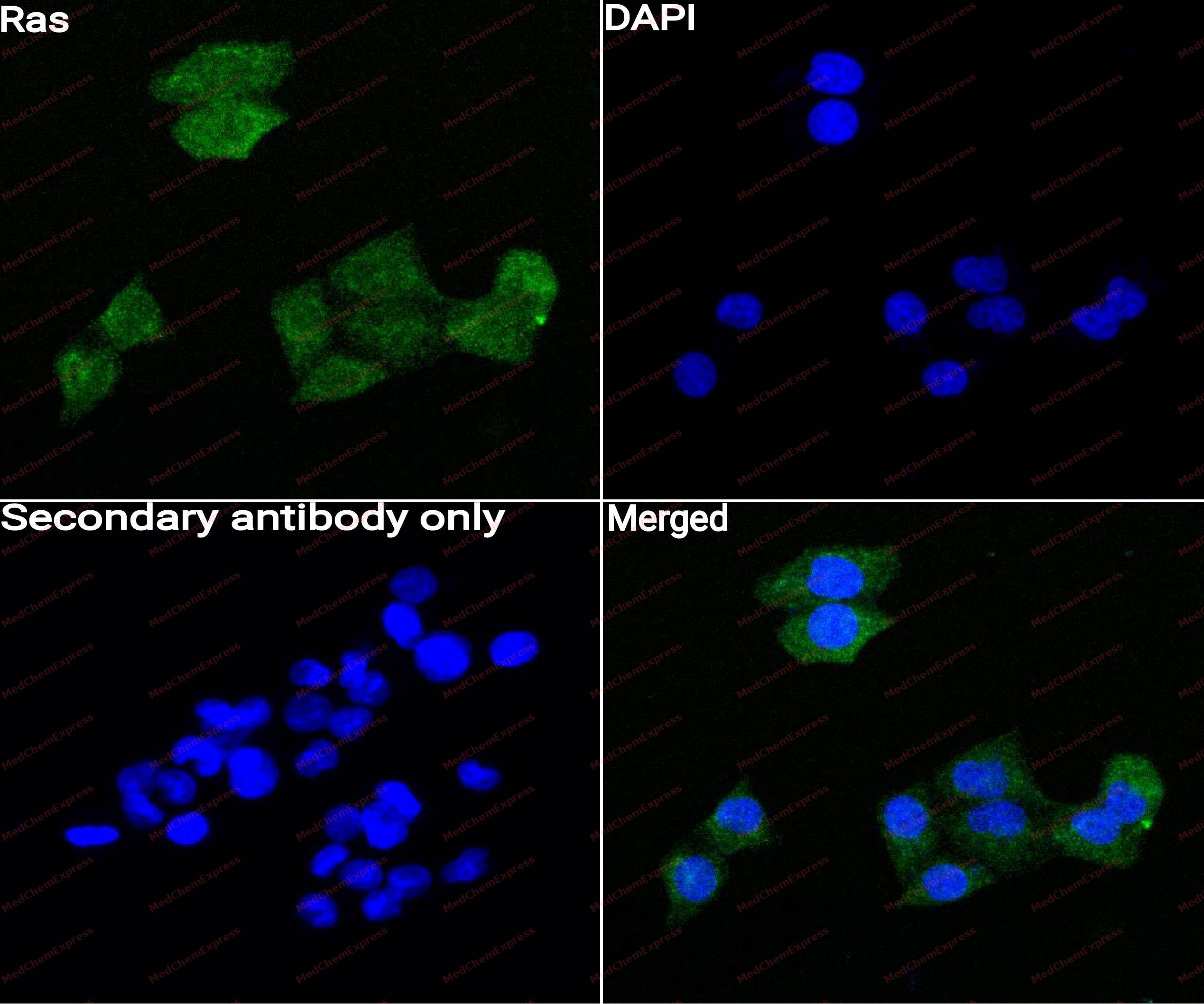 Immunocytochemistry analysis of MCF-7 cells labeling Ras with Ras Antibody (HY-P80298) at 1/50 dilution. Cells were fixed in 4% paraformaldehyde for 15 minutes at room temperature, permeabilized with 0.1% Triton X-100 for 10 minutes at room temperature, then blocked with QuickBlock™ Blocking Buffer for Immunol Staining for 10 min at room temperature. Cells were then incubated with Ras Antibody (HY-P80298) at 1/50 dilution in QuickBlock™ Blocking Buffer for Immunol Staining at 4 ℃. Alexa Fluor® 488-conjugated AffiniPure Goat Anti-Rabbit IgG H&L(HY-P8002, Green) was used as the secondary antibody at 1/1,000 dilution. PBS instead of the primary antibody was used as the secondary antibody only control. The Nuclear counterstain was DAPI (Blue).
 -
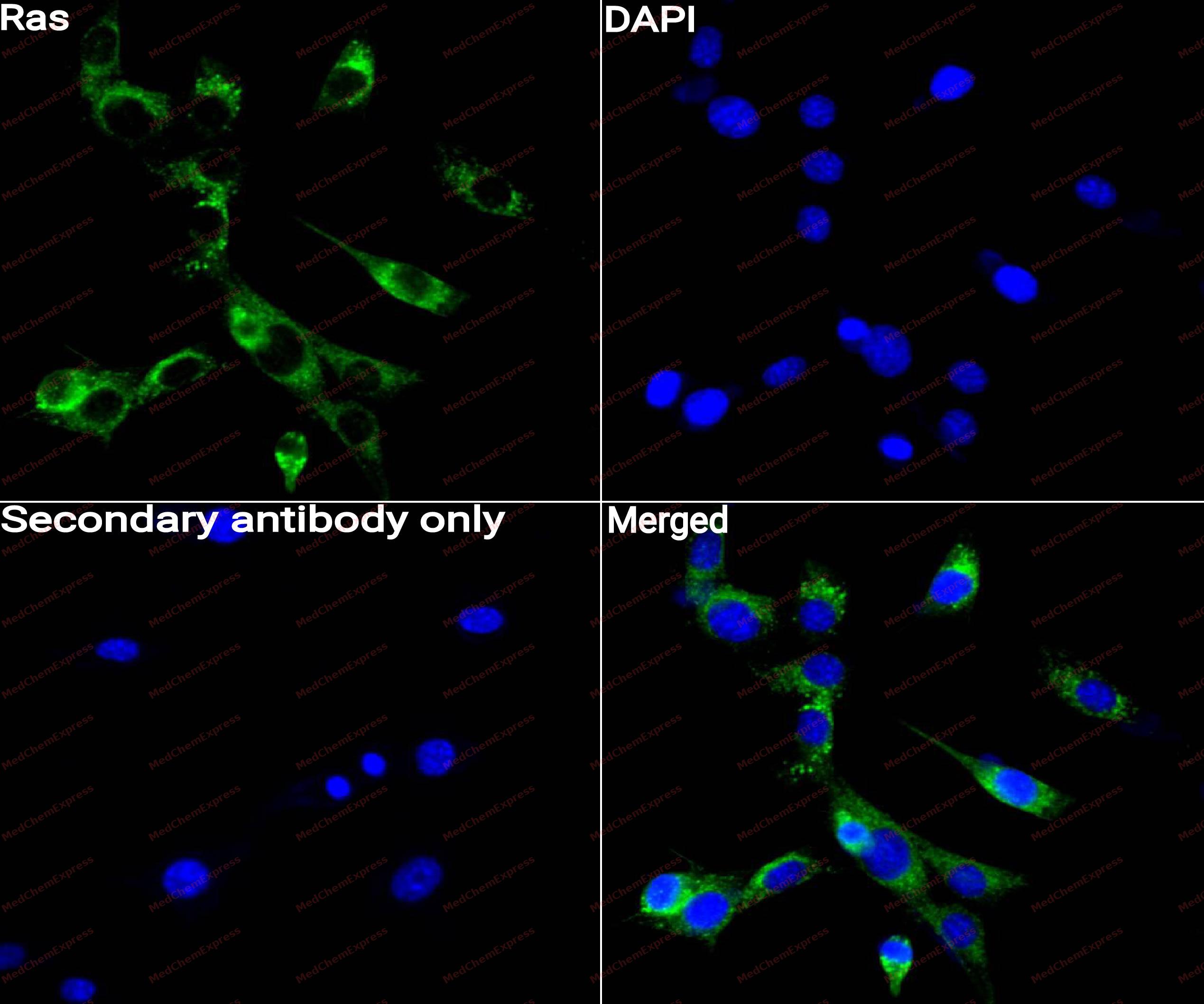 Immunocytochemistry analysis of NIH3T3 cells labeling Ras with Ras Antibody (HY-P80298) at 1/50 dilution. Cells were fixed in 4% paraformaldehyde for 15 minutes at room temperature, permeabilized with 0.1% Triton X-100 for 10 minutes at room temperature, then blocked with QuickBlock™ Blocking Buffer for Immunol Staining for 10 min at room temperature. Cells were then incubated with Ras Antibody (HY-P80298)at 1/50 dilution in QuickBlock™ Blocking Buffer for Immunol Staining at 4 ℃. Alexa Fluor® 488-conjugated AffiniPure Goat Anti-Rabbit IgG H&L(HY-P8002,Green) was used as the secondary antibody at 1/1,000 dilution. PBS instead of the primary antibody was used as the secondary antibody only control. The Nuclear counterstain was DAPI (Blue).


Background
-
Function
Ras is the name given to a family of related proteins which is ubiquitously expressed in all cell lineages and organs. All Ras protein family members belong to a class of protein called small GTPase, and are involved in transmitting signals within cells. Ras is the prototypical member of the Ras superfamily of proteins, which are all related in 3D structure and regulate diverse cell behaviours. When Ras is 'switched on' by incoming signals, it subsequently switches on other proteins, which ultimately turn on genes involved in cell growth, differentiation and survival. As a result, mutations in ras genes can lead to the production of permanently activated Ras proteins. This can cause unintended and overactive signalling inside the cell, even in the absence of incoming signals. Because these signals result in cell growth and division, overactive Ras signaling can ultimately lead to cancer. The 3 Ras genes in humans (HRAS,KRAS, and NRAS) are the most common oncogenes in human cancer; Ras inhibitors are being studied as a treatment for cancer, and other diseases with Ras overexpression.
-
Subcellular Localization
Cell membrane; Lipid-anchor; Cytoplasmic side; Golgi apparatus membrane; Lipid-anchor
-
Subunit
Interacts (active GTP-bound form preferentially) with RGS14 (By similarity). Interacts (active GTP-bound form) with RASSF7 (PubMed:21278800). Interacts (active GTP-bound form) with both SHOC2 and PP1c (all isoforms) to form a tertiary complex; SHOC2 and PP1c preferably bind M-Ras/MRAS, but they also bind K-Ras/KRAS, N-Ras/NRAS and H-Ras/HRAS (PubMed:36175670, PubMed:35768504, PubMed:35831509, PubMed:35830882)
-
SwissProt ID
-
Synonyms
NRAS; HRAS; KRAS
-
Research Field
Cell Biology
Documentation
-
Data Sheet (262 KB)
-
SDS (251 KB)
- English - EN (251 KB)
- Français - FR (251 KB)
- Deutsch - DE (251 KB)
- Norwegian - NO (251 KB)
- Español - ES (251 KB)
- Swedish - SV (251 KB)
- Italian - IT (251 KB)
- Korean - KR (251 KB)
- Portuguese - PT (251 KB)
-
User Guide for Antibodies (1077 KB)
References
[1]. Wan X, et al. The Prognostic Value of HRAS mRNA Expression in Cutaneous Melanoma. Biomed Res Int. 2017;2017:5356737. [Content Brief]
[2]. Wikipedia.
[3]. Parker JA, et al. The K-Ras, N-Ras, and H-Ras Isoforms: Unique Conformational Preferences and Implications for Targeting Oncogenic Mutants. Cold Spring Harb Perspect Med. 2018 Aug 1;8(8):a031427. [Content Brief]
[7]. Hobbs GA, et al. RAS isoforms and mutations in cancer at a glance. J Cell Sci. 2016 Apr 1;129(7):1287-92. [Content Brief]

 Powered by Bioz
