



MGST1 Antibody (YA1636)
(Synonyms: Microsomal GST-I)
Based on 1 Customer Validation
MGST1 Antibody (YA1636) is a Rabbit-derived and non-conjugated IgG monoclonal antibody, targeting to MGST1.
-
Host:
Rabbit
-
Isotype:
IgG
-
Application:
WB, IHC-P
-
Reactivity :
Human, Rat
-
Formulation:
Supplied in 50mM Tris-Glycine(pH 7.4), 0.15M NaCl, 40% Glycerol, 0.01% Sodium azide and 0.05% BSA
-
Conjugation:
Non-conjugated


To place orders, for customer services and technical support, please contact: MedChemExpress USA
Tel: 609-228-6898 E-mail: [email protected] [email protected]





Applications
| Application |
WB

WB: Western Blot
|
IHC-P
IHC-P: Immunohistochemistry-Paraffin
|
|---|---|---|
| Dilution Ratio | 1:500-1:1000 | 1:50-1:100 |


Product Details
MGST1 Antibody (YA1636) is a Rabbit-derived and non-conjugated IgG monoclonal antibody, targeting to MGST1.
-
Host Rabbit

-
Clonality Recombinant,Monoclonal

-
Species ReactivityHuman, Rat
-
Observed Molecular WeightObserved band size: 18 kDa
Note: Due to possible protein modifications or aggregation, the molecular weight should be confirmed by actual measurement, and the predicted value is for reference only.

-
Calculated Molecular Weight Predicted band size: 18 kDa

Entrez Gene: 4257 Human ; 56615 Mouse ; 171341 Rat
SwissProt: P10620 Human ; Q91VS7 Mouse ; P08011 Rat
OMIM: 138330 Human
A synthetic peptide of human Microsomal Glutathione S-transferase 1
Endogenous
Affinity Purified
Non-conjugated
Unmodified
IgG


Product Properties
-
Appearance
Solution
-
Formulation
Supplied in 50mM Tris-Glycine(pH 7.4), 0.15M NaCl, 40% Glycerol, 0.01% Sodium azide and 0.05% BSA
-
Concentration
Batch-dependent, Please check the COA for the concentration of each lot. Check Lot Concentration
-
Storage & Stability
Stored at -20°C for 1 year. Avoid repeated freeze / thaw cycles.
-
Shipping
Shipping with blue ice.


Verification Images
-
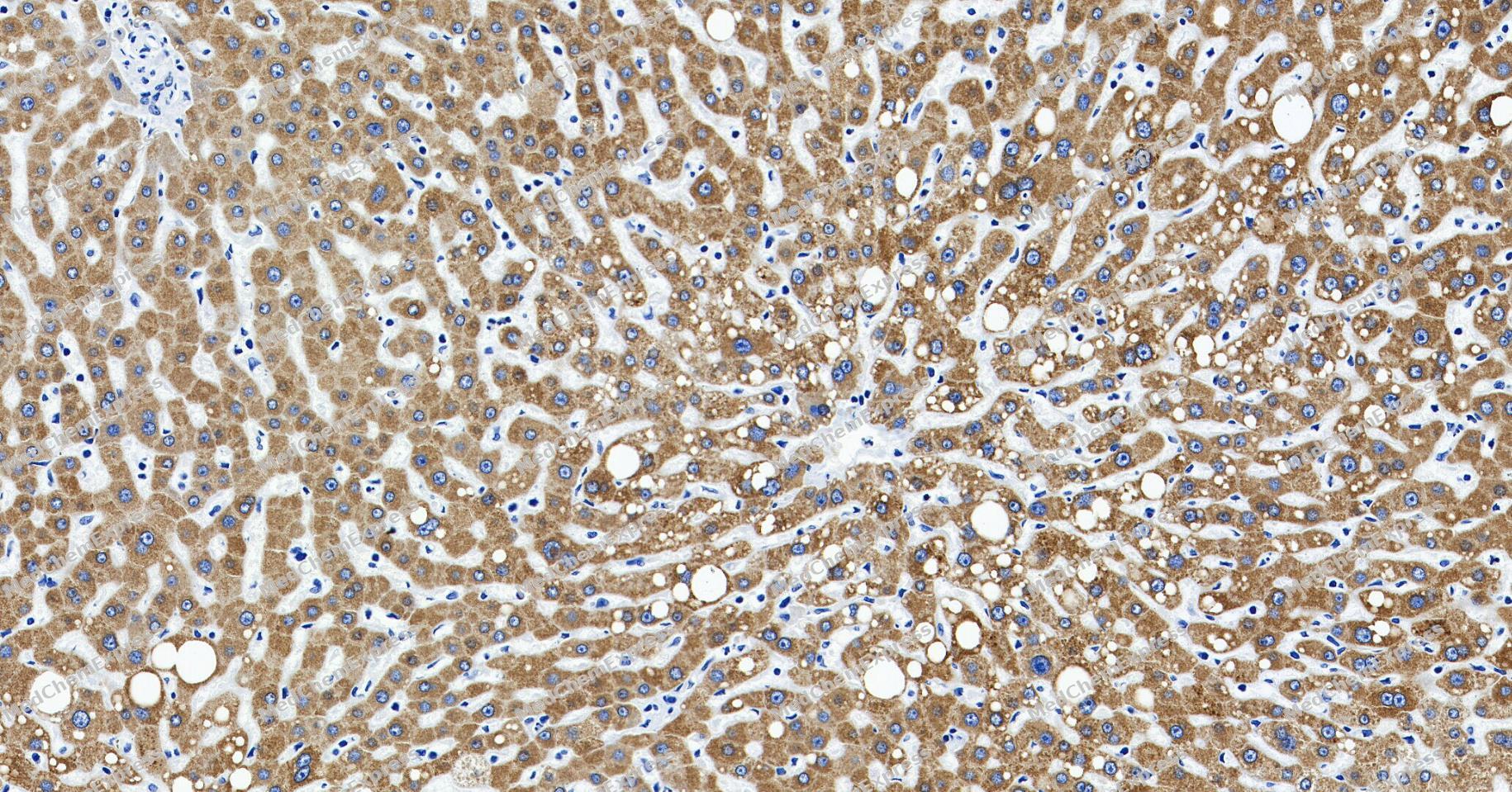 Immunohistochemical analysis of paraffin-embedded human liver tissue using MGST1 Antibody (YA1636). The section was pre-treated using heat mediated antigen retrieval with Tris/EDTA buffer (pH 9.0) for 20 minutes. The tissues were probed with the primary antibody (HY-P81891, 1/100) overnight at 4℃. The detection was performed using an HRP conjugated compact polymer system. DAB was used as the chromogen. Tissues were counterstained with hematoxylin and mounted with neutral balsam.
 -
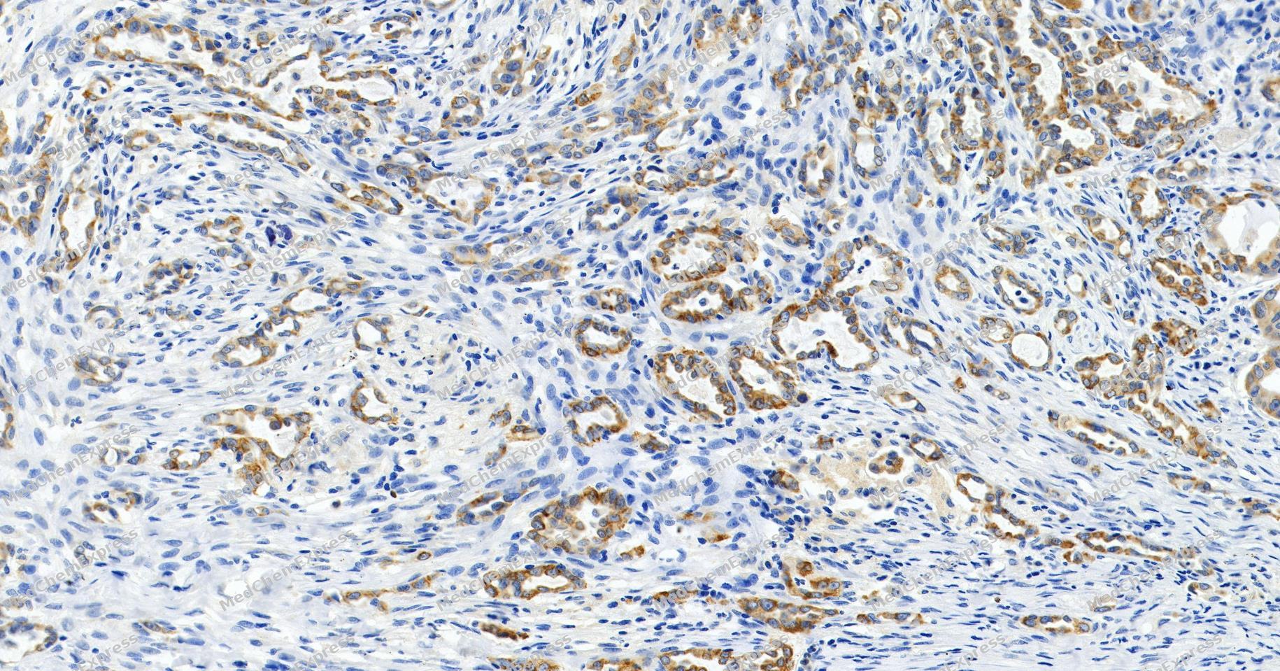 Immunohistochemical analysis of paraffin-embedded human lung adenocarcinoma tissue using MGST1 Antibody (YA1636). The section was pre-treated using heat mediated antigen retrieval with Tris/EDTA buffer (pH 9.0) for 20 minutes. The tissues were probed with the primary antibody (HY-P81891, 1/100) overnight at 4℃. The detection was performed using an HRP conjugated compact polymer system. DAB was used as the chromogen. Tissues were counterstained with hematoxylin and mounted with neutral balsam.
 -
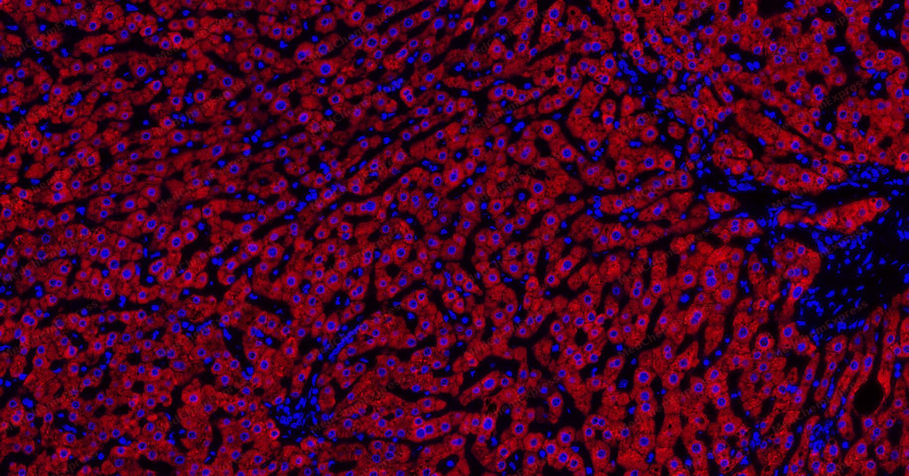 Tyramide signaling amplification based immunofluorescence analysis of paraffin-embedded human liver tissue using MGST1 Antibody (YA1636). The section was pre-treated using heat mediated antigen retrieval with Tris/EDTA buffer (pH 9.0) for 20 minutes. The tissues were probed with the primary antibody (HY-P81891, 1/300) overnight at 4℃. The detection was performed using an HRP conjugated compact polymer and fluorescent tyramide signal amplification system.Immunostaining was performed with Vari Fluor 594 TSA (200×)(HY-D1835). Tissues were counterstained with DAPI (blue) and mounted with Anti-fade fluorescence mounting medium.
 -
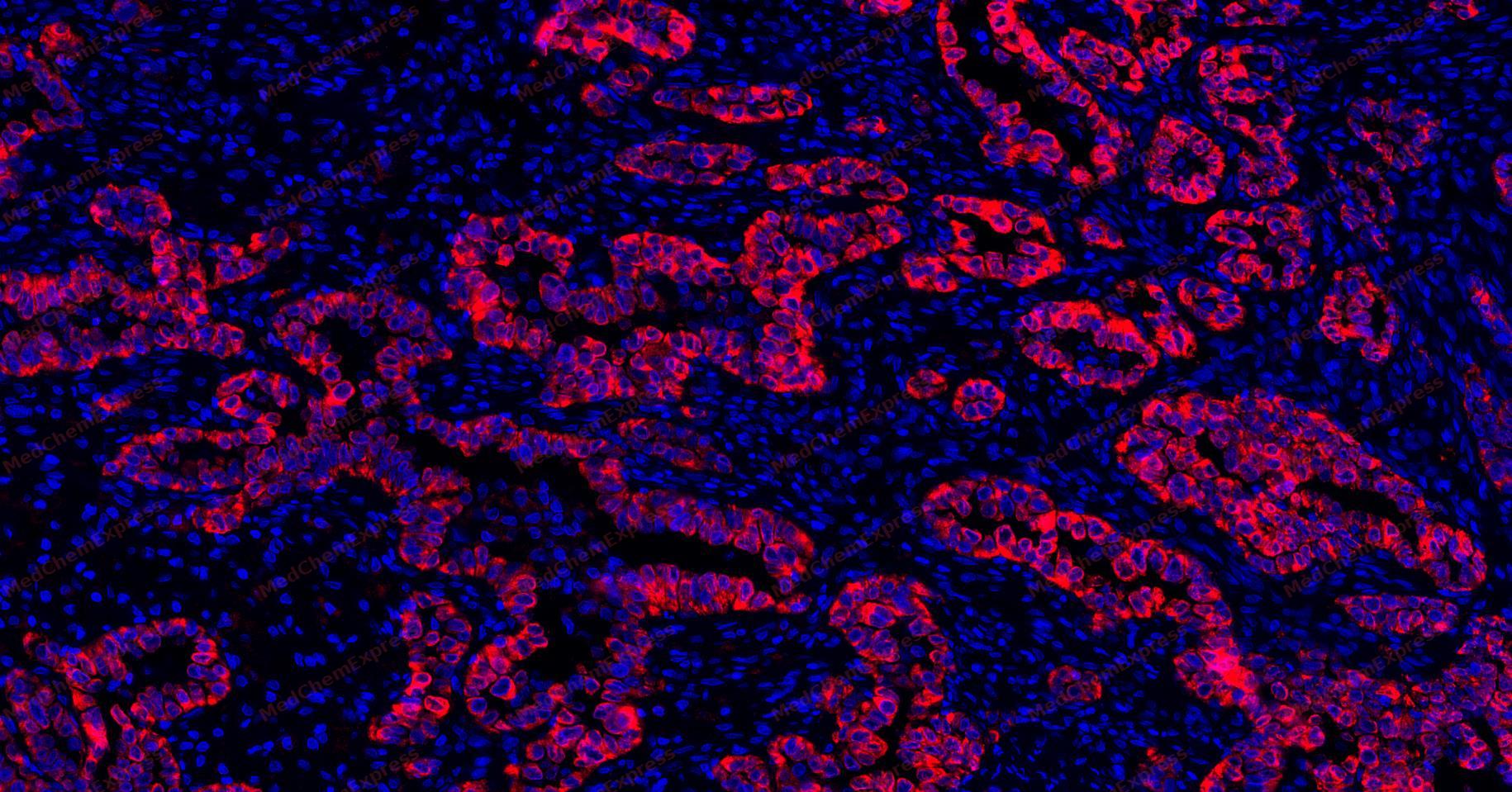 Tyramide signaling amplification based immunofluorescence analysis of paraffin-embedded human lung adenocarcinoma tissue using MGST1 Antibody (YA1636). The section was pre-treated using heat mediated antigen retrieval with Tris/EDTA buffer (pH 9.0) for 20 minutes. The tissues were probed with the primary antibody (HY-P81891, 1/300) overnight at 4℃. The detection was performed using an HRP conjugated compact polymer and fluorescent tyramide signal amplification system.Immunostaining was performed with Vari Fluor 594 TSA (200×)(HY-D1835). Tissues were counterstained with DAPI (blue) and mounted with Anti-fade fluorescence mounting medium.
 -
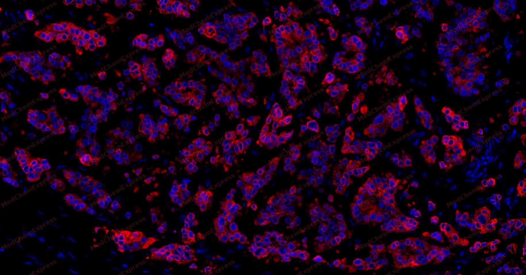 Tyramide signaling amplification based immunofluorescence analysis of paraffin-embedded human breast cancer tissue using MGST1 Antibody (YA1636). The section was pre-treated using heat mediated antigen retrieval with Tris/EDTA buffer (pH 9.0) for 20 minutes. The tissues were probed with the primary antibody (HY-P81891, 1/300) overnight at 4℃. The detection was performed using an HRP conjugated compact polymer and fluorescent tyramide signal amplification system.Immunostaining was performed using a Bicolor signal amplification fluorescence staining kit. Tissues were counterstained with DAPI (blue) and mounted with Anti-fade fluorescence mounting medium.
 -
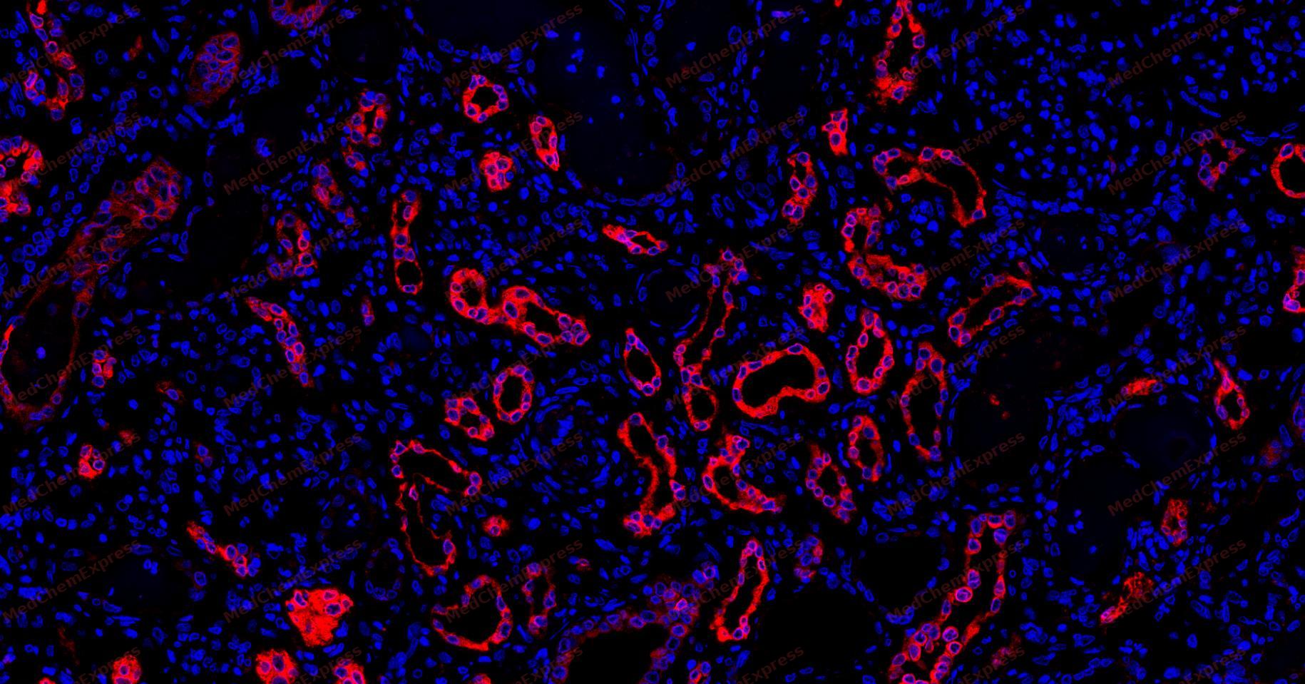 Tyramide signaling amplification based immunofluorescence analysis of paraffin-embedded human kidney tissue using MGST1 Antibody (YA1636). The section was pre-treated using heat mediated antigen retrieval with Tris/EDTA buffer (pH 9.0) for 20 minutes. The tissues were probed with the primary antibody (HY-P81891, 1/300) overnight at 4℃. The detection was performed using an HRP conjugated compact polymer and fluorescent tyramide signal amplification system.Immunostaining was performed using a Bicolor signal amplification fluorescence staining kit. Tissues were counterstained with DAPI (blue) and mounted with Anti-fade fluorescence mounting medium.


Background
-
Function
MGST1 is a Conjugation of reduced glutathione to a wide number of exogenous and endogenous hydrophobic electrophiles
-
Subcellular Localization
Endoplasmic reticulum membrane; Multi-pass membrane protein; Mitochondrion outer membrane
-
Expression
Tissue_specificity:Highly expressed in liver -
Isoforms & Post-Translational Modification
P10620 has 2 isomers: P10620-1: 17599 Da (predicted); P10620-2: 9984 Da (predicted).
-
Subunit
Homotrimer; The trimer binds only one molecule of glutathione
-
SwissProt ID
-
Synonyms
Microsomal GST-I
-
Research Field
Cell Biology


Documentation
-
Data Sheet (234 KB)
-
SDS (251 KB)
- English - EN (251 KB)
- Français - FR (251 KB)
- Deutsch - DE (251 KB)
- Norwegian - NO (251 KB)
- Español - ES (251 KB)
- Swedish - SV (251 KB)
- Italian - IT (251 KB)
- Korean - KR (251 KB)
- Portuguese - PT (251 KB)
-
User Guide for Antibodies (1077 KB)

 Powered by Bioz
