



MMP8 Antibody (YA3030)
(Synonyms: HNC; CLG1; MMP-8; PMNL-CL; COLLAGENASE I; NEUTROPHIL; matrix metalloproteinase 8)
Based on 1 Customer Validation
MMP8 Antibody (YA3030) is a Rabbit-derived and non-conjugated IgG monoclonal antibody, targeting to MMP8.
-
Host:
Rabbit
-
Isotype:
IgG
-
Application:
WB, IHC-P, ICC/IF, IP, FC
-
Reactivity :
Human, Mouse, Rat
-
Formulation:
Supplied in Rabbit IgG in 10mM phosphate buffered saline , pH 7.4, 150mM sodium chloride, 0.05% BSA, 0.02% sodium azide and 50% glycerol.
-
Conjugation:
Non-conjugated


To place orders, for customer services and technical support, please contact: MedChemExpress USA
Tel: 609-228-6898 E-mail: [email protected] [email protected]





Applications
| Application |
WB

WB: Western Blot
|
IHC-P
IHC-P: Immunohistochemistry-Paraffin
|
ICC/IF
ICC/IF: Immunocytochemistry/
Immunofluorescence |
IP
IP: Immunoprecipitation
|
FC
FC: Flow Cytometry
|
|---|---|---|---|---|---|
| Dilution Ratio | 1:500-1:1000 | 1:50-1:100 | 1:50-1:200 | 1:50 | 1:50-1:100 |


Product Details
MMP8 Antibody (YA3030) is a Rabbit-derived and non-conjugated IgG monoclonal antibody, targeting to MMP8.
-
Host Rabbit

-
Clonality Recombinant,Monoclonal

-
Species ReactivityHuman, Mouse, Rat
-
Observed Molecular WeightObserved band size: 53-70 kDa
Note: Due to possible protein modifications or aggregation, the molecular weight should be confirmed by actual measurement, and the predicted value is for reference only.

-
Calculated Molecular Weight Predicted band size: 53 kDa

Entrez Gene: 4317 Human ; 17394 Mouse ; 63849 Rat
SwissProt: P22894 Human ; O70138 Mouse ; O88766 Rat
OMIM: 120355 Human
A synthesized peptide derived from human MMP8 aa150-200.
Endogenous
Affinity Chromatography
Non-conjugated
Unmodified
IgG


Product Properties
-
Appearance
Solution
-
Formulation
Supplied in Rabbit IgG in 10mM phosphate buffered saline , pH 7.4, 150mM sodium chloride, 0.05% BSA, 0.02% sodium azide and 50% glycerol.
-
Concentration
Batch-dependent, Please check the COA for the concentration of each lot. Check Lot Concentration
-
Storage & Stability
Stored at -20°C for 1 year. Avoid repeated freeze / thaw cycles.
-
Shipping
Shipping with blue ice.


Verification Images
-
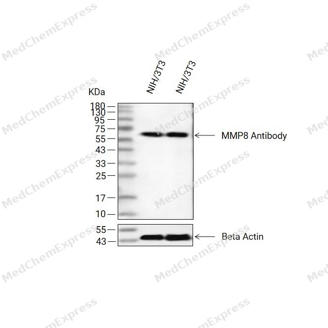 Western blot analysis of extracts from NIH/3T3(lane 2(20ug) and NIH/3T3(lane 3(40ug) using MMP8 Antibody (HY-P83285) Rabbit mAb. Proteins were transferred to a PVDF membrane and blocked with 5% non-fat milk in TBST for 2 hour at room temperature. The primary antibody (1/1000) and Loading control antibody (Beta Actin, HY-P83730, 1/10000) was used in 5% non-fat milk in TBST at 4°C overnight. Goat Anti-Mouse/Rabbit IgG-HRP Secondary Antibody (1/10000) was used for 1 hour at room temperature.
 -
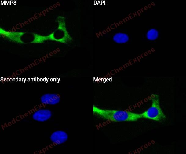 Immunocytochemistry analysis of Hela cells labeling MMP8 with MMP8 Antibody (HY-P83285)at 1/50 dilution. Cells were fixed in 4% paraformaldehyde for 15 minutes at room temperature, permeabilized with 0.1% Triton X-100 for 10 minutes at room temperature, then blocked with QuickBlock™ Blocking Buffer for Immunol Staining for 10 min at room temperature. Cells were then incubated with MMP8 Antibody (HY-P83285) at 1/50 dilution in QuickBlock™ Blocking Buffer for Immunol Staining at 4 ℃. Alexa Fluor® 488-conjugated AffiniPure Goat Anti-Rabbit IgG H&L(HY-P8002, Green) was used as the secondary antibody at 1/1,000 dilution. PBS instead of the primary antibody was used as the secondary antibody only control. The Nuclear counterstain was DAPI (Blue).
 -
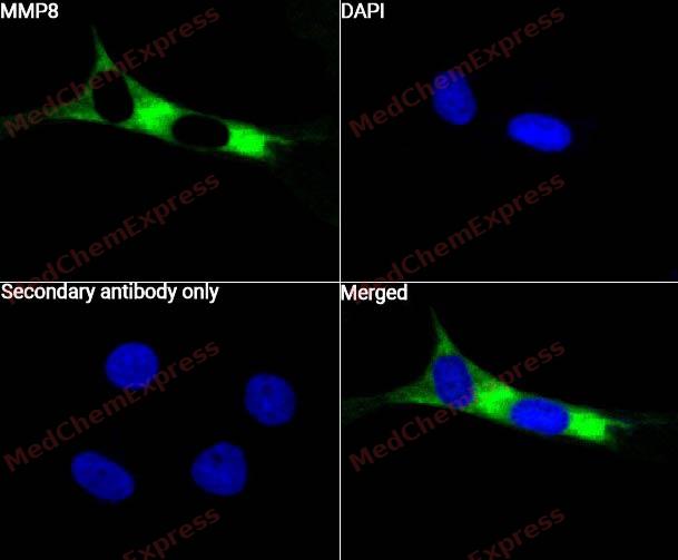 Immunocytochemistry analysis of Hela cells labeling MMP8 with MMP8 Antibody (HY-P83285) at 1/100 dilution. Cells were fixed in 4% paraformaldehyde for 15 minutes at room temperature, permeabilized with 0.1% Triton X-100 for 10 minutes at room temperature, then blocked with QuickBlock™ Blocking Buffer for Immunol Staining for 10 min at room temperature. Cells were then incubated with MMP8 Antibody (HY-P83285) at 1/100 dilution in QuickBlock™ Blocking Buffer for Immunol Staining at 4 ℃. Alexa Fluor® 488-conjugated AffiniPure Goat Anti-Rabbit IgG H&L(HY-P8002, Green) was used as the secondary antibody at 1/1,000 dilution. PBS instead of the primary antibody was used as the secondary antibody only control. The Nuclear counterstain was DAPI (Blue).
 -
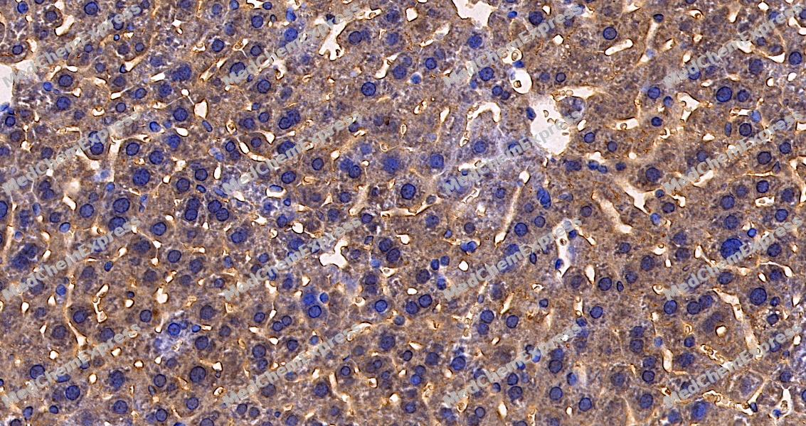 Immunohistochemical analysis of paraffin-embedded mouse liver tissue using MMP8 Antibody. The section was pre-treated using heat mediated antigen retrieval with sodium citrate buffer (pH 6.0) for 8 minutes. The tissues were blocked in QuickBlock for 20 minutes at room temperature, washed with ddH2O and PBS, and then probed with the primary antibody at 1/100 dilution in 4℃ overnight. The detection was performed using an HRP conjugated compact polymer system. DAB was used as the chromogen. Tissues were counterstained with hematoxylin and mounted with DPX.
 -
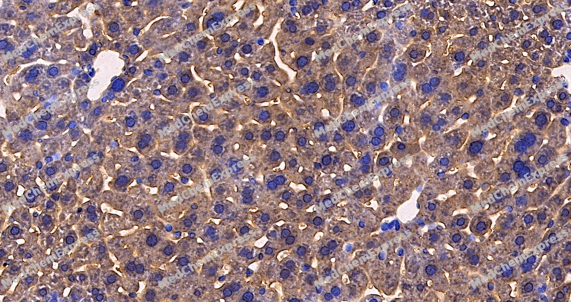 Immunohistochemical analysis of paraffin-embedded mouse liver tissue using MMP8 Antibody. The section was pre-treated using heat mediated antigen retrieval with sodium citrate buffer (pH 6.0) for 8 minutes. The tissues were blocked in QuickBlock for 20 minutes at room temperature, washed with ddH2O and PBS, and then probed with the primary antibody at 1/100 dilution in 4℃ overnight. The detection was performed using an HRP conjugated compact polymer system. DAB was used as the chromogen. Tissues were counterstained with hematoxylin and mounted with DPX.


Background
-
Function
MMP-8 (matrix metalloproteinase-8), also known as neutrophil collagenase or collagenase-2, is a Zn2+-dependent extracellular matrix protease that primarily cleaves interstitial collagens and contributes to tissue remodeling and inflammatory responses[1][2]. Mechanistically, MMP-8 regulates extracellular matrix turnover through degradation of collagen substrates and can influence inflammatory cell trafficking by generating bioactive collagen-derived peptides that promote neutrophil migration within collagen-rich tissues[3][4]. Beyond structural matrix degradation, MMP-8 has been implicated in acute and chronic inflammatory processes, where excessive proteolytic activity contributes to tissue injury and disease progression[2][5]. In disease settings, elevated MMP-8 activity is strongly associated with periodontal and peri-implant tissue destruction, and active MMP-8 serves as a marker of ongoing collagen degradation in inflammatory lesions[5][6]. Experimental and clinical studies have also linked MMP-8 to sepsis, aortic aneurysm development, and cancer-related processes, highlighting its context-dependent roles in pathological tissue remodeling and host responses[7][8][9]. Compared with related collagenases such as MMP-1 and MMP-13, MMP-8 is distinguished by its predominant storage in neutrophil granules and its specialized contribution to neutrophil-driven inflammatory responses, although all three enzymes share collagenolytic activity[2][10]. For experimental applications, selective MMP-8 inhibitors have been developed and extensively characterized, providing useful tools for investigating collagenase-dependent mechanisms and evaluating therapeutic modulation of inflammatory tissue destruction[11][12].
-
Subcellular Localization
Cytoplasmic granule; Secreted, extracellular space, extracellular matrix
-
Expression
Tissue_specificity:Neutrophils -
SwissProt ID
-
Synonyms
HNC; CLG1; MMP-8; PMNL-CL; COLLAGENASE I; NEUTROPHIL; matrix metalloproteinase 8
-
Research Field
Cardiovascular


Documentation
-
Data Sheet (262 KB)
-
SDS (251 KB)
- English - EN (251 KB)
- Français - FR (251 KB)
- Deutsch - DE (251 KB)
- Norwegian - NO (251 KB)
- Español - ES (251 KB)
- Swedish - SV (251 KB)
- Italian - IT (251 KB)
- Korean - KR (251 KB)
- Portuguese - PT (251 KB)
-
User Guide for Antibodies (1077 KB)
References
[1]. Maymon E, et al. Human neutrophil collagenase (matrix metalloproteinase 8) in parturition, premature rupture of the membranes, and intrauterine infection. Am J Obstet Gynecol. 2000 Jul;183(1):94-9. [Content Brief]
[2]. Lee CM, et al. Using positron emission tomography to facilitate CNS drug development. Trends Pharmacol Sci. 2006 Jun;27(6):310-6. [Content Brief]
[3]. Lin M, et al. Matrix metalloproteinase-8 facilitates neutrophil migration through the corneal stromal matrix by collagen degradation and production of the chemotactic peptide Pro-Gly-Pro. Am J Pathol. 2008 Jul;173(1):144-53. [Content Brief]
[5]. Al-Majid A, et al. Matrix Metalloproteinase-8 as an Inflammatory and Prevention Biomarker in Periodontal and Peri-Implant Diseases. Int J Dent. 2018 Sep 16;2018:7891323. [Content Brief]
[6]. Romanelli R, et al. Activation of neutrophil collagenase in periodontitis. Infect Immun. 1999 May;67(5):2319-26. [Content Brief]
[7]. Liu H, et al. Baseline serum matrix metalloproteinase-8 and 28-day mortality in sepsis at ICU admission. Crit Care. 2026 Jan 31;30(1):51. [Content Brief]
[8]. Wilson WR, et al. Matrix metalloproteinase 8 (neutrophil collagenase) in the pathogenesis of abdominal aortic aneurysm. Br J Surg. 2005 Jul;92(7):828-33. [Content Brief]
[9]. Juurikka K, et al. The Role of MMP8 in Cancer: A Systematic Review. Int J Mol Sci. 2019 Sep 11;20(18):4506. [Content Brief]
[10]. Medicine-and-dentistry/neutrophil-collagenase. sciencedirect.topics.
[11]. Matter H, et al. Quantitative structure-activity relationship of human neutrophil collagenase (MMP-8) inhibitors using comparative molecular field analysis and X-ray structure analysis. J Med Chem. 1999 Jun 3;42(11):1908-20. [Content Brief]

 Powered by Bioz
