



MYH9 Antibody (YA3113)
(Synonyms: MHA; FTNS; EPSTS; BDPLT6; DFNA17; MATINS; NMMHCA; NMHC-II-A; NMMHC-IIA)
Based on 1 Customer Validation
MYH9 Antibody (YA3113) is a Rabbit-derived and non-conjugated IgG monoclonal antibody, targeting to MYH9.
-
Host:
Rabbit
-
Isotype:
IgG
-
Application:
WB, IHC-P, IP
-
Reactivity :
Human, Rat
-
Formulation:
Supplied in 50mM Tris-Glycine(pH 7.4), 0.15M NaCl, 40% Glycerol, 0.01% Sodium azide and 0.05% BSA
-
Conjugation:
Non-conjugated


To place orders, for customer services and technical support, please contact: MedChemExpress USA
Tel: 609-228-6898 E-mail: [email protected] [email protected]





Applications
| Application |
WB

WB: Western Blot
|
IHC-P
IHC-P: Immunohistochemistry-Paraffin
|
IP
IP: Immunoprecipitation
|
|---|---|---|---|
| Dilution Ratio | 1:500-1:1000 | 1:50-1:100 | 1:20 |


Product Details
MYH9 Antibody (YA3113) is a Rabbit-derived and non-conjugated IgG monoclonal antibody, targeting to MYH9.
-
Host Rabbit

-
Clonality Recombinant,Monoclonal

-
Species ReactivityHuman, Rat
-
Observed Molecular WeightObserved band size: 227 kDa
Note: Due to possible protein modifications or aggregation, the molecular weight should be confirmed by actual measurement, and the predicted value is for reference only.

-
Calculated Molecular Weight Predicted band size: 227 kDa

Entrez Gene: 4627 Human ; 17886 Mouse ;
SwissProt: P35579 Human ; Q8VDD5 Mouse ; Q62812 Rat
OMIM: 155100 Human
A synthetic peptide of human non-muscle Myosin IIA
Endogenous
Affinity Purified
Non-conjugated
Unmodified
IgG


Product Properties
-
Appearance
Solution
-
Formulation
Supplied in 50mM Tris-Glycine(pH 7.4), 0.15M NaCl, 40% Glycerol, 0.01% Sodium azide and 0.05% BSA
-
Concentration
Batch-dependent, Please check the COA for the concentration of each lot. Check Lot Concentration
-
Storage & Stability
Stored at -20°C for 1 year. Avoid repeated freeze / thaw cycles.
-
Shipping
Shipping with blue ice.


Verification Images
-
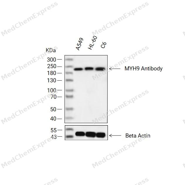 Western blot analysis of extracts from A549(lane 2(20ug) , HL-60(lane 3(20ug) and C6(lane 4(20ug) using MYH9 Antibody (HY-P83368) Rabbit mAb. Proteins were transferred to a PVDF membrane and blocked with 5% non-fat milk in TBST for 2 hour at room temperature. The primary antibody (1/1000) and Loading control antibody (Beta Actin, HY-P80438, 1/10000) was used in 5% non-fat milk in TBST at 4°C overnight. Goat Anti-Mouse/Rabbit IgG-HRP Secondary Antibody (1/10000) was used for 1 hour at room temperature.
 -
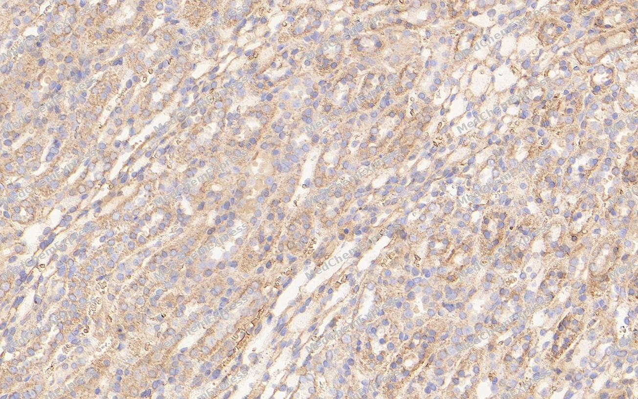 Immunohistochemical analysis of paraffin-embedded Rat kidney tissue using MYH9 Antibody (YA3113). The section was pre-treated using heat mediated antigen retrieval with Tris-EDTA buffer (pH 9.0) for 8 minutes. The tissues were blocked in QuickBlock for 20 minutes at room temperature, washed with ddH2O and PBS, and then probed with the primary antibody (HY-P83368, 1/100) in 4℃ overnight. The detection was performed using an HRP conjugated compact polymer system. DAB was used as the chromogen. Tissues were counterstained with hematoxylin and mounted with DPX.
 -
Immunohistochemical analysis of paraffin-embedded Rat kidney tissue using MYH9 Antibody (YA3113). The section was pre-treated using heat mediated antigen retrieval with Tris-EDTA buffer (pH 9.0) for 8 minutes. The tissues were blocked in QuickBlock for 20 minutes at room temperature, washed with ddH2O and PBS, and then probed with the primary antibody (HY-P83368, 1/100) in 4℃ overnight. The detection was performed using an HRP conjugated compact polymer system. DAB was used as the chromogen. Tissues were counterstained with hematoxylin and mounted with DPX.


Background
-
Function
MYH9 is a Cellular myosin that appears to play a role in cytokinesis, cell shape, and specialized functions such as secretion and capping. Required for cortical actin clearance prior to oocyte exocytosis. Promotes cell motility in conjunction with S100A4. During cell spreading, plays an important role in cytoskeleton reorganization, focal contact formation (in the margins but not the central part of spreading cells), and lamellipodial retraction; this function is mechanically antagonized by MYH10; (Microbial infection) Acts as a receptor for herpes simplex virus 1/HHV-1 envelope glycoprotein B[1][2].
-
Subcellular Localization
Cytoplasm, cytoskeleton; Cytoplasm, cell cortex; Cytoplasmic vesicle, secretory vesicle, Cortical granule; Cell membrane; Cell membrane
-
Expression
Tissue_specificity:In the kidney, expressed in the glomeruli. Also expressed in leukocytes -
Isoforms & Post-Translational Modification
P35579 has 2 isomers: P35579-1: 226532 Da (predicted); P35579-2: 159864 Da (predicted).
ISGylated;Ubiquitination -
Subunit
Myosin is a hexameric protein that consists of 2 heavy chain subunits (MHC), 2 alkali light chain subunits (MLC) and 2 regulatory light chain subunits (MLC-2). Interacts with RASIP1 (By similarity). Interacts with DDR1 (By similarity). Interacts with PDLIM2 (By similarity). Interacts with SVIL (PubMed:12917436, PubMed:17925381). Interacts with HTRA3 (PubMed:22229724). Interacts with Myo7a (By similarity). Interacts with CFAP95 (PubMed:28345668). Interacts with LIMCH1; independently of the integration of MYH9 into the myosin complex (PubMed:28228547). Interacts with RAB3A (PubMed:27325790). Interacts with ZBED4 (PubMed:22693546). Interacts with S100A4; this interaction increases cell motility (PubMed:16707441)
-
SwissProt ID
-
Synonyms
MHA; FTNS; EPSTS; BDPLT6; DFNA17; MATINS; NMMHCA; NMHC-II-A; NMMHC-IIA
-
Research Field
Signal Transduction


Documentation
-
Data Sheet (260 KB)
-
SDS (251 KB)
- English - EN (251 KB)
- Français - FR (251 KB)
- Deutsch - DE (251 KB)
- Norwegian - NO (251 KB)
- Español - ES (251 KB)
- Swedish - SV (251 KB)
- Italian - IT (251 KB)
- Korean - KR (251 KB)
- Portuguese - PT (251 KB)
-
User Guide for Antibodies (1077 KB)
References
[1]. Li ZH, et al. The S100A4 metastasis factor regulates cellular motility via a direct interaction with myosin-IIA. Cancer Res. 2006 May 15;66(10):5173-80. [Content Brief]
[2]. Betapudi V, et al. Myosin II motor proteins with different functions determine the fate of lamellipodia extension during cell spreading. PLoS One. 2010 Jan 5;5(1):e8560. [Content Brief]

 Powered by Bioz
