



PKM2 Antibody
(Synonyms: PKM; OIP3; PK2; PK3; PKM2; Pyruvate kinase isozymes M1/M2; Cytosolic thyroid hormone-binding protein; CTHBP; Opa-interacting protein 3; OIP-3; Pyruvate kinase 2/3; Pyruvate kinase muscle isozyme; Thyroid hormone-binding protein 1; THBP1; Tu)Based on 2 publication(s) in Google Scholar
PKM2 Antibody is a Rabbit-derived and non-conjugated IgG polyclonal antibody, targeting to PKM2.
-
Host:
Rabbit
-
Isotype:
IgG
-
Application:
WB, IHC-P, ICC/IF, FC
-
Reactivity :
Human, Mouse, Rat
-
Formulation:
Supplied in 1*PBS (pH 7.3), 50% glycerol and 0.5% BSA. Preservative: 0.02% sodium azide.
-
Conjugation:
Non-conjugated


To place orders, for customer services and technical support, please contact: MedChemExpress USA
Tel: 609-228-6898 E-mail: [email protected] [email protected]



Publications Citing Use of MedChemExpress (MCE) PKM2 Antibody
More

Applications
| Application |
WB

WB: Western Blot
|
IHC-P
IHC-P: Immunohistochemistry-Paraffin
|
ICC/IF
ICC/IF: Immunocytochemistry/
Immunofluorescence |
FC
FC: Flow Cytometry
|
|---|---|---|---|---|
| Dilution Ratio | 1:500-1:1000 | 1:50-1:200 | 1:50-1:200 | 1:50 |


Product Details
PKM2 Antibody is a Rabbit-derived and non-conjugated IgG polyclonal antibody, targeting to PKM2.
-
Host Rabbit

-
Clonality Polyclonal

-
Species ReactivityHuman, Mouse, Rat
-
Observed Molecular WeightObserved band size: 58 kDa
Note: Due to possible protein modifications or aggregation, the molecular weight should be confirmed by actual measurement, and the predicted value is for reference only.

-
Calculated Molecular Weight Predicted band size: 58 kDa

Synthetic peptide corresponding to Human PKM2 AA range 50-100.
Endogenous
affinity purified
Non-conjugated
Unmodified
IgG


Product Properties
-
Appearance
Solution
-
Formulation
Supplied in 1*PBS (pH 7.3), 50% glycerol and 0.5% BSA. Preservative: 0.02% sodium azide.
-
Concentration
Batch-dependent, Please check the COA for the concentration of each lot. Check Lot Concentration
-
Storage & Stability
Stored at -20°C for 1 year. Avoid repeated freeze / thaw cycles.
-
Shipping
Shipping with blue ice.


Publications (2)
-
Journal Impact Factor
-
Most Recent
-
J Exp Clin Cancer Res
Disruption of HSPA8-GEMIN5 interaction suppresses colorectal cancer by impaired splicing-translation coupling-mediated proteostasis imbalance. [Abstract]2026 Jan 16;45(1):47. PMID: 41545989 -
Clin Transl Oncol
Apigenin 7-glucoside reprograms tumor metabolism and enhances immunotherapy efficacy in colorectal cancer via DLX5. [Abstract]2026 Feb 25. PMID: 41739401


Verification Images
-
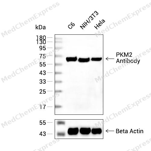 Western blot analysis of extracts from C6(lane 2(20μg) ,NIH/3T3(lane 3(20μg) and Hela(lane 4(20ug)using PKM2 Antibody (HY-P80870) Rabbit mAb. Proteins were transferred to a PVDF membrane and blocked with 5% non-fat milk in TBST for 2 hour at room temperature. The primary antibody (1/1000) and Loading control antibody (Beta Actin, HY-P80438, 1/10000) was used in 5% non-fat milk in TBST at 4°C overnight. Goat Anti-Mouse/Rabbit IgG-HRP Secondary Antibody (1/10000) was used for 1 hour at room temperature.
 -
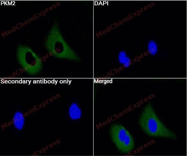 Immunocytochemistry analysis of A549 cells labeling PKM2 with beta PKM2 Antibody (HY-P80870) at 1/50 dilution. Cells were fixed in 4% paraformaldehyde for 15 minutes at room temperature, then blocked with QuickBlock™ Blocking Buffer for Immunol Staining for 10 min at room temperature. Cells were then incubated with PKM2 Antibody (HY-P80870) at 1/50 dilution in QuickBlock™ Blocking Buffer for Immunol Staining at 4 ℃. Alexa Fluor® 488-conjugated AffiniPure Goat Anti-Rabbit IgG H&L(HY-P8002, Green) was used as the secondary antibody at 1/1,000 dilution. PBS instead of the primary antibody was used as the secondary antibody only control. The Nuclear counterstain was DAPI (Blue).
 -
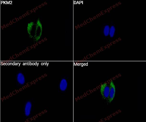 Immunocytochemistry analysis of PC-3 cells labeling PKM2 with beta PKM2 Antibody (HY-P80870) at 1/50 dilution. Cells were fixed in 4% paraformaldehyde for 15 minutes at room temperature, then blocked with QuickBlock™ Blocking Buffer for Immunol Staining for 10 min at room temperature. Cells were then incubated with PKM2 Antibody (HY-P80870) at 1/50 dilution in QuickBlock™ Blocking Buffer for Immunol Staining at 4 ℃. Alexa Fluor® 488-conjugated AffiniPure Goat Anti-Rabbit IgG H&L(HY-P8002, Green) was used as the secondary antibody at 1/1,000 dilution. PBS instead of the primary antibody was used as the secondary antibody only control. The Nuclear counterstain was DAPI (Blue).
 -
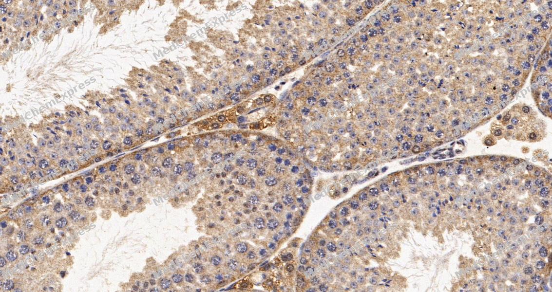 Immunohistochemical analysis of paraffin-embedded mouse testis tissue using PKM2 Antibody. The section was pre-treated using heat mediated antigen retrieval with Tris-EDTA buffer (pH 9.0) for 8 minutes. The tissues were blocked in QuickBlock for 20 minutes at room temperature, washed with ddH2O and PBS, and then probed with the primary antibody at 1/100 dilution in 4℃ overnight. The detection was performed using an HRP conjugated compact polymer system. DAB was used as the chromogen. Tissues were counterstained with hematoxylin and mounted with DPX.
 -
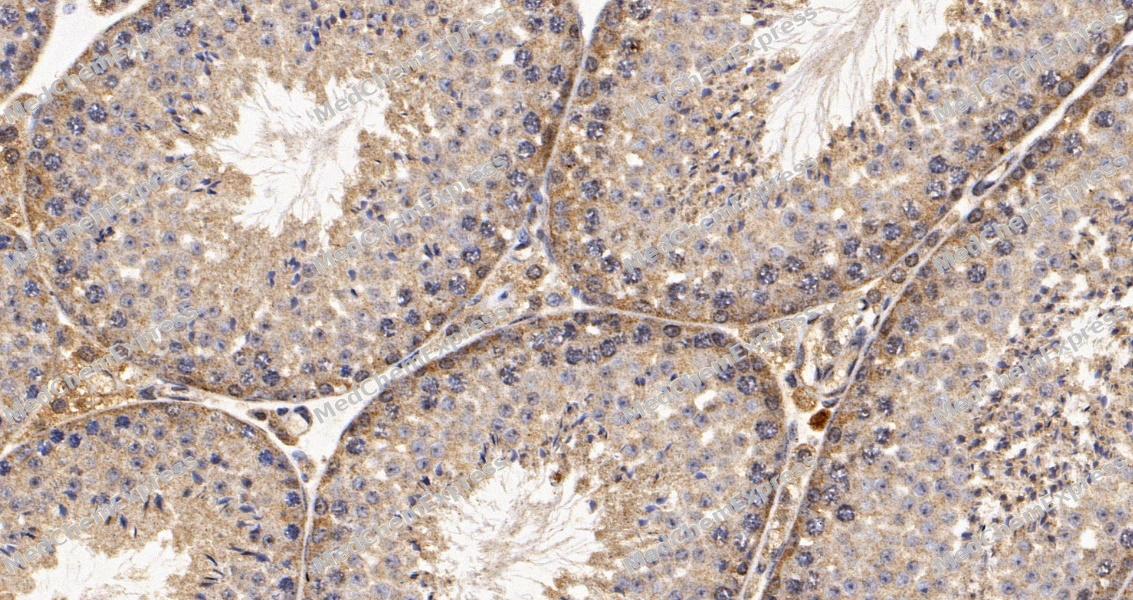 Immunohistochemical analysis of paraffin-embedded mouse testis tissue using PKM2 Antibody. The section was pre-treated using heat mediated antigen retrieval with Tris-EDTA buffer (pH 9.0) for 8 minutes. The tissues were blocked in QuickBlock for 20 minutes at room temperature, washed with ddH2O and PBS, and then probed with the primary antibody at 1/100 dilution in 4℃ overnight. The detection was performed using an HRP conjugated compact polymer system. DAB was used as the chromogen. Tissues were counterstained with hematoxylin and mounted with DPX.
 -
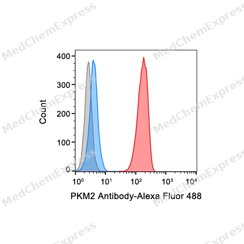 Flow cytometric analysis of 1X10^6 Hela cells labeling PKM2 Antibody (HY-P80870, red). Cells were fixed with 4% paraformaldehyde and permeabilised with 90% methanol. Then stained with the primary antibody at 1/50 dilution for an hour at 4℃. Alexa Fluor® 488-conjugated AffiniPure Goat Anti-Rabbit IgG H&L (HY-P8002) was used as the secondary antibody at 1/1,000 dilution for 30 minutes at 4℃. Rabbit IgG Isotype Control (HY-P80879, blue) was used as the isotype control, cells without incubation with primary antibody were used as the unlabeled control (black).


Background
-
Function
Pyruvate kinase M2 (PKM2) is a glycolytic enzyme that catalyzes the final rate-limiting step of glycolysis, converting phosphoenolpyruvate to pyruvate and ATP, and it is highly expressed in proliferating cells and many tumor types[1]. PKM2 contributes to metabolic reprogramming by maintaining a relatively low catalytic state that redirects glucose-derived intermediates from energy production toward anabolic biosynthesis required for rapid cell growth[1][2]. Mechanistically, PKM2 functions not only as a metabolic enzyme but also as a regulator of gene expression and signaling, with nuclear PKM2 supporting transcriptional programs associated with proliferation, tumor growth, and metabolic adaptation[2][3]. In disease contexts, PKM2 expression has been closely associated with cancer metabolism, embryonic development, tissue repair, and multiple models of tumor progression[4][5]. Compared with the related isoform PKM1, which forms constitutively active tetramers with high catalytic activity, PKM2 exists in multiple oligomeric states and exhibits distinct regulatory properties that enable dynamic control of glycolytic flux and cellular biosynthetic pathways[1][6]. This functional distinction is considered a key determinant of the metabolic phenotype observed in proliferating cells and tumors[4][6]. For experimental applications, PKM2 has become a widely studied therapeutic target because modulation of its oligomeric state can alter both metabolic and non-metabolic functions[2]. Small-molecule activators that promote PKM2 tetramer formation, including TEPP-46, have been used extensively to investigate the relationship between PKM2 activity, glycolytic regulation, and disease-associated cellular phenotypes[2][7].
-
Subcellular Localization
Cytoplasm; Nucleus; Cytoplasm
-
Expression
Tissue_specificity:It is specifically expressed in proliferating cells, such as embryonic stem cells, embryonic cancer cells, and cancer cells; it is expressed in adult tissues (PubMed:18337823) . It is not expressed in tumor cells (PubMed:18337823) . -
Isoforms & Post-Translational Modification
P14618 has 3 isomers: P14618-1: 57937 Da (predicted); P14618-2: 58062 Da (predicted); P14618-3: 56273 Da (predicted).
ISGylated;Under hypoxia, hydroxylated by EGLN3;Acetylation at Lys-305 is stimulated by high glucose concentration, it decreases enzyme activity and promotes its lysosomal-dependent degradation via chaperone-mediated autophagy;Acetylated at Lys-433 by EP300, leading to impair phosphoenolpyruvate substrate-binding and promote its homodimerization and subsequent translocation to the nucleus (PubMed:24120661). Deacetylation at Lys-433 by SIRT6 promotes its nuclear export into the cytoplasm, leading to suppress its nuclear localization and oncogenic function (PubMed:26787900);S-nitrosylation at Cys-423 and Cys-424 inhibits homotetramerization and pyruvate kinase activity (PubMed:30487609). S-nitrosylation is indirectly inhibited by AKR1A1 which degrades S-nitroso-CoA, a cofactor required to S-nitrosylate proteins (PubMed:30487609);FGFR1-dependent tyrosine phosphorylation is reduced by interaction with TRIM35 -
Subunit
Monomer and homotetramer; exists as a monomer in the absence of D-fructose 1,6-bisphosphate (FBP), and reversibly associates to form a homotetramer in the presence of FBP (PubMed:15996096, PubMed:18298799, PubMed:18337815, PubMed:1854723, PubMed:23064226, PubMed:2813362). The monomeric form binds 3,3',5-triiodo-L-thyronine (T3) (PubMed:15996096). Tetramer formation induces pyruvate kinase activity (PubMed:15996096, PubMed:18298799, PubMed:18337815, PubMed:1854723, PubMed:23064226, PubMed:2813362). The tetrameric form has high affinity for the substrate and is associated within the glycolytic enzyme complex (PubMed:15996096, PubMed:18298799, PubMed:18337815, PubMed:1854723, PubMed:23064226, PubMed:2813362). FBP stimulates the formation of tetramers from dimers (PubMed:15996096, PubMed:18298799, PubMed:18337815, PubMed:1854723, PubMed:23064226, PubMed:2813362). Homodimer; exists in a dimeric form in tumor cells and the dimeric form has less affinity for the phosphoenolpyruvate substrate (PubMed:22306293, PubMed:24120661). The homodimer converts into a protein kinase (PubMed:22306293, PubMed:24120661). Interacts with HERC1, POU5F1 and PML (PubMed:12650930, PubMed:18191611). Interacts with EGLN3; the interaction hydroxylates PKM under hypoxia and enhances binding to HIF1A (PubMed:21483450, PubMed:21620138). Interacts with HIF1A; the interaction is enhanced by binding of EGLN3, promoting enhanced transcription activity under hypoxia (PubMed:21620138). Interacts with TRIM35; this interaction prevents FGFR1-dependent tyrosine phosphorylation (PubMed:25263439). Interacts with JMJD8 (PubMed:27199445). Interacts with TRAF4 (PubMed:32268273). Interacts with (phosphorylated) CTNNB1; leading to activate transcription (PubMed:22056988). Interacts with TSC22D2; the interaction results in reduced nuclear levels of PKM isoform M2, leading to repression of cyclin CCND1 transcription and reduced cell growth (PubMed:27573352)
-
SwissProt ID
-
Synonyms
PKM; OIP3; PK2; PK3; PKM2; Pyruvate kinase isozymes M1/M2; Cytosolic thyroid hormone-binding protein; CTHBP; Opa-interacting protein 3; OIP-3; Pyruvate kinase 2/3; Pyruvate kinase muscle isozyme; Thyroid hormone-binding protein 1; THBP1; Tu
-
Research Field
Signal Transduction
Documentation
-
Data Sheet (261 KB)
-
SDS (252 KB)
- English - EN (252 KB)
- Français - FR (252 KB)
- Deutsch - DE (252 KB)
- Norwegian - NO (252 KB)
- Español - ES (252 KB)
- Swedish - SV (252 KB)
- Italian - IT (252 KB)
- Korean - KR (252 KB)
- Portuguese - PT (252 KB)
-
User Guide for Antibodies (1077 KB)
References
[1]. Wang RH, et al. Hydrogen sulfide coordinates glucose metabolism switch through destabilizing tetrameric pyruvate kinase M2. Nat Commun. 2024 Aug 29;15(1):7463. [Content Brief]
[2]. Huang Y, et al. The adjuvant treatment role of ω-3 fatty acids by regulating gut microbiota positively in the acne vulgaris. J Dermatolog Treat. 2024 Dec;35(1):2299107. [Content Brief]
[3]. Snaebjornsson MT, et al. Non-canonical functions of enzymes facilitate cross-talk between cell metabolic and regulatory pathways. Exp Mol Med. 2018 Apr 16;50(4):1-16. [Content Brief]
[4]. Dayton TL, et al. PKM2, cancer metabolism, and the road ahead. EMBO Rep. 2016 Dec;17(12):1721-1730. [Content Brief]
[5]. Wei Y, et al. Pyruvate kinase type M2 promotes tumour cell exosome release via phosphorylating synaptosome-associated protein 23. Nat Commun. 2017 Jan 9;8:14041. [Content Brief]
[6]. Chen X, et al. Protein kinase function of pyruvate kinase M2 and cancer. Cancer Cell Int. 2020 Oct 29;20(1):523. [Content Brief]
[7]. Yu C, et al. Pyruvate kinase M2 -mediated histone lactylation alters three-dimensional genomic architecture in polycystic ovary syndrome. Signal Transduct Target Ther. 2025 Nov 19;10(1):376. [Content Brief]

 Powered by Bioz
