



PPT1 Antibody (YA7885)
(Synonyms: CLN1; INCL; PPT)
Based on 1 Customer Validation
PPT1 Antibody (YA7885) is a Mouse-derived and non-conjugated IgG2b monoclonal antibody, targeting to PPT1.
-
Host:
Mouse
-
Isotype:
IgG
-
Application:
WB, IHC-P
-
Reactivity :
Human
-
Formulation:
Spplied in PBS (pH 7.3) containing 1% BSA, 50% glycerol and 0.02% sodium azide.
-
Conjugation:
Non-conjugated


To place orders, for customer services and technical support, please contact: MedChemExpress USA
Tel: 609-228-6898 E-mail: [email protected] [email protected]





Applications
| Application |
WB

WB: Western Blot
|
IHC-P
IHC-P: Immunohistochemistry-Paraffin
|
|---|---|---|
| Dilution Ratio | 1:500-2000 | 1:150-500 |


Product Details
PPT1 Antibody (YA7885) is a Mouse-derived and non-conjugated IgG2b monoclonal antibody, targeting to PPT1.
-
Host Mouse

-
Clonality Monoclonal

-
Species ReactivityHuman
-
Calculated Molecular Weight Predicted band size: 31.2 kDa

Human recombinant protein fragment corresponding to amino acids 100-306 of human PPT1 produced in E.coli.
Endogenous
Affinity purified
Non-conjugated
Unmodified
IgG


Product Properties
-
Appearance
Solution
-
Formulation
Spplied in PBS (pH 7.3) containing 1% BSA, 50% glycerol and 0.02% sodium azide.
-
Concentration
Batch-dependent, Please check the COA for the concentration of each lot. Check Lot Concentration
-
Storage & Stability
Stored at -20°C for 1 year. Avoid repeated freeze / thaw cycles.
-
Shipping
Shipping with blue ice.


Verification Images
-
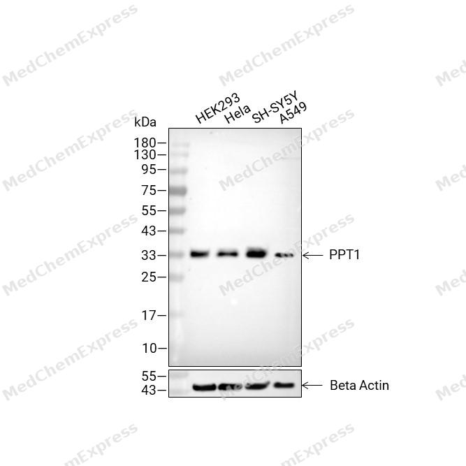 Western blot analysis of extracts from HEK293 (lane2(20μg), Hela (lane3(20μg), SH-SY5Y (lane4(20μg) and A549 (lane5(20μg) using PPT1 Antibody (HY-P88201). Proteins were transferred to a PVDF membrane and blocked with 5% non-fat milk in TBST for 2 hour at room temperature. The primary antibody (1/1000) and Loading control antibody (Beta Actin, HY-P80993, 1/10,000) was used in 5% non-fat milk in TBST at 4°C overnight. Goat Anti-Mouse IgG-HRP Secondary Antibody (HY-P8004 ,1/10,000) was used for 1 hour at room temperature.
 -
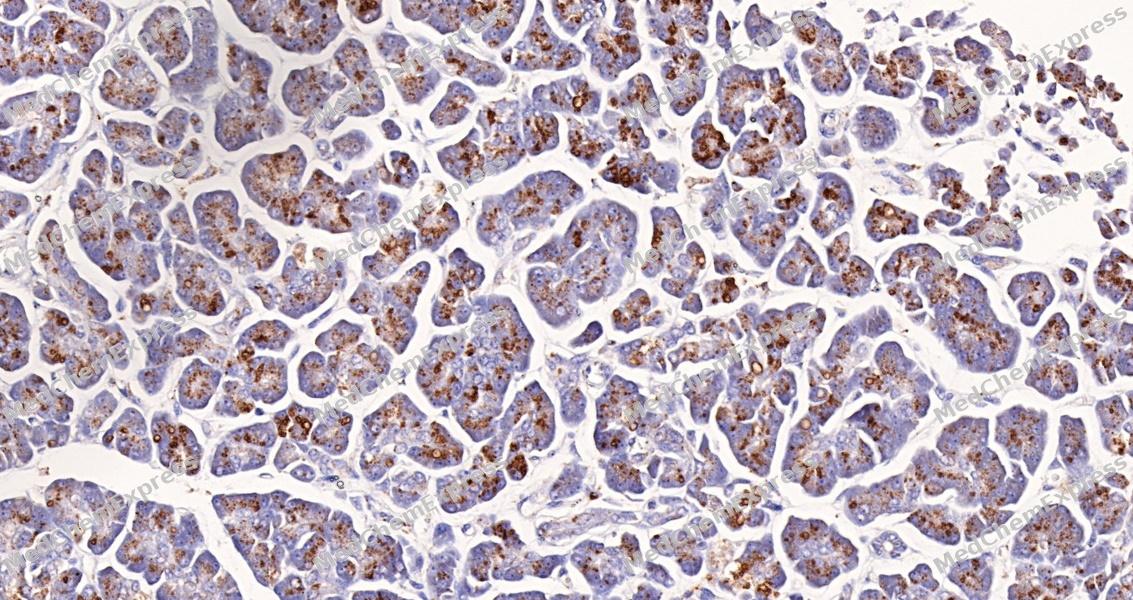 Immunohistochemical analysis of paraffin-embedded human pancreas using PPT1 Antibody (HY-P88201, 1/500). The section was pretreated using heat mediated antigen retrieval with sodium citrate buffer (pH 6.0) for 8 minutes. The tissues were blocked with quick block buffer for 0.5 hours at room temperature, washed with PBS and PBST, and then incubated with the primary antibody overnight at 4℃. The detection was performed using an HRP conjugated compact polymer system. DAB was used as the chromogen. Tissues were counterstained with hematoxylin and mounted with DPX.
 -
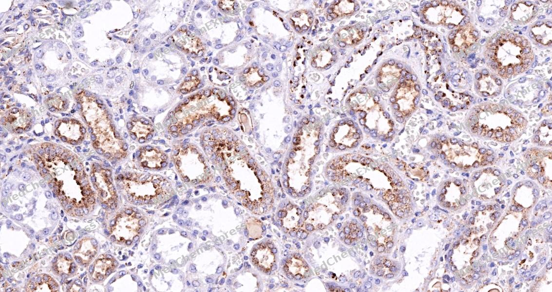 Immunohistochemical analysis of paraffin-embedded human kidney tissue using PPT1 Antibody (HY-P88201, 1/500). The section was pretreated using heat mediated antigen retrieval with sodium citrate buffer (pH 6.0) for 8 minutes. The tissues were blocked with quick block buffer for 0.5 hours at room temperature, washed with PBS and PBST, and then incubated with the primary antibody overnight at 4℃. The detection was performed using an HRP conjugated compact polymer system. DAB was used as the chromogen. Tissues were counterstained with hematoxylin and mounted with DPX.
 -
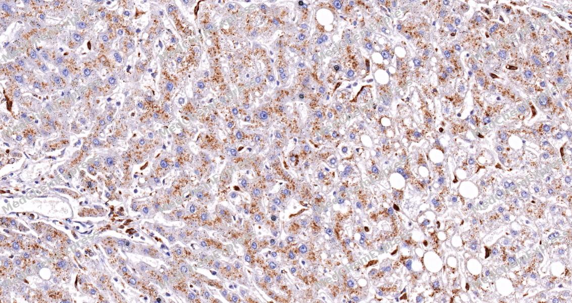 Immunohistochemical analysis of paraffin-embedded human liver tissue using PPT1 Antibody (HY-P88201, 1/500). The section was pretreated using heat mediated antigen retrieval with sodium citrate buffer (pH 6.0) for 8 minutes. The tissues were blocked with quick block buffer for 0.5 hours at room temperature, washed with PBS and PBST, and then incubated with the primary antibody overnight at 4℃. The detection was performed using an HRP conjugated compact polymer system. DAB was used as the chromogen. Tissues were counterstained with hematoxylin and mounted with DPX.
 -
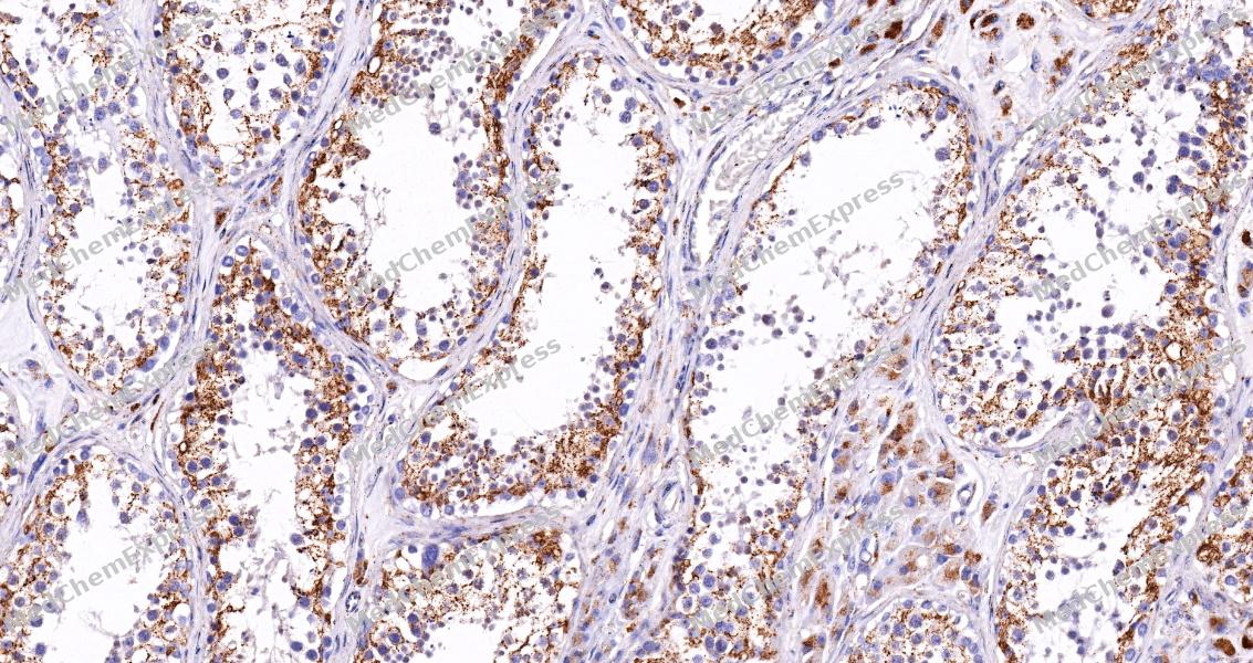 Immunohistochemical analysis of paraffin-embedded human testis tissue using PPT1 Antibody (HY-P88201, 1/500). The section was pretreated using heat mediated antigen retrieval with sodium citrate buffer (pH 6.0) for 8 minutes. The tissues were blocked with quick block buffer for 0.5 hours at room temperature, washed with PBS and PBST, and then incubated with the primary antibody overnight at 4℃. The detection was performed using an HRP conjugated compact polymer system. DAB was used as the chromogen. Tissues were counterstained with hematoxylin and mounted with DPX.
 -
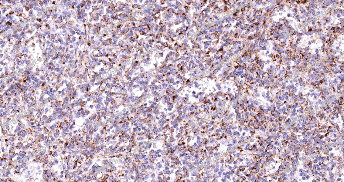 Immunohistochemical analysis of paraffin-embedded human spleen tissue using PPT1 Antibody (HY-P88201, 1/500). The section was pretreated using heat mediated antigen retrieval with sodium citrate buffer (pH 6.0) for 8 minutes. The tissues were blocked with quick block buffer for 0.5 hours at room temperature, washed with PBS and PBST, and then incubated with the primary antibody overnight at 4℃. The detection was performed using an HRP conjugated compact polymer system. DAB was used as the chromogen. Tissues were counterstained with hematoxylin and mounted with DPX.
 -
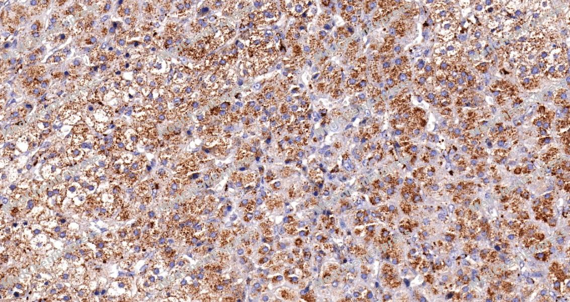 Immunohistochemical analysis of paraffin-embedded human adrenal gland tissue using PPT1 Antibody (HY-P88201, 1/500). The section was pretreated using heat mediated antigen retrieval with sodium citrate buffer (pH 6.0) for 8 minutes. The tissues were blocked with quick block buffer for 0.5 hours at room temperature, washed with PBS and PBST, and then incubated with the primary antibody overnight at 4℃. The detection was performed using an HRP conjugated compact polymer system. DAB was used as the chromogen. Tissues were counterstained with hematoxylin and mounted with DPX.


Background
-
Function
Palmitoyl protein thioesterase 1 (PPT1) is a lysosomal depalmitoylating enzyme that removes thioester-linked fatty acids, predominantly palmitate, from S-palmitoylated proteins and thereby promotes the degradation and turnover of lipid-modified proteins during lysosomal catabolism[1][2]. PPT1 functions within the broader protein depalmitoylation network and contributes to cellular processes associated with lysosomal degradation, autophagy-lysosome function, endocytosis, neuronal morphogenesis, and synaptic maintenance[3][4][5]. Mechanistically, loss of PPT1 activity impairs the clearance of palmitoylated proteins, leading to intracellular accumulation of ceroid lipofuscin and disruption of lysosomal homeostasis[4][6]. Mutations in PPT1 cause neuronal ceroid lipofuscinosis type 1 (CLN1), also known as infantile neuronal ceroid lipofuscinosis, a severe neurodegenerative lysosomal storage disorder characterized by progressive neuronal dysfunction and degeneration[4][6][7]. Experimental studies using PPT1-deficient cells and mouse models have demonstrated defects in endocytosis, synaptic vesicle recycling, neuronal survival, and regional brain integrity, supporting the utility of these models for investigating disease mechanisms and therapeutic strategies[5][8][7][9]. Compared with the closely related lysosomal thioesterase PPT2, PPT1 uniquely hydrolyzes palmitoylated proteins and palmitoylcysteine substrates, providing a key biochemical distinction that underlies its specialized role in protein depalmitoylation and neurobiology[10]. For experimental applications, PPT1 has also emerged as a pharmacological target in lysosome-centered studies, and inhibition of PPT1 has been used to investigate lysosomal signaling, autophagy regulation, and disease-associated cellular pathways[11].
-
Subcellular Localization
Lysosome,Secreted,Golgi apparatus,Endoplasmic reticulum
-
Isoforms & Post-Translational Modification
P50897 has two isomers: P50897-1: 34193 Da (predicted); P50897-2: 23094 Da (predicted).
Glycosylated -
Subunit
Interacts with CLN5 (PubMed:19941651)
-
SwissProt ID
-
Synonyms
CLN1; INCL; PPT


Documentation
References
[1]. Bellizzi JJ 3rd, et al. The crystal structure of palmitoyl protein thioesterase 1 and the molecular basis of infantile neuronal ceroid lipofuscinosis. Proc Natl Acad Sci U S A. 2000 Apr 25;97(9):4573-8. [Content Brief]
[2]. Koster KP, et al. Depalmitoylation by Palmitoyl-Protein Thioesterase 1 in Neuronal Health and Degeneration. Front Synaptic Neurosci. 2019 Aug 29;11:25. [Content Brief]
[3]. Kiros M, et al. Trends in HIV-1 pretreatment drug resistance and HIV-1 variant dynamics among antiretroviral therapy-naive Ethiopians from 2003 to 2018: a pooled sequence analysis. Virol J. 2023 Oct 25;20(1):243. [Content Brief]
[4]. Unsworth HC, et al. Tissue-specific effects of wild-type and mutant connexin 31: a role in neurite outgrowth. Hum Mol Genet. 2007 Jan 15;16(2):165-72. [Content Brief]
[5]. Gorenberg EL, et al. Identification of substrates of palmitoyl protein thioesterase 1 highlights roles of depalmitoylation in disulfide bond formation and synaptic function. PLoS Biol. 2022 Mar 31;20(3):e3001590. [Content Brief]
[6]. Kim SJ, et al. Palmitoyl protein thioesterase-1 deficiency impairs synaptic vesicle recycling at nerve terminals, contributing to neuropathology in humans and mice. J Clin Invest. 2008 Sep;118(9):3075-86. [Content Brief]
[7]. Bible E, et al. Regional and cellular neuropathology in the palmitoyl protein thioesterase-1 mutant mouse model of infantile neuronal ceroid lipofuscinosis. Neurobiol Dis. 2004 Jul;16(2):346-59. [Content Brief]
[8]. Atiskova Y, et al. Mice deficient in the lysosomal enzyme palmitoyl-protein thioesterase 1 (PPT1) display a complex retinal phenotype. Sci Rep. 2019 Oct 2;9(1):14185. [Content Brief]

 Powered by Bioz
