



SHP2 Antibody (YA1643)
(Synonyms: PTPN11; PTP2C; SHPTP2; Tyrosine-protein phosphatase non-receptor type 11; Protein-tyrosine phosphatase 1D; PTP-1D; Protein-tyrosine phosphatase 2C; PTP-2C; SH-PTP2; SHP-2; Shp2; SH-PTP3)Based on 1 Customer Validation
SHP2 Antibody (YA1643) is a Rabbit-derived and non-conjugated IgG monoclonal antibody, targeting to SHP2.
-
Host:
Rabbit
-
Isotype:
IgG
-
Application:
WB, IHC-P, IP
-
Reactivity :
Human
-
Formulation:
Supplied in 50mM Tris-Glycine(pH 7.4), 0.15M NaCl, 40% Glycerol, 0.01% Sodium azide and 0.05% BSA
-
Conjugation:
Non-conjugated


To place orders, for customer services and technical support, please contact: MedChemExpress USA
Tel: 609-228-6898 E-mail: [email protected] [email protected]





Applications
| Application |
WB

WB: Western Blot
|
IHC-P
IHC-P: Immunohistochemistry-Paraffin
|
IP
IP: Immunoprecipitation
|
|---|---|---|---|
| Dilution Ratio | 1:500-1:1000 | 1:50-1:100 | 1:20 |


Product Details
SHP2 Antibody (YA1643) is a Rabbit-derived and non-conjugated IgG monoclonal antibody, targeting to SHP2.
-
Host Rabbit

-
Clonality Recombinant,Monoclonal

-
Species ReactivityHuman
-
Observed Molecular WeightObserved band size: 68 kDa
Note: Due to possible protein modifications or aggregation, the molecular weight should be confirmed by actual measurement, and the predicted value is for reference only.

-
Calculated Molecular Weight Predicted band size: 68 kDa

A synthetic peptide of human SHP2
Endogenous
Affinity Purified
Non-conjugated
Unmodified
IgG


Product Properties
-
Appearance
Solution
-
Formulation
Supplied in 50mM Tris-Glycine(pH 7.4), 0.15M NaCl, 40% Glycerol, 0.01% Sodium azide and 0.05% BSA
-
Concentration
Batch-dependent, Please check the COA for the concentration of each lot. Check Lot Concentration
-
Storage & Stability
Stored at -20°C for 1 year. Avoid repeated freeze / thaw cycles.
-
Shipping
Shipping with blue ice.


Verification Images
-
 Western blot analysis of extracts from Hela (lane 2(20μg), Hela (lane 3(40μg), using SHP2 Antibody. Proteins were transferred to a PVDF membrane and blocked with 5% BSA in TBST for 2 hour at room temperature. The primary antibody and Loading control antibody (Beta Actin, HY-P80438, 1/3000) was used in 5% BSA in TBST at 4°C overnight. Goat Anti-Mouse/Rabbit IgG-HRP Secondary Antibody (HY-P8004/HY-P8001, 1/10,000) was used for 1 hour at room temperature.
 -
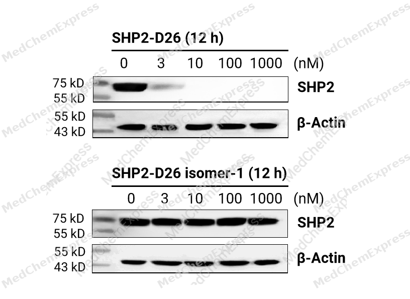 KYSE-520 cells were seeded at 1*106 cells/well in a 6-well plate and were cultured overnight. Then, cells were treated with 0-1000 nM SHP2-D26 (HY-145162) or SHP2-D26 isomer-1 (HY-145703) for 12 hours to assess the expression of SHP2. The results demonstrated that SHP2-D26 effectively degraded SHP2 protein, but SHP2-D26 isomer-1 had no such effect. Primary antibody: SHP2 antibody (HY-P81898).
 -
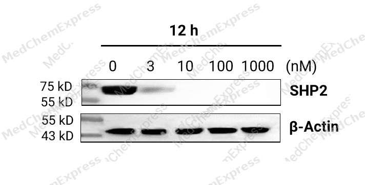 KYSE-520 cells were seeded at 1*106 cells/well in a 6-well plate and were cultured overnight. Then, cells were treated with 0-1000 nM SHP2-D26 (HY-145162) for 12 hours to assess the expression of SHP2. The results demonstrated that SHP2-D26 effectively degraded SHP2 protein. Primary antibody: SHP2 antibody (HY-P81898).
 -
 Immunohistochemical analysis of paraffin-embedded human tonsil tissue using SHP2 Antibody (HY-P81898, 1/100). The section was pretreated using heat mediated antigen retrieval with sodium citrate buffer (pH 6.0) for 8 minutes. The tissues were blocked with quick block buffer for 0.5 hours at room temperature, washed with PBS and PBST, and then incubated with the primary antibody overnight at 4℃. The detection was performed using an HRP conjugated compact polymer system. DAB was used as the chromogen. Tissues were counterstained with hematoxylin and mounted with DPX.
 -
 Immunohistochemical analysis of paraffin-embedded human thyroid gland tissue using SHP2 Antibody (HY-P81898, 1/100). The section was pretreated using heat mediated antigen retrieval with sodium citrate buffer (pH 6.0) for 8 minutes. The tissues were blocked with quick block buffer for 0.5 hours at room temperature, washed with PBS and PBST, and then incubated with the primary antibody overnight at 4℃. The detection was performed using an HRP conjugated compact polymer system. DAB was used as the chromogen. Tissues were counterstained with hematoxylin and mounted with DPX.
 -
 Immunohistochemical analysis of paraffin-embedded human breast tissue using SHP2 Antibody (HY-P81898, 1/100). The section was pretreated using heat mediated antigen retrieval with sodium citrate buffer (pH 6.0) for 8 minutes. The tissues were blocked with quick block buffer for 0.5 hours at room temperature, washed with PBS and PBST, and then incubated with the primary antibody overnight at 4℃. The detection was performed using an HRP conjugated compact polymer system. DAB was used as the chromogen. Tissues were counterstained with hematoxylin and mounted with DPX.
 -
 Immunohistochemical analysis of paraffin-embedded human breast cancer tissue using SHP2 Antibody (HY-P81898, 1/100). The section was pretreated using heat mediated antigen retrieval with sodium citrate buffer (pH 6.0) for 8 minutes. The tissues were blocked with quick block buffer for 0.5 hours at room temperature, washed with PBS and PBST, and then incubated with the primary antibody overnight at 4℃. The detection was performed using an HRP conjugated compact polymer system. DAB was used as the chromogen. Tissues were counterstained with hematoxylin and mounted with DPX.
 -
 Immunohistochemical analysis of paraffin-embedded human cervical cancer tissue using SHP2 Antibody (HY-P81898, 1/100). The section was pretreated using heat mediated antigen retrieval with sodium citrate buffer (pH 6.0) for 8 minutes. The tissues were blocked with quick block buffer for 0.5 hours at room temperature, washed with PBS and PBST, and then incubated with the primary antibody overnight at 4℃. The detection was performed using an HRP conjugated compact polymer system. DAB was used as the chromogen. Tissues were counterstained with hematoxylin and mounted with DPX.
 -
 Immunohistochemical analysis of paraffin-embedded human glioma tissue using SHP2 Antibody (HY-P81898, 1/100). The section was pretreated using heat mediated antigen retrieval with sodium citrate buffer (pH 6.0) for 8 minutes. The tissues were blocked with quick block buffer for 0.5 hours at room temperature, washed with PBS and PBST, and then incubated with the primary antibody overnight at 4℃. The detection was performed using an HRP conjugated compact polymer system. DAB was used as the chromogen. Tissues were counterstained with hematoxylin and mounted with DPX.


Background
-
Function
Src-homology-2-containing protein tyrosine phosphatase 2 (SHP2), encoded by the PTPN11 gene, is a 593 amino acid classical non-receptor protein tyrosine phosphatase (PTP) . SHP2 is involved in diverse signalling pathways such as RAS-MAPK, PI3K-AKT, JAK-STAT and PD-1/PD-L1. Besides, SHP2 negatively regulates the activation of recombinant NLRP3 (NLR family, pyrin domain containing protein 3) inflammasome via mitochondrial homeostasis. SHP2 has been regarded as an extremely attractive target for human diseases therapies, particular in cancer.
-
Subcellular Localization
Cytoplasm; Nucleus
-
Expression
Tissue_specificity:It is widely expressed, with the highest concentrations found in the heart, brain, and skeletal muscle. -
Isoforms & Post-Translational Modification
Q06124 has 3 isomers: Q06124-2: 68011 Da (predicted); Q06124-1: 68436 Da (predicted); Q06124-3: 52828 Da (predicted).
Phosphorylated on Tyr-542 and Tyr-580 upon receptor protein tyrosine kinase activation; which creates a binding site for GRB2 and other SH2-containing proteins. Phosphorylated upon activation of the receptor-type kinase FLT3. Phosphorylated upon activation of the receptor-type kinase PDGFRA (By similarity). Phosphorylated by activated PDGFRB -
Subunit
Interacts with phosphorylated LIME1 and BCAR3. Interacts with SHB and INPP5D/SHIP1 (By similarity). Interacts with MILR1 (tyrosine-phosphorylated). Interacts with FLT1 (tyrosine-phosphorylated), FLT3 (tyrosine-phosphorylated), FLT4 (tyrosine-phosphorylated), KIT and GRB2. Interacts with PDGFRA (tyrosine phosphorylated). Interacts (via SH2 domain) with TEK/TIE2 (tyrosine phosphorylated) (By similarity). Interacts with PTPNS1 and CD84. Interacts with phosphorylated SIT1 and MPZL1. Interacts with FCRL4, FCRL6 and ANKHD1. Interacts with KIR2DL1; the interaction is enhanced by ARRB2. Interacts with GAB2. Interacts with TERT; the interaction retains TERT in the nucleus. Interacts with PECAM1 and FER. Interacts with EPHA2 (activated); participates in PTK2/FAK1 dephosphorylation in EPHA2 downstream signaling. Interacts with ROS1; mediates PTPN11 phosphorylation. Interacts with PDGFRB (tyrosine phosphorylated); this interaction increases the PTPN11 phosphatase activity. Interacts with GAREM1 isoform 1 (tyrosine phosphorylated); the interaction increases MAPK/ERK activity and does not affect the GRB2/SOS complex formation. Interacts with CDC73 (PubMed:26742426). Interacts with CEACAM1 (via cytoplasmic domain); this interaction depends on the monomer/dimer equilibrium and is phosphorylation-dependent (By similarity). Interacts with MPIG6B (via ITIM motif) (PubMed:23112346). Interacts with SIGLEC10 (By similarity). Interacts with FCRL3 (via phosphorylated ITIM motifs) (PubMed:11162587, PubMed:19843936). Interacts with CLEC12B (via ITIM motif); this interaction triggers dephosphorylation and activation of PTPN11. Interacts (via SH2 domains) with NEDD9/CAS-L; the interaction is enhanced when NEDD9/CAS-L is tyrosine phosphorylated (PubMed:19275884)
-
SwissProt ID
-
Synonyms
PTPN11; PTP2C; SHPTP2; Tyrosine-protein phosphatase non-receptor type 11; Protein-tyrosine phosphatase 1D; PTP-1D; Protein-tyrosine phosphatase 2C; PTP-2C; SH-PTP2; SHP-2; Shp2; SH-PTP3
-
Research Field
Signal Transduction


Documentation
-
Data Sheet (234 KB)
-
SDS (252 KB)
- English - EN (252 KB)
- Français - FR (252 KB)
- Deutsch - DE (252 KB)
- Norwegian - NO (252 KB)
- Español - ES (252 KB)
- Swedish - SV (252 KB)
- Italian - IT (252 KB)
- Korean - KR (252 KB)
- Portuguese - PT (252 KB)
-
User Guide for Antibodies (1077 KB)

 Powered by Bioz
