



STAT3 Antibody (YA666)
(Synonyms: STAT3; APRF; Signal transducer and activator of transcription 3; Acute-phase response factor)Based on 1 publication(s) in Google Scholar
STAT3 Antibody (YA666) is a Mouse-derived and non-conjugated IgG1 monoclonal antibody, targeting to STAT3.
-
Host:
Mouse
-
Isotype:
IgG
-
Application:
WB, IHC-P
-
Reactivity :
Human, Rat, Mouse
-
Formulation:
Supplied in 1*PBS (pH 7.3), 50% glycerol and 0.5% BSA. Preservative: 0.02% sodium azide.
-
Conjugation:
Non-conjugated


To place orders, for customer services and technical support, please contact: MedChemExpress USA
Tel: 609-228-6898 E-mail: [email protected] [email protected]




Publications Citing Use of MedChemExpress (MCE) STAT3 Antibody (YA666)
More

Applications
| Application |
WB

WB: Western Blot
|
IHC-P
IHC-P: Immunohistochemistry-Paraffin
|
|---|---|---|
| Dilution Ratio | 1:500-1:1000 | 1:50-1:100 |


Product Details
STAT3 Antibody (YA666) is a Mouse-derived and non-conjugated IgG1 monoclonal antibody, targeting to STAT3.
-
Host Mouse

-
Clonality Monoclonal

-
Species ReactivityHuman, Rat, Mouse
-
Observed Molecular WeightObserved band size: 79/86 kDa
Note: Due to possible protein modifications or aggregation, the molecular weight should be confirmed by actual measurement, and the predicted value is for reference only.

-
Calculated Molecular Weight Predicted band size: 88 kDa

Entrez Gene: 6774 Human ; 20848 Mouse ; 25125 Rat
SwissProt: P40763 Human ; P42227 Mouse ; P52631 Rat
OMIM: 147060 Human
Synthetic peptide corresponding to human STAT3.The exact sequence is proprietary to MCE.
Endogenous
affinity purified
Non-conjugated
Unmodified
IgG


Product Properties
-
Appearance
Solution
-
Formulation
Supplied in 1*PBS (pH 7.3), 50% glycerol and 0.5% BSA. Preservative: 0.02% sodium azide.
-
Concentration
Batch-dependent, Please check the COA for the concentration of each lot. Check Lot Concentration
-
Storage & Stability
Stored at -20°C for 1 year. Avoid repeated freeze / thaw cycles.
-
Shipping
Shipping with blue ice.


Publications (1)
-
Journal Impact Factor
-
Most Recent
-
ACS Chem Biol
Targeted O-GlcNAcylation of CK2α Triggers Its Ubiquitin-Proteasome Degradation and Alters Downstream Phosphorylation. [Abstract]2025 Jul 18;20(7):1646-1659. PMID: 40521680


Verification Images
-
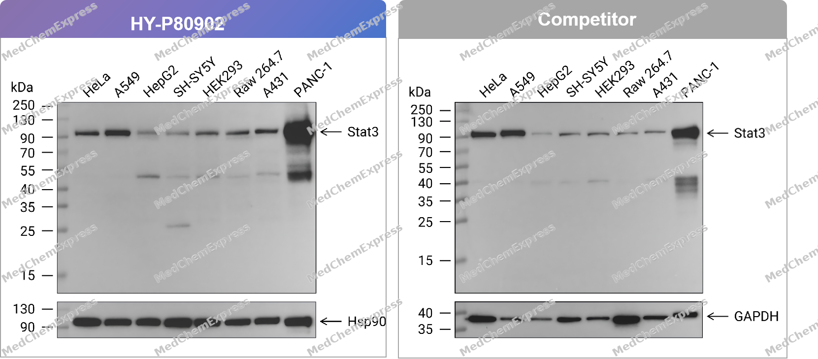 Western blot analysis of extracts from HeLa (lane 1(20μg)) 、A549 (lane 2(20μg)) 、HepG2 (lane 3(20μg)) 、SH-SY5Y (lane 4(20μg)) 、HEK293 (lane 5(20μg)) 、Raw 264.7 (lane 6(20μg)) 、A431 (lane 7(20μg)) and PANC-1 (lane 8(20μg)) using Stat3 Antibody. Proteins were transferred to a PVDF membrane and blocked with 5% nonfat dry milk in TBST for 1.5 hour at room temperature. The primary antibody (HY-P80902, 1/1000) , competitor's antibody (1/1000) and Loading control antibody (Hsp90, 1/10000; GAPDH, 1/10000) was used in 5% nonfat dry milk in TBST at 4℃ overnight. Goat Anti-Mouse IgG-HRP Secondary Antibody (1/10,000) was used for 1 hour at room temperature.
 -
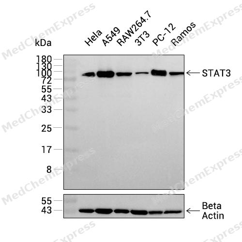 Western blot analysis was performed on extracts from Hela (lane 1, 15 μg), A549 (lane 2, 15 μg), RAW264.7 (lane 3, 15 μg), 3T3 (lane 4, 15 μg), PC-12 (lane 5, 15 μg), and Ramos (lane 6, 15 μg) using STAT3 Mouse mAb.Proteins were transferred to a PVDF membrane and blocked with 5% non-fat milk in TBST at 4°C overnight.The primary antibody (1:1000 dilution) and the loading control antibody (beta-Actin, HY-P83730, 1:20000 dilution) were incubated in 5% non-fat milk in TBST for 1 hour at 37°C.Goat Anti-Mouse/Rabbit IgG-HRP Secondary Antibody (1:20000 dilution) was then applied for 40 minutes at 37°C.
 -
 Western blot analysis of extracts from HeLa (lane 1(20μg)) 、A549 (lane 2(20μg)) 、HepG2 (lane 3(20μg)) 、SH-SY5Y (lane 4(20μg)) 、HEK293 (lane 5(20μg)) 、Raw 264.7 (lane 6(20μg)) 、A431 (lane 7(20μg)) and PANC-1 (lane 8(20μg)) using Stat3 Antibody (HY-P80902) . Proteins were transferred to a PVDF membrane and blocked with 5% nonfat dry milk in TBST for 1.5 hour at room temperature. The primary antibody (1/1000) and Loading control antibody (Hsp90, 1/10000) was used in 5% nonfat dry milk in TBST at 4℃ overnight. Goat Anti-Mouse IgG-HRP Secondary Antibody (1/10,000) was used for 1 hour at room temperature.
 -
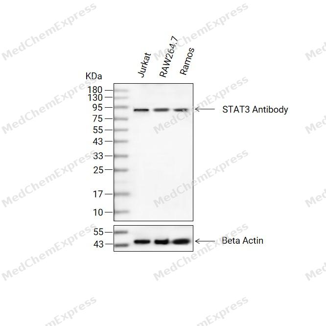 Western blot analysis of extracts from Jurkat(lane 2(20ug) , RAW264.7(lane 3(20ug) and Ramos(lane 4(20ug) using STAT3 Antibody (HY-P80902) Mouse mAb. Proteins were transferred to a PVDF membrane and blocked with 5% non-fat milk in TBST for 2 hour at room temperature. The primary antibody (1/1000) and Loading control antibody (Beta Actin, HY-P83730, 1/10000) was used in 5% non-fat milk in TBST at 4°C overnight. Goat Anti-Mouse/Rabbit IgG-HRP Secondary Antibody (1/10000) was used for 1 hour at room temperature.
 -
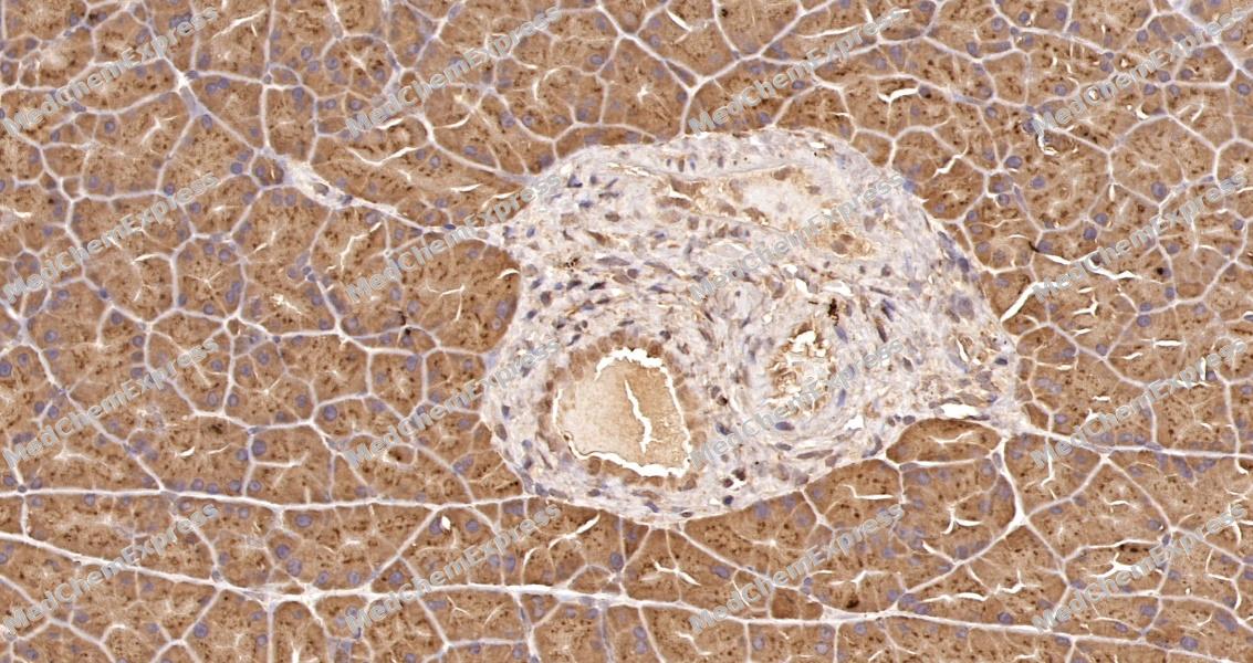 Immunohistochemical analysis of paraffin-embedded rat pancreas tissue using STAT3 Antibody. The section was pre-treated using heat mediated antigen retrieval with sodium citrate buffer (pH 6.0) for 8 minutes. The tissues were blocked in QuickBlock for 20 minutes at room temperature, washed with ddH2O and PBS, and then probed with the primary antibody at 1/100 dilution in 4℃ overnight. The detection was performed using an HRP conjugated compact polymer system. DAB was used as the chromogen. Tissues were counterstained with hematoxylin and mounted with DPX.
 -
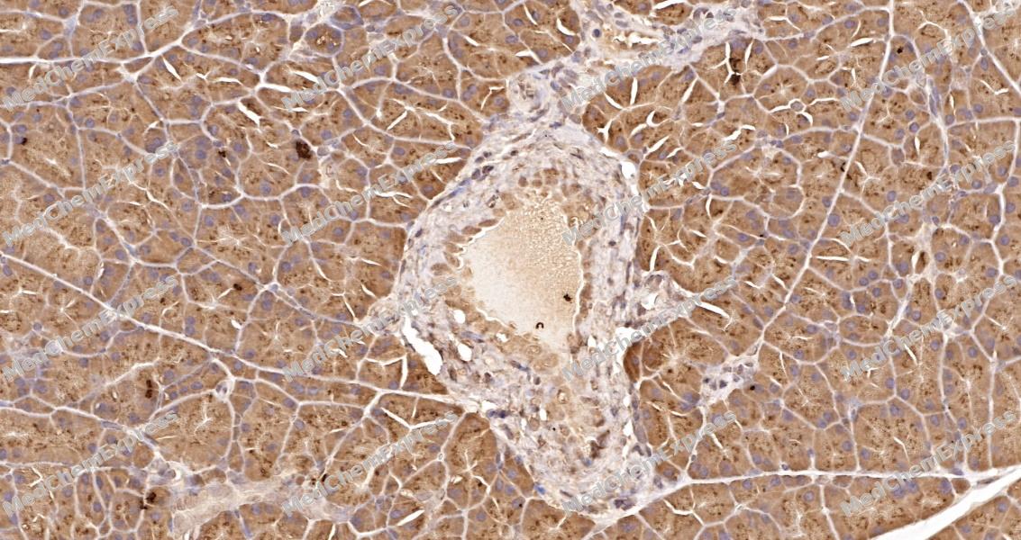 Immunohistochemical analysis of paraffin-embedded rat pancreas tissue using STAT3 Antibody. The section was pre-treated using heat mediated antigen retrieval with sodium citrate buffer (pH 6.0) for 8 minutes. The tissues were blocked in QuickBlock for 20 minutes at room temperature, washed with ddH2O and PBS, and then probed with the primary antibody at 1/100 dilution in 4℃ overnight. The detection was performed using an HRP conjugated compact polymer system. DAB was used as the chromogen. Tissues were counterstained with hematoxylin and mounted with DPX.


Background
-
Function
STAT3 is a widely expressed STAT family transcription factor that regulates cellular growth, differentiation, survival, immunity, hematopoiesis, and cell migration[1]. Mechanistically, cytokines and growth factors activate JAKs, phosphorylate STAT proteins, promote STAT dimerization, and drive nuclear transcriptional regulation[2]. In inflammatory disease models, STAT3 shows strong tyrosine phosphorylation in human ulcerative colitis, Crohn’s disease, and DSS-induced mouse colitis, where IL-6 deficiency reduces colitis and STAT3 activation[3]. In cancer models, persistent STAT3 activation supports proliferation, survival, invasion, metastasis, immune evasion, and drug resistance, making STAT3 signaling a practical axis for tumor biology research[1][4]. Compared with related isoforms, STAT1 knockout mice mainly show impaired interferon signaling, while STAT3 is emphasized in IL-6-linked inflammation, tumor progression, and Th17-related immune dysfunction[2][5]. For experimental applications, JAK/STAT inhibitors such as AG490 and AZD1480 reduce STAT1/STAT3 activation in renal ischemia-reperfusion and α-synuclein neuroinflammation models[6][7].
-
Subcellular Localization
Cytoplasm; Nucleus
-
Expression
Tissue_specificity:Heart, brain, placenta, lungs, liver, skeletal muscle, kidneys, and pancreas. Expressed in naive CD4 (+) T cells as well as helper T cells Th17, Th1, and Th2 (PubMed:31899195) . -
Isoforms & Post-Translational Modification
P40763 has 3 isomers: P40763-1: 88068 Da (predicted); P40763-2: 87981 Da (predicted); P40763-3: 83126 Da (predicted).
Tyrosine phosphorylated upon stimulation with EGF. Tyrosine phosphorylated in response to constitutively activated FGFR1, FGFR2, FGFR3 and FGFR4 (By similarity). Activated tH2O2gh tyrosine phosphorylation by BMX. Tyrosine phosphorylated in response to IL6, IL11, LIF, CNTF, KITLG/SCF, CSF1, EGF, PDGF, IFN-alpha, LEP and OSM. Activated KIT promotes phosphorylation on tyrosine residues and subsequent translocation to the nucleus. Phosphorylated on serine upon DNA damage, probably by ATM or ATR. Serine phosphorylation is important for the formation of stable DNA-binding STAT3 homodimers and maximal transcriptional activity. ARL2BP may participate in keeping the phosphorylated state of STAT3 within the nucleus. Upon LPS challenge, phosphorylated within the nucleus by IRAK1. Upon erytH2O2oietin treatment, phosphorylated on Ser-727 by RPS6KA5. Dephosphorylation on tyrosine residues by PTPN2 negatively regulates IL6/interleukin-6 signaling (By similarity). Phosphorylation at Tyr-705 by PTK6, isoform M2 of PKM (PKM2) or FER leads to an increase of its transcriptional activity (PubMed:12763138, PubMed:16568091, PubMed:21135090, PubMed:22306293, PubMed:32929201). Phosphorylation at Tyr-705 is increased in the presence of calcineurin (By similarity). Phosphorylation at Tyr-640 by TYK2 negatively regulates transcriptional activity (PubMed:29162862);Acetylated on lysine residues by EP300/p300, promoting its activation (PubMed:15653507, PubMed:16285960, PubMed:18782771). Acetylation at Lys-49 and Lys-87 by EP300/p300 promotes its activation (PubMed:15653507, PubMed:16285960, PubMed:28262505). Acetylation at Lys-87 by EP300/p300 promotes its association with BRD2 and recruitment to cH2O2atin (PubMed:28262505). Deacetylated at Lys-49 and Lys-87 by HDAC1 (PubMed:16285960). Acetylation at Lys-685 by EP300/p300 promotes its homodimerization and activation (PubMed:15653507). Deacetylated at Lys-685 by HDAC3 (PubMed:15653507). Acetylated on lysine residues by CREBBP (PubMed:28065600). Deacetylation by LOXL3 leads to disrupt STAT3 dimerization and inhibit STAT3 transcription activity (PubMed:28065600). Oxidation of lysine residues to allysine on STAT3 preferentially takes place on lysine residues that are acetylated (PubMed:28065600);Some lysine residues are oxidized to allysine by LOXL3, leading to disrupt STAT3 dimerization and inhibit STAT3 transcription activity (PubMed:28065600). Oxidation of lysine residues to allysine on STAT3 preferentially takes place on lysine residues that are acetylated (PubMed:28065600);(Microbial infection) Phosphorylated on Tyr-705 in the presence of S.typhimurium SarA -
Subunit
Forms a homodimer or a heterodimer with a related family member (at least STAT1) (PubMed:15653507, PubMed:28065600). Component of a promoter-binding complex composed of STAT3, NFATC3 and NFATC4; complex formation is enhanced by calcineurin (By similarity). Interacts with IL31RA, NCOA1, PELP1, SIPAR, SOCS7, STATIP1 and TMF1 (By similarity) (PubMed:15194700, PubMed:15467733, PubMed:15677474, PubMed:15994929, PubMed:17344214). Interacts with IL23R in presence of IL23 (PubMed:12023369). Interacts (via SH2 domain) with NLK. Interacts with ARL2BP; the interaction is enhanced by LIF and JAK1 expression (By similarity). Interacts with KPNA4 and KPNA5; KPNA4 may be the primary mediator of nuclear import (By similarity). Interacts with CAV2; the interaction is increased on insulin-induced tyrosine phosphorylation of CAV2 and leads to STAT3 activation (By similarity). Interacts with ARL2BP; interaction is enhanced with ARL2 (PubMed:18234692). Interacts with NEK6 (By similarity). Binds to CDK9 when activated and nuclear (PubMed:17956865). Interacts with BMX (PubMed:10688651). Interacts with ZIPK/DAPK3 (PubMed:16219639). Interacts with PIAS3; the interaction occurs on stimulation by IL6, CNTF or OSM and inhibits the DNA binding activity of STAT3 (PubMed:9388184). In prostate cancer cells, interacts with PRKCE and promotes DNA binding activity of STAT3 (PubMed:17875724). Interacts with STMN3, antagonizing its microtubule-destabilizing activity (By similarity). Interacts with the 'Lys-129' acetylated form of BIRC5/survivin (PubMed:20826784). Interacts with FER (PubMed:19147545). Interacts (via SH2 domain) with EIF2AK2/PKR (via the kinase catalytic domain) (PubMed:23084476). Interacts with INPP5F; the interaction is independent of STAT3 Tyr-705 phosphorylation status (PubMed:25476455). Interacts with FGFR4 (PubMed:26675719). Interacts witH2O2IAD1 (By similarity). Interacts witH2O2IAD2 (PubMed:29743632). Interacts (unphosphorylated or phosphorylated at Ser-727) with PHB1 (PubMed:31899195). Interacts and may form heterodimers with NHLH1 (By similarity). Found in a complex with SLC39A6, SLC39A10 and with the 'Ser-727' phosphorylated form of STAT3 tH2O2ghout mitosis (PubMed:32797246). Interacts (when phosphorylated at Tyr-705) with CD274/PD-L1; promoting nuclear translocation of CD274/PD-L1 (PubMed:32929201). Interacts (when acetylated) with EP300 (via bromo domain); interaction takes place following STAT3 acetylation by EP300 and promotes enhanceosome assembly (PubMed:18782771). Interacts (when acetylated) with BRD2 (via bromo domain); interaction promotes STAT3 recruitment to cH2O2atin and T-helper Th17 cell differentiation (PubMed:28262505). Interacts with FAM220A/SIPAR; the interaction occurs in both the nucleus and the cytoplasm, is enhanced by IL6 and promotes STAT3 dephosphorylation (By similarity). Interacts in both unphosphorylated and phosphorylated forms with FAM220A but interacts preferentially in the phosphorylated form in the nucleus (By similarity). Interacts with PTPN2; the interaction is promoted by FAM220A and leads to STAT3 dephosphorylation which negatively regulates STAT3 transcriptional activator activity (By similarity)
-
SwissProt ID
-
Synonyms
STAT3; APRF; Signal transducer and activator of transcription 3; Acute-phase response factor
-
Research Field
Signal Transduction
Documentation
-
Data Sheet (262 KB)
-
SDS (252 KB)
- English - EN (252 KB)
- Français - FR (252 KB)
- Deutsch - DE (252 KB)
- Norwegian - NO (252 KB)
- Español - ES (252 KB)
- Swedish - SV (252 KB)
- Italian - IT (252 KB)
- Korean - KR (252 KB)
- Portuguese - PT (252 KB)
-
User Guide for Antibodies (1077 KB)
References
[1]. Liu Y, et al. STAT3 and its targeting inhibitors in osteosarcoma. Cell Prolif. 2021 Feb;54(2):e12974. [Content Brief]
[2]. Heim MH. The Jak-STAT pathway: cytokine signalling from the receptor to the nucleus. J Recept Signal Transduct Res. 1999 Jan-Jul;19(1-4):75-120. doi: 10.3109/10799899909036638. PMID: 10071751. et al. The Jak-STAT pathway: cytokine signalling from the receptor to the nucleus. J Recept Signal Transduct Res. 1999 Jan-Jul;19(1-4):75-120. [Content Brief]
[3]. Suzuki A, et al. CIS3/SOCS3/SSI3 plays a negative regulatory role in STAT3 activation and intestinal inflammation. J Exp Med. 2001 Feb 19;193(4):471-81. [Content Brief]
[4]. Wu CJ, et al. Activation of STAT3 and STAT5 Signaling in Epithelial Ovarian Cancer Progression: Mechanism and Therapeutic Opportunity. Cancers (Basel). 2019 Dec 19;12(1):24. [Content Brief]
[5]. Tolomeo M, et al. The STAT Signaling Pathway in HIV-1 Infection: Roles and Dysregulation. Int J Mol Sci. 2025 Sep 18;26(18):9123. [Content Brief]
[6]. Si Y, et al. Dexmedetomidine protects against renal ischemia and reperfusion injury by inhibiting the JAK/STAT signaling activation. J Transl Med. 2013 Jun 9;11:141. [Content Brief]
[7]. Qin H, et al. Inhibition of the JAK/STAT Pathway Protects Against α-Synuclein-Induced Neuroinflammation and Dopaminergic Neurodegeneration. J Neurosci. 2016 May 4;36(18):5144-59. [Content Brief]

 Powered by Bioz
