



Tyrosine Hydroxylase Antibody (YA023)
(Synonyms: TYH, TH, Tyrosine 3-monooxygenase, Tyrosine 3-hydroxylase)Based on 2 publication(s) in Google Scholar
Tyrosine Hydroxylase Antibody (YA023) is a Rabbit-derived and non-conjugated IgG monoclonal antibody, targeting to Tyrosine Hydroxylase.
-
Host:
Rabbit
-
Isotype:
IgG
-
Application:
WB, IHC-P, ICC/IF, FC, IF-Tissue
-
Reactivity :
Human, Mouse, Rat
-
Formulation:
Supplied in 1*TBS (pH7.4), 0.05% BSA and 40% Glycerol. Preservative: 0.05% Sodium Azide.
-
Conjugation:
Non-conjugated


To place orders, for customer services and technical support, please contact: MedChemExpress USA
Tel: 609-228-6898 E-mail: [email protected] [email protected]



Publications Citing Use of MedChemExpress (MCE) Tyrosine Hydroxylase Antibody (YA023)
More
 2 Publications Citing Use of MCE Tyrosine Hydroxylase Antibody (YA023) (2)
-
IHC

-
IF


Applications
| Application |
WB

WB: Western Blot
|
ICC/IF
ICC/IF: Immunocytochemistry/
Immunofluorescence |
IHC-P
IHC-P: Immunohistochemistry-Paraffin
|
FC
FC: Flow Cytometry
|
IF-Tissue
IF-Tissue: Immunofluorescence-Tissue
|
|---|---|---|---|---|---|
| Dilution Ratio | 1:1000-1:10000 | 1:50-1:200 | 1:2000 | 1:50-1:100 | 1:200 |


Product Details
Tyrosine Hydroxylase Antibody (YA023) is a Rabbit-derived and non-conjugated IgG monoclonal antibody, targeting to Tyrosine Hydroxylase.
-
Host Rabbit

-
Clonality Recombinant,Monoclonal

-
Species ReactivityHuman, Mouse, Rat
-
Observed Molecular WeightObserved band size: 59 kDa
Note: Due to possible protein modifications or aggregation, the molecular weight should be confirmed by actual measurement, and the predicted value is for reference only.

-
Calculated Molecular Weight Predicted band size: 59 kDa

Entrez Gene: 7054 Human ; 21823 Mouse ; 25085 Rat
SwissProt: P07101 Human ; P24529 Mouse ; P04177 Rat
OMIM: 605407 Human
Synthetic peptide within C-terminal human Tyrosine Hydroxylase.
Endogenous
Protein A affinity purified.
Non-conjugated
Unmodified
IgG


Product Properties
-
Appearance
Solution
-
Formulation
Supplied in 1*TBS (pH7.4), 0.05% BSA and 40% Glycerol. Preservative: 0.05% Sodium Azide.
-
Concentration
Batch-dependent, Please check the COA for the concentration of each lot. Check Lot Concentration
-
Storage & Stability
Stored at -20°C for 1 year. Avoid repeated freeze / thaw cycles.
-
Shipping
Shipping with blue ice.


Publications (2)
-
Journal Impact Factor
-
Most Recent
-
Sci Rep
Nardosinone improves levodopa-induced dyskinesia in Parkinsonian rats through the microbiota-gut-brain axis. [Abstract]2026 Feb 16;16(1):9364. PMID: 41699080
Tyrosine Hydroxylase Antibody (YA023) purchased from MedChemExpress. Usage Cited in: Sci Rep. 2026 Feb 16;16(1):9364. [Abstract]
Effect of treatments on the density of TH-positive neurons in PD rats. NDS exerted a protective effect on dopaminergic neurons. IHC staining for Tyrosine Hydroxylase (TH) in the SN of each group (schematic images for morphological observation). Primary antibody: Tyrosine Hydroxylase Antibody (YA023) (1:1000; incubated overnight).
-
J Pharmacol Exp Ther
Metabolic characteristics and antidepressant mechanism of cerebroprotein hydrolysate oral liquid via regulating tyrosine hydroxylase and neurotransmitter balance. [Abstract]2025 Dec 8;393(1):103791. PMID: 41475181
Tyrosine Hydroxylase Antibody (YA023) purchased from MedChemExpress. Usage Cited in: J Pharmacol Exp Ther. 2025 Dec 8;393(1):103791. [Abstract]
Immunofluorescence images of TH in the prefrontal cortex (n = 3). Brain tissue was sectioned into 20 μm thick slices using a cryostat. The sections were then incubated at room temperature in a blocking solution containing Triton X-100 for 20 minutes, followed by incubation with primary anti-TH antibody (Tyrosine Hydroxylase Antibody (YA023), HY-P80362) at 4 ◦C for 12 hours. Subsequently, the sections were labeled with the Cy3-conjugated secondary antibodies (Cy3-conjugated Goat Anti-Rabbit IgG H&L, HY-P81017) at room temperature for 2 hours.


Verification Images
-
 Western blot analysis of extracts from Neuro-2a (lane 2, 20ug), SH-SY5Y (lane 3, 20ug), HEK293 (lane 4, 20ug) and C6 (lane 5, 20ug) using Tyrosine Hydroxylase Antibody (HY-P80362). Proteins were transferred to a PVDF membrane and blocked with 5% non-fat milk in TBST at 4°C overnight. The primary antibody (1/1000) and Loading control antibody (Beta Actin, HY-P80993, 1/10,000) was used in 5% non-fat milk in TBST for 2 hour at room temperature. Goat Anti-Rabbit IgG-HRP Secondary Antibody (HY-P8001, 1/10,000) was used for 1 hour at room temperature.
 -
 Immunohistochemical analysis of paraffin-embedded Mouse brain tissue using Tyrosine Hydroxylase Antibody. The section was pre-treated using heat mediated antigen retrieval with Tris-EDTA buffer (pH 9.0) for 8 minutes. The tissues were blocked in QuickBlock for 20 minutes at room temperature, washed with ddH2O and PBS, and then probed with the primary antibody (HY-P80362, 1/2000) in 4℃ overnight. The detection was performed using an HRP conjugated compact polymer system. DAB was used as the chromogen. Tissues were counterstained with hematoxylin and mounted with DPX.
 -
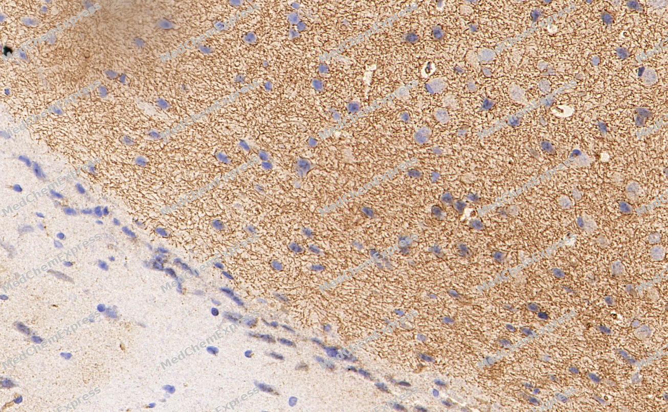 Immunohistochemical analysis of paraffin-embedded Mouse brain tissue using Tyrosine Hydroxylase Antibody. The section was pre-treated using heat mediated antigen retrieval with Tris-EDTA buffer (pH 9.0) for 8 minutes. The tissues were blocked in QuickBlock for 20 minutes at room temperature, washed with ddH2O and PBS, and then probed with the primary antibody (HY-P80362, 1/2000) in 4℃ overnight. The detection was performed using an HRP conjugated compact polymer system. DAB was used as the chromogen. Tissues were counterstained with hematoxylin and mounted with DPX.
 -
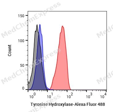 Flow cytometric analysis of 1X10^6 HEK293 cells labeling Tyrosine Hydroxylase Antibody (HY-P80362, red). Cells were fixed with 4% paraformaldehyde and permeabilised with 90% methanol. Then stained with the primary antibody at 1μg/mL dilution for an hour at 4℃. Alexa Fluor® 488-conjugated AffiniPure Goat Anti-Rabbit IgG H&L (HY-P8002) was used as the secondary antibody at 1/1,000 dilution for 30 minutes at 4℃. Rabbit IgG Isotype Control (HY-P80879, blue) was used as the isotype control, cells without incubation with primary antibody were used as the unlabeled control (black).
 -
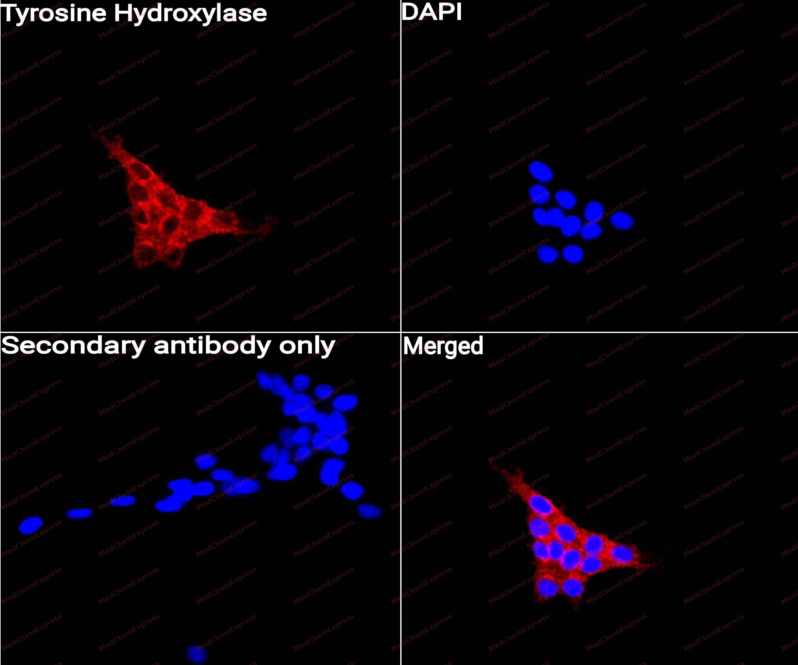 Immunocytochemistry analysis of C6 cells labeling Tyrosine Hydroxylase with Tyrosine Hydroxylase Antibody (HY-P80362) at 1/50 dilution. Cells were fixed in 4% paraformaldehyde for 15 minutes at room temperature, permeabilized with 0.1% Triton X-100 for 10 minutes at room temperature, then blocked with QuickBlock™ Blocking Buffer for Immunol Staining for 10 min at room temperature. Cells were then incubated with Tyrosine Hydroxylase Antibody (HY-P80362) at 1/50 dilution in QuickBlock™ Blocking Buffer for Immunol Staining at 4 ℃. Alexa Fluor® 594-conjugated AffiniPure Goat Anti-Rabbit IgG H&L(HY-P8003, Green) was used as the secondary antibody at 1/1,000 dilution. PBS instead of the primary antibody was used as the secondary antibody only control. The Nuclear counterstain was DAPI (Blue).
 -
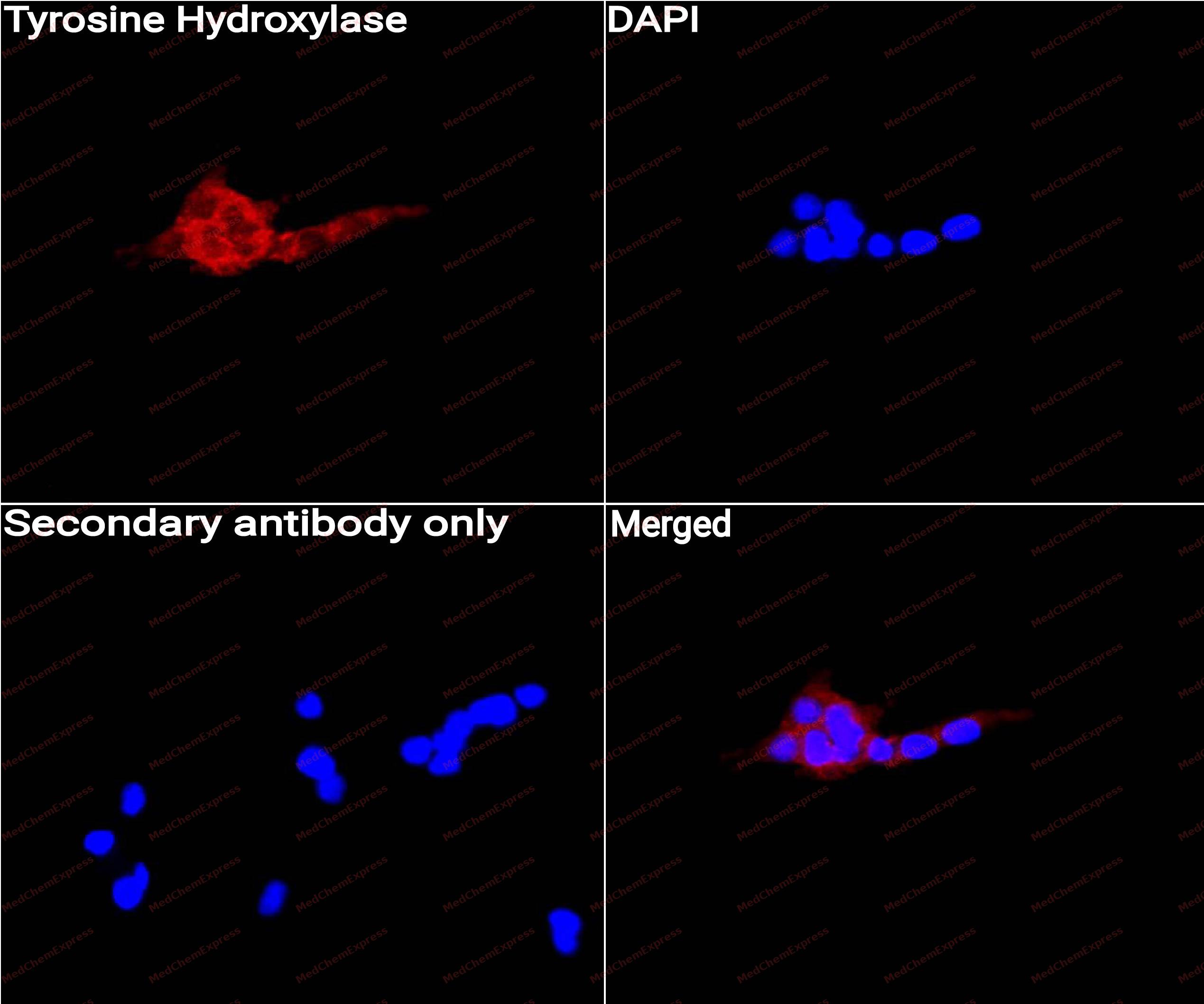 Immunocytochemistry analysis of C6 cells labeling Tyrosine Hydroxylase with Tyrosine Hydroxylase Antibody (HY-P80362) at 1/100 dilution. Cells were fixed in 4% paraformaldehyde for 15 minutes at room temperature, permeabilized with 0.1% Triton X-100 for 10 minutes at room temperature, then blocked with QuickBlock™ Blocking Buffer for Immunol Staining for 10 min at room temperature. Cells were then incubated with Tyrosine Hydroxylase Antibody (HY-P80362)at 1/100 dilution in QuickBlock™ Blocking Buffer for Immunol Staining at 4 ℃. Alexa Fluor® 594-conjugated AffiniPure Goat Anti-Rabbit IgG H&L(HY-P8003,Green) was used as the secondary antibody at 1/1,000 dilution. PBS instead of the primary antibody was used as the secondary antibody only control. The Nuclear counterstain was DAPI (Blue).


Background
-
Function
Tyrosine hydroxylase (TyrH, TH) is a rate-limiting enzyme in catecholamine biosynthesis and belongs to the family of aromatic amino acid hydroxylases. Tyrosine hydroxylase uses tetrahydrobiopterin and molecular oxygen to convert tyrosine to dopamine. The amino-terminal 150 amino acids of tyrosine hydroxylase form a domain whose structure is involved in regulating the activity of the enzyme. Regulation involves phosphorylation by multiple kinases on 4 different serine residues and dephosphorylation by 2 phosphatases. Tyrosine hydroxylase is inhibited by catecholamine neurotransmitters in a feedback manner. Dopamine competes with tetrahydrobiopterin for binding to tyrosine hydroxylase and interacts with the R domain. Tyrosine hydroxylase activity is regulated by protein-protein interactions with enzymes in the same pathway or the tetrahydrobiopterin pathway, structural proteins that are thought to be chaperones in mediating the oxidative state of neurons, and proteins that transfer dopamine to secretory vesicles. Tyrosine hydroxylase is modified in the presence of NO. Tyrosine hydroxylase expression maintains dopamine synthesis. Tyrosine hydroxylase is severely lost in Parkinson's disease and slightly lost in Alzheimer's disease.
-
Subcellular Localization
Cytoplasm, perinuclear region; Nucleus; Cell projection, axon; Cytoplasm; Cytoplasmic vesicle, secretory vesicle, synaptic vesicle
-
Expression
Tissue_specificity:Primarily expressed in the brain and adrenal glands -
Subunit
Homotetramer (PubMed:24947669, Ref.18). Interacts (when phosphorylated at Ser-19) with YWHAG; one YWHAG dimer binds to one TH tetramer and this interaction may influence the phosphorylation and dephosphorylation of other sites (PubMed:24947669).
-
SwissProt ID
-
Synonyms
TYH, TH, Tyrosine 3-monooxygenase, Tyrosine 3-hydroxylase
-
Research Field
Neuroscience
Documentation
-
Data Sheet (261 KB)
-
SDS (251 KB)
- English - EN (251 KB)
- Français - FR (251 KB)
- Deutsch - DE (251 KB)
- Norwegian - NO (251 KB)
- Español - ES (251 KB)
- Swedish - SV (251 KB)
- Italian - IT (251 KB)
- Korean - KR (251 KB)
- Portuguese - PT (251 KB)
-
User Guide for Antibodies (1077 KB)
References
[1]. Daubner SC, et al. Tyrosine hydroxylase and regulation of dopamine synthesis. Arch Biochem Biophys. 2011 Apr 1;508(1):1-12. [Content Brief]
[2]. Zhu Y, et al. Overview of tyrosine hydroxylase in Parkinson's disease. CNS Neurol Disord Drug Targets. 2012 Jun 1;11(4):350-8. [Content Brief]
[3]. Torack RM, et al. Tyrosine hydroxylase-like (TH) immunoreactivity in Parkinson's disease and Alzheimer's disease. J Neural Transm Park Dis Dement Sect. 1992;4(2):165-71. [Content Brief]

 Powered by Bioz
