



Tricalcium phosphate

Based on 1 Customer Validation
Tricalcium phosphate is a calcium-phosphorus compound mainly composed of calcium ions and phosphate (PO43-). Tricalcium phosphate promotes osteogenic differentiation, inhibits adipogenic and chondrogenic differentiation, and serves as a scaffold for spinal fusion implants. Tricalcium phosphate supports reparative dentin formation, mineralized nodule formation, and calcium ion release. Tricalcium phosphate is commonly used in fields such as biomaterials, bone repair, food additives, and pharmaceutical preparations.
Nos produits utilisent uniquement pour la recherche. Nous ne vendons pas aux patients.
- Pureté : 98.00%
- CAS No.: 7758-87-4
- Formule: Ca3(PO4)2
- Masse moléculaire:310.18
-
Stockage:Powder -20°C, 3 years , 4°C, 2 years ; In solvent -80°C, 6 months , -20°C, 1 month


To place orders, for customer services and technical support, please contact: MedChemExpress USA
Tel: 609-228-6898 E-mail: [email protected] [email protected]


-
 Activité biologique
-
Chemical Information
- Protocole
- Pureté et documentation
- Références
-
 Help & FAQs


-

HY-W243408D
Calcium carbonate, for cell cultureAcidity Regulator -

HY-21170B
Trifluoromethanesulfonate lithiumCatalyst -
HY-W011727A
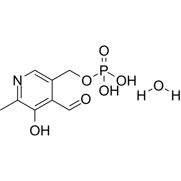
Pyridoxal 5'-phosphate monohydrateVitamin B6 Derivative -
HY-Y0332B
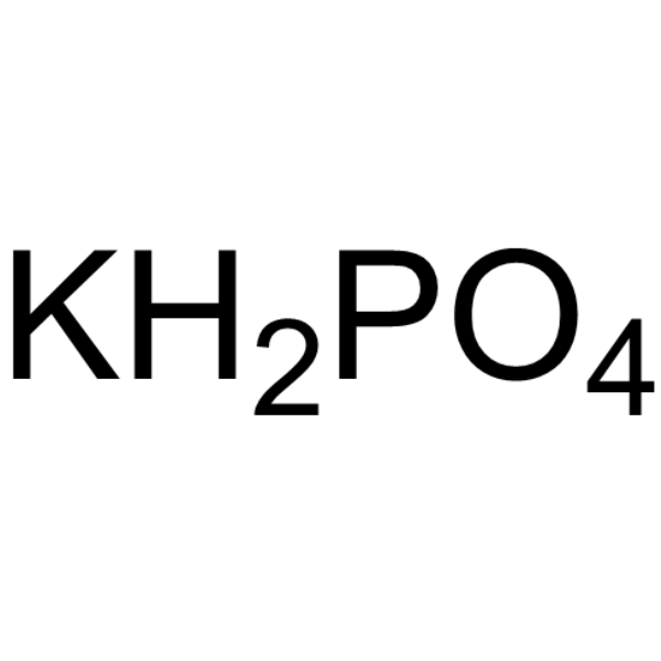
Potassium dihydrogen phosphate, AR,99.5%Cell Culture Reagent -
HY-Y0332K
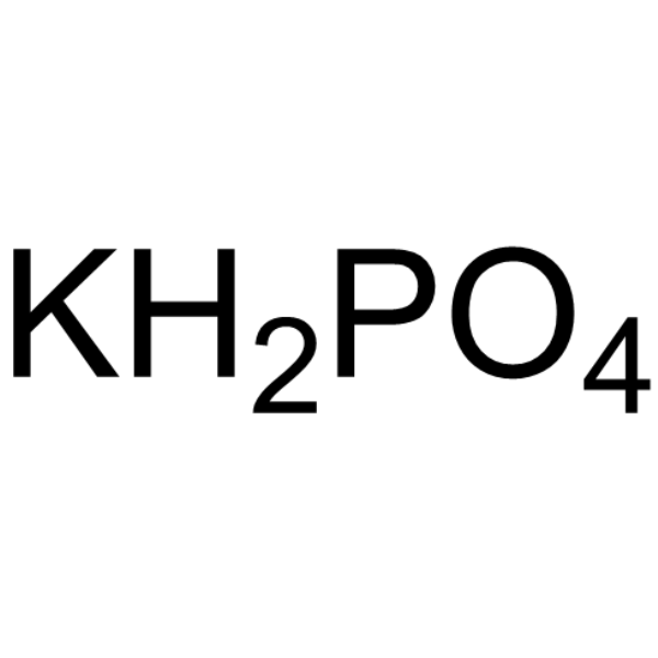
Potassium dihydrogen phosphate, for cell cultureCell Culture Reagent -
HY-Y0308E
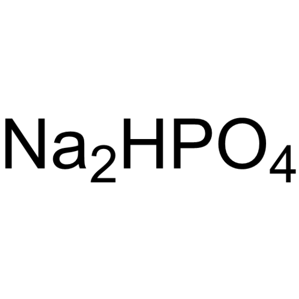
Sodium phosphate dibasic, for cell cultureBuffer -

HY-Y1267D
Magnesium sulfate, for cell cultureAnti-Inflammatory Agent -

HY-Y1267
Magnesium sulfateAnti-Inflammatory Agent -
HY-Y0308
Disodium phosphatePharmaceutical Excipient


Activité biologique
Description
In Vitro
Tricalcium phosphate (5 mg/mL; 8-25 days) increase ALP activity, a marker of osteogenic differentiation, in hBMSCs by 1.4-fold after 8 days and 2.2-fold after 25 days[2].
Tricalcium phosphate (5 mg/mL; 8 days) induce osteogenic gene expression in hBMSCs, increasing Runx2 by 2.3-fold and BMP2 by 4.7-fold after 8 days[2].
Tricalcium phosphate (10-20%; 4, 7, 10 days) conditioned medium from light-cured composites causes human dental pulp stem cells to exhibit comparable mineralized nodule formation to each other, with lower mineralization than controls under differentiation conditions on day 10[3].
MedChemExpress (MCE) has not independently confirmed the accuracy of these methods. They are for reference only. Further protocols information, click here.
In Vivo
Tricalcium phosphate (5 mg/mL; added to hBMSC culture media; 8 days; surgically implanted; single administration) scaffold combined with untreated hBMSCs supports spinal fusion in immunodeficient rats, achieving 75% fusion with 0.5% bone surface area in fusion mass after 8 weeks[2].
Tricalcium phosphate (10%; local application; light-cured for 40 s) resolves pulp inflammation by week 3 and supports moderate to abundant mineralized tissue formation by weeks 3 and 6, respectively, in Wistar rats with exposed dental pulp[3].
MedChemExpress (MCE) has not independently confirmed the accuracy of these methods. They are for reference only.
Essai clinique
| NCT Number | Sponsor | Condition | Start Date |
Phase

|
|---|---|---|---|---|
| NCT01329991 | Plexxikon| | 2011-05 | PHASE1 |


Chemical Information
-
CAS No. 7758-87-4
-
Appearance Solid
-
Masse moléculaire 310.18
-
Formule Ca3(PO4)2
-
Color White to off-white
-
SMILES
[Ca3(PO4)2]
-
Livraison
Room temperature in continental US; may vary elsewhere.
-
Stockage
Powder -20°C 3 years 4°C 2 years In solvent -80°C 6 months -20°C 1 month

Protocole
-
Mesenchymal stromal/stem cell chondrogenic differentiation
MSC chondrogenic differentiation is commonly induced by culturing bone marrow-derived mesenchymal stromal/stem cells as high-density three-dimensional pellets or micromass aggregates in defined chondrogenic medium containing TGF-β family stimulation; the readout is formation of cartilage-like extracellular matrix, especially sulfated proteoglycans, aggrecan, and type II collagen. The assay detects chondrogenesis by pellet enlargement, metachromatic or Alcian blue/Safranin O staining of proteoglycan-rich matrix, immunodetection of type II collagen and aggrecan, and gene-expression changes in cartilage matrix markers; hypertrophic or fibrocartilaginous drift can be assessed by collagen X and collagen I readouts when included.
-
Mesenchymal stromal/stem cell osteogenic differentiation
Mesenchymal stromal/stem cells can be induced toward an osteoblast-like lineage in vitro by culture in osteogenic medium containing dexamethasone, ascorbic acid or ascorbate-2-phosphate, and β-glycerophosphate; the differentiation process is commonly evaluated by alkaline phosphatase activity, osteogenic marker expression, collagenous matrix formation, and calcium-rich matrix mineralization. The main readouts are alkaline phosphatase activity as an early osteogenic marker and Alizarin Red S staining as a calcium-deposit readout for mineralized extracellular matrix; Alizarin Red S can be inspected microscopically or extracted and measured colorimetrically at 405 nm.


Pureté et documentation
-
Fiche technique (281 KB)
-
SDS (393 KB)
- English - EN (393 KB)
- Français - FR (393 KB)
- Deutsch - DE (393 KB)
- Norwegian - NO (393 KB)
- Español - ES (393 KB)
- Swedish - SV (393 KB)
- Italian - IT (393 KB)
- Korean - KR (393 KB)
- Portuguese - PT (393 KB)
-
Instruction de manipulation (2659 KB)
Références

Calculators
Concentration (start) × Volume (start) = Concentration (final) × Volume (final)


Tricalcium phosphate Related Classifications
HY-D0833 Related Classifications
 Powered by Bioz