



CAP 3
CAP 3 is a cholic acid-peptide conjugate (CAP) antimicrobial agent. CAP 3 effectively inhibits Gram-negative bacteria, with MIC99 (minimum inhibitory concentration for 99% bacterial killing) values of 8 μM, 16 μM, and 16 μM against E. coli, Klebsiella pneumoniae and Acinetobacter baumannii, respectively. CAP 3 exerts its antibacterial effects by disrupting the structural integrity of the bacterial lipopolysaccharide (LPS) outer membrane. CAP 3 rapidly kills bacteria, inhibits biofilm formation, and effectively combats drug-resistant strains and persistent bacterial infections.
For research use only. We do not sell to patients.
- CAS No.: 2292123-03-4
- Formula: C52H82N6O11
- Molecular Weight:967.24
-
Storage:
Please store the product under the recommended conditions in the Certificate of Analysis.


To place orders, for customer services and technical support, please contact: MedChemExpress USA
Tel: 609-228-6898 E-mail: [email protected] [email protected]


-
 Biological Activity
-
Chemical Information
- Protocols
- Purity & Documentation
- References
-
 Help & FAQs


-
HY-117660
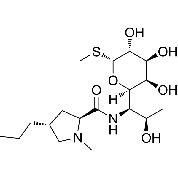
LincomycinRibosome Inhibitor -
HY-113829
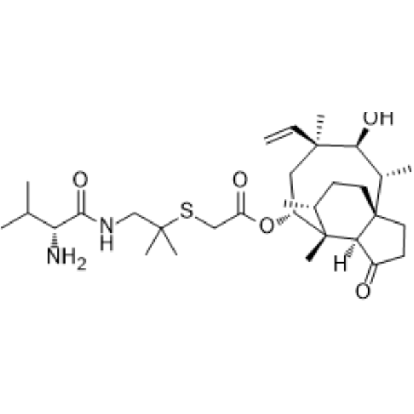
ValnemulinAntibacterial Agent -
HY-138075
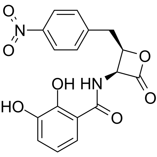
ObafluorinAntibiotic -

HY-117660R
Lincomycin (Standard)Standard of Lincomycin -

HY-120256
Corynecin II -

HY-Z7555
Cefazolin impurity HCefazolin Impurity -

HY-P3119
DesotamideAntibiotic -

HY-126746
Corynecin IAntibiotic


Biological Activity
Description


Chemical Information
-
CAS No. 2292123-03-4
-
Molecular Weight 967.24
-
Formula C52H82N6O11
-
SMILES
CC(C)[C@H](N)C(NCC(O[C@H]1[C@]2([H])[C@@](C[C@@H]([C@]3([C@@]2([H])CC[C@]3([H])[C@H](C)CCC(OCC4=CC=CC=C4)=O)C)OC(CNC([C@@H](N)C(C)C)=O)=O)([H])[C@@]5([C@](C[C@@H](CC5)OC(CNC([C@@H](N)C(C)C)=O)=O)([H])C1)C)=O)=O
-
Shipping
Room temperature in continental US; may vary elsewhere.
-
Storage
Please store the product under the recommended conditions in the Certificate of Analysis.


Purity & Documentation
References

Calculators
Concentration (start) × Volume (start) = Concentration (final) × Volume (final)


CAP 3 Related Classifications
HY-115693 Related Classifications
 Powered by Bioz