



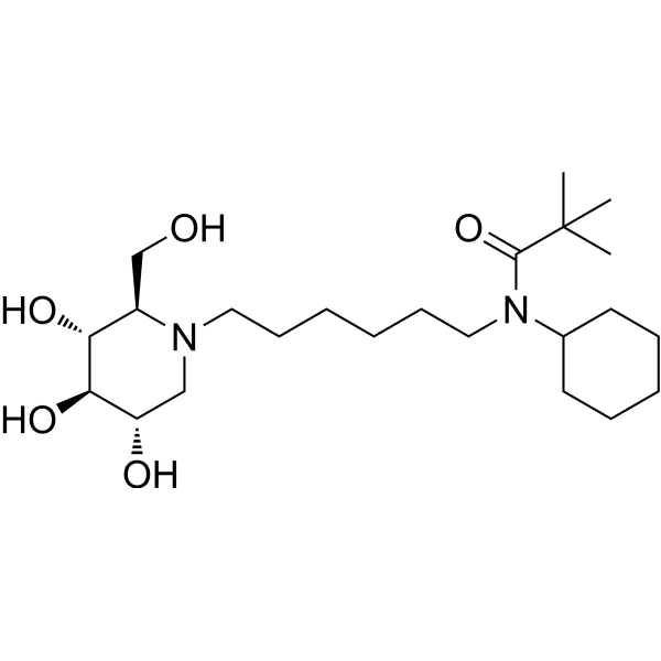
IHVR-17028

Based on 1 Customer Validation
IHVR-17028 is a potent and broad-spectrum antiviral agent. IHVR-17028 exhibits antiviral activity against BVDV, TCRV and DENV with EC50 values of 0.4 μM, 0.26 μM, 0.3 μM, respectively. IHVR-17028 is a potent ER α-glucosidase I inhibitor with an IC50 of 0.24 μM. IHVR-17028 can be used for infectious diseases research.
For research use only. We do not sell to patients.
- Purity : 99.61%
- CAS No.: 1428247-78-2
- Formula: C23H44N2O5
- Molecular Weight:428.61
-
Storage:Pure form -20°C, 3 years ; In solvent -80°C, 6 months , -20°C, 1 month

To place orders, for customer services and technical support, please contact: MedChemExpress USA
Tel: 609-228-6898 E-mail: [email protected] [email protected]


-
 Biological Activity
-
Chemical Information
- Protocols
- Purity & Documentation
- References
-
 Help & FAQs


-
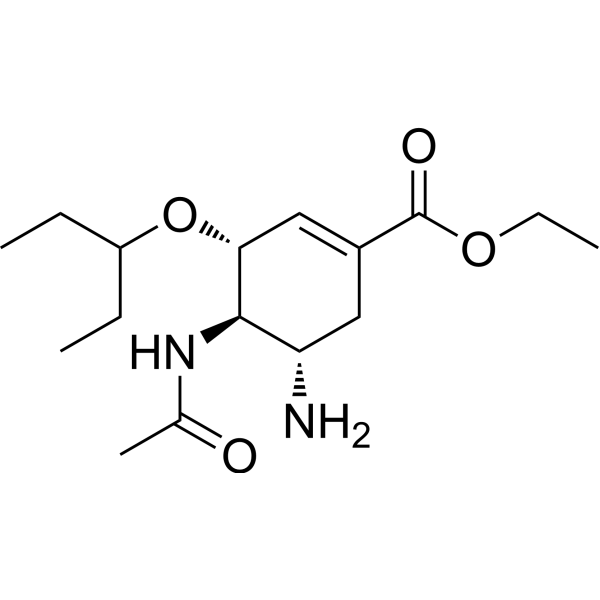
HY-13317
OseltamivirInfluenza Virus Inhibitor -
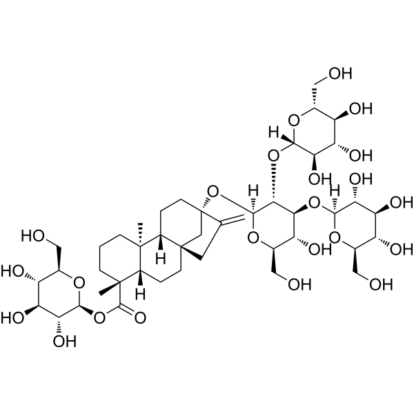
HY-N0466
Rebaudioside Aα-Glucosidase Inhibitor -
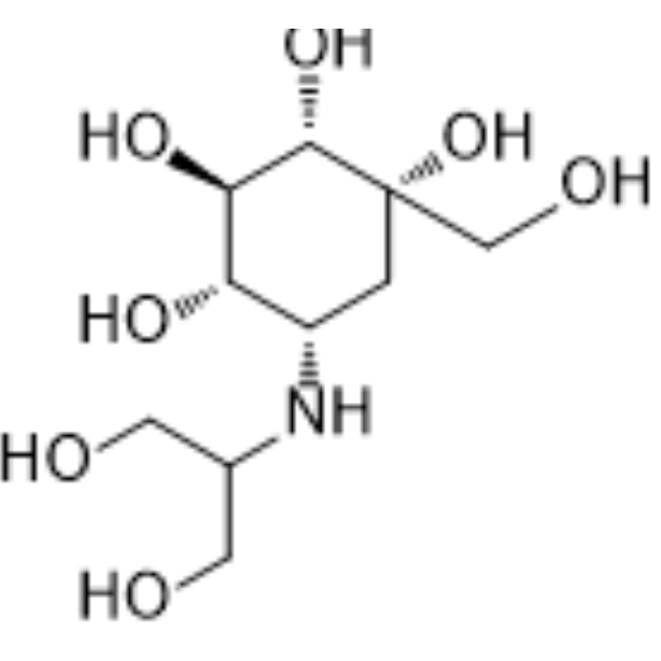
HY-B0025
VogliboseGlucosidase Inhibitor -
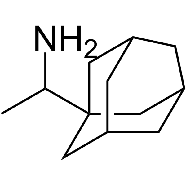
HY-B0338
RimantadineAnti-Influenza Virus Agent -
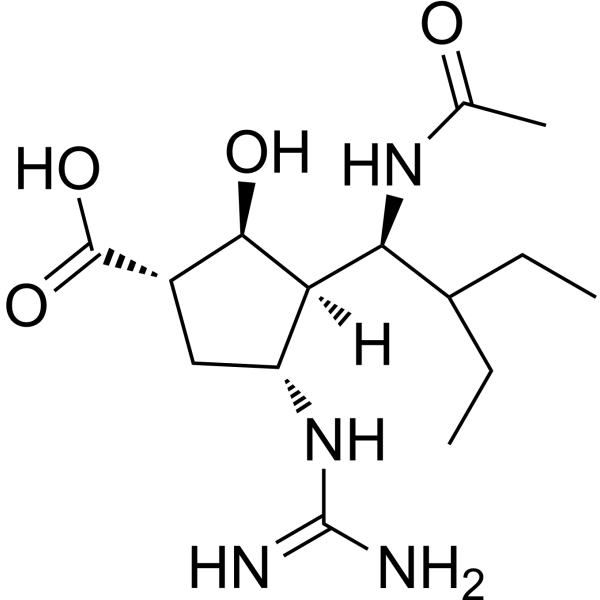
HY-17015A
PeramivirCyclopentane neuraminidase Inhibitor -
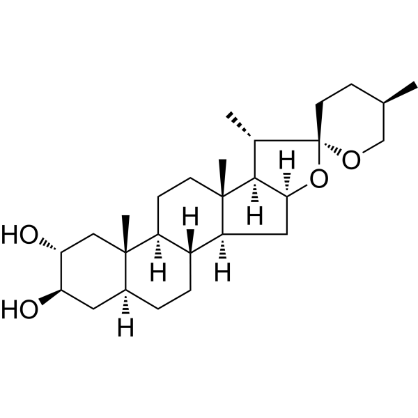
HY-N2574
GitogeninGlucosidase Inhibitor -
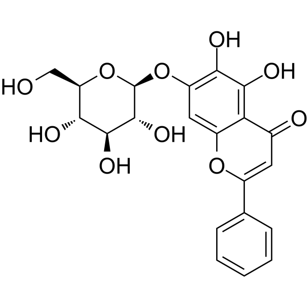
HY-N2025
Oroxin APartial PPARγ Agonist -
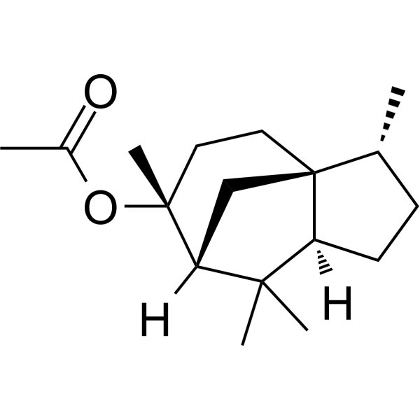
HY-W009417
Cedryl acetateα-glucosidase Inhibitor -
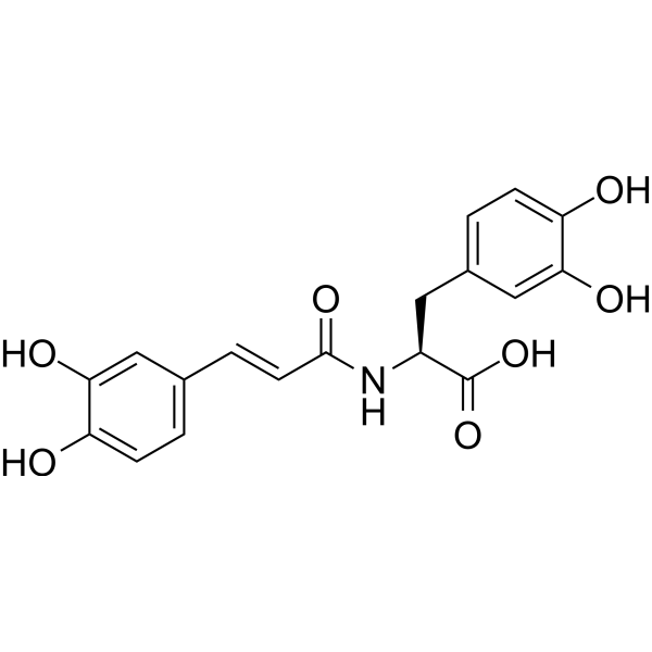
HY-122267
ClovamideAntioxidant Agent -
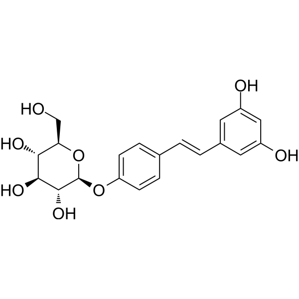
HY-N4195
Resveratrolosideα-Glucosidase Inhibitior -
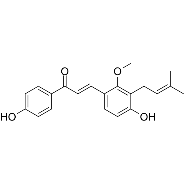
HY-N0374
Licochalcone Cα-Glucosidase Inhibitor -

HY-W005598
2-Hydroxyquinolineα-Glucosidase/α-Amylase Inhibitor


Biological Activity
Description
Cellular Effect
|
Cell Line

|
Type | Value | Description | References |
|---|---|---|---|---|
| Huh-7 | CC50 |
500 μM
Compound: 24
|
Cytotoxicity against human Huh7.5 cells by MTT assay
Cytotoxicity against human Huh7.5 cells by MTT assay
|
[PMID: 23453839] |
| Huh-7 | CC50 |
500 μM
Compound: 3
|
Cytotoxicity against human Huh7.5 cells by MTT assay
Cytotoxicity against human Huh7.5 cells by MTT assay
|
[PMID: 23747225] |
| Huh-7 | EC50 |
0.2 μM
Compound: 3
|
Antiviral activity against Tacaribe virus infected in human Huh7.5 cells assessed as reduction in viral yield after 3 days by neutral-red staining-based microscopic analysis
Antiviral activity against Tacaribe virus infected in human Huh7.5 cells assessed as reduction in viral yield after 3 days by neutral-red staining-based microscopic analysis
|
[PMID: 23747225] |
| Huh-7 | EC50 |
0.2 μM
Compound: 24
|
Antiviral activity against Tacaribe virus infected in human Huh7.5 cells
Antiviral activity against Tacaribe virus infected in human Huh7.5 cells
|
[PMID: 23453839] |
In Vitro
In virus yield reduction assays, IHVR-17028 inhibits viral activities with EC50 values of 0.4 μM, 0.26 μM, 0.3 μM for bovine viral diarrhea virus (BVDV) (NADL strain), tacaribe virus (TCRV) (11573 strain), DENV (serotype 2, New Guinea C), respectively. And in MTT assays, IHVR-17028 exhibits IC50 values of all >500 μM in?MDBK, Huh7.5 or BHK cells[1].
MedChemExpress (MCE) has not independently confirmed the accuracy of these methods. They are for reference only. Further protocols information, click here.
In Vivo
MedChemExpress (MCE) has not independently confirmed the accuracy of these methods. They are for reference only.
-
Animal Model:BALB/c mice are challenged with 1,000 PFU mouse adapted MARV via IP injection[1]
-
Dosage:50 mg/kg
-
Administration:Treatment 1 day prior to virus challenging
-
Result:Exhibited protection in mouse when the treatment is initiated 1 day prior to virus challenging.
-
Animal Model:C57B1/6 mice challenged with 1,000 PFU mouse adapted EBOV[1]
-
Dosage:25 mg/kg
-
Administration:Twice daily at 12 h interval; starting 4 h post infection for 10 days
-
Result:Inhibited EBOV infection in mice.


Chemical Information
-
CAS No. 1428247-78-2
-
Appearance Oil
-
Molecular Weight 428.61
-
Formula C23H44N2O5
-
Color Colorless to light yellow
-
SMILES
OC[C@@H]1[C@@H](O)[C@H](O)[C@@H](O)CN1CCCCCCN(C(C(C)(C)C)=O)C2CCCCC2
-
Shipping
Room temperature in continental US; may vary elsewhere.
-
Storage
Pure form -20°C 3 years In solvent -80°C 6 months -20°C 1 month

Protocols
-
Research Protocol for Infectious Diseases
Infectious-disease experiments test how pathogens interact with host barriers, innate immune receptors, inflammatory signaling, pathogen replication, and tissue injury; pattern-recognition receptors such as TLRs, RIG-I-like receptors, NOD-like receptors, and inflammasomes detect microbial molecules and activate NF-κB, interferon, and cytokine responses. The central hypothesis is that infection severity reflects the balance between pathogen burden and host response: protective inflammation restricts pathogen growth, whereas excessive or mislocalized inflammation contributes to tissue damage and disease phenotype. Unresolved questions include which host pathways are protective versus pathogenic, why some infection models fail to translate to human disease, and which combined readouts best predict clinically relevant infection outcomes.
-
Cell Cytotoxicity Assay
Cytotoxicity assays are usually based on the assessment of cell membrane damage, which can also be indirectly detected by measuring cell viability. Detection methods include MTT assay, CKK-8 assay, LDH assay and ATP assay, etc.


Purity & Documentation
-
Data Sheet (272 KB)
-
SDS (252 KB)
- English - EN (252 KB)
- Français - FR (252 KB)
- Deutsch - DE (252 KB)
- Norwegian - NO (252 KB)
- Español - ES (252 KB)
- Swedish - SV (252 KB)
- Italian - IT (252 KB)
- Korean - KR (252 KB)
- Portuguese - PT (252 KB)
-
Handling Instructions (2659 KB)
References

Calculators
Concentration (start) × Volume (start) = Concentration (final) × Volume (final)


IHVR-17028 Related Classifications
HY-139663 Related Classifications
 Powered by Bioz