



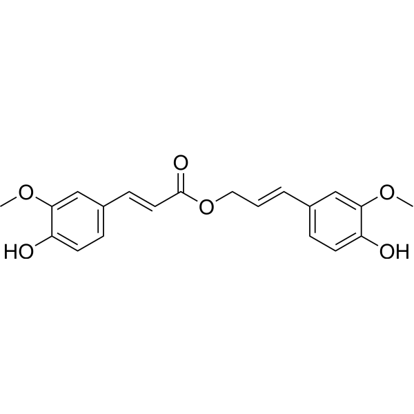
Coniferyl ferulate
Based on 1 publication(s) in Google Scholar
Coniferyl ferulate is an orally active phenolic acid compound. Coniferyl ferulate is a potent inhibitor of glutathione S-transferase (GST) (IC50 = 0.3 μM), which downregulates P-gp expression, induces apoptosis in B-MD-C1 (ADR+/+) cells, and reverses multidrug resistance. Coniferyl ferulate blocks the NMDAR/NR2B-CaMKII-MAPKs signaling pathway, inhibits ROS production and mitochondrial apoptosis, while reshapes the intestinal microbiota and microbial metabolism, ameliorates colonic inflammation and alleviates depressive symptoms in mice. Coniferyl ferulate can alleviate the toxicity of xylene to hematopoietic stem and progenitor cells by targeting Mgst2. Coniferyl ferulate exhibits antibacterial activity against the Gram-positive Bacillus subtilis and Staphylococcus aureus.
For research use only. We do not sell to patients.
- Purity : 98.73%
- CAS No.: 63644-62-2
- Formula: C20H20O6
- Molecular Weight:356.37
-
Storage:
-20°C, protect from light
* In solvent : -80°C, 6 months; -20°C, 1 month (protect from light)

To place orders, for customer services and technical support, please contact: MedChemExpress USA
Tel: 609-228-6898 E-mail: [email protected] [email protected]


-
 Biological Activity
-
Chemical Information
-
Solvent & Solubility
- Protocols
- Purity & Documentation
- References
-
 Help & FAQs


-
HY-B1640
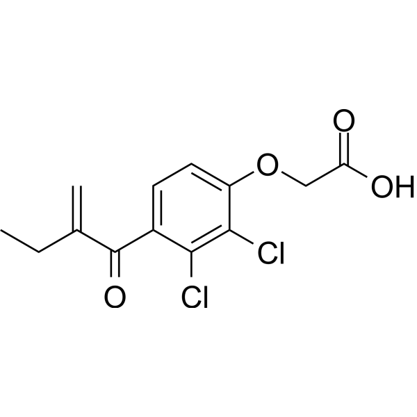
Ethacrynic acidDiuretic, Win Inhibitor, GSTs Inhibitor, Radiosensitizer -
HY-50671
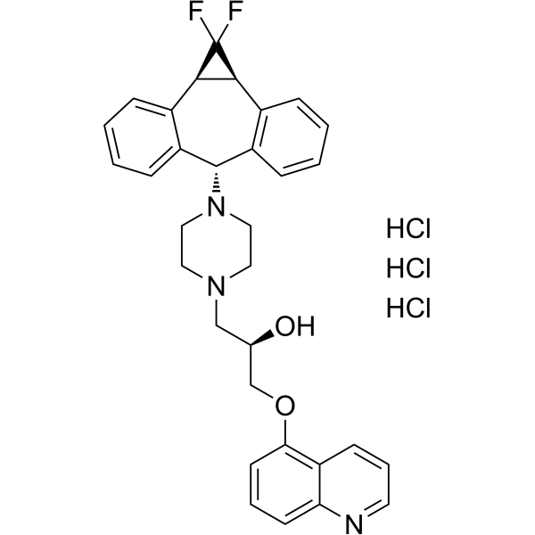
Zosuquidar trihydrochlorideP-gp Inhibitor -
HY-101413
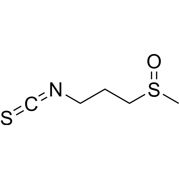
IberinApoptosis Inducer -
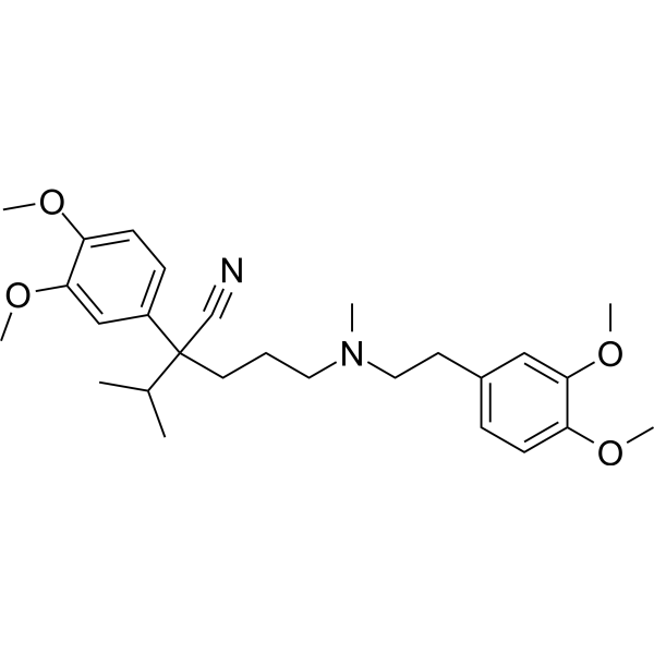
HY-14275
VerapamilCalcium Channel Blocker -
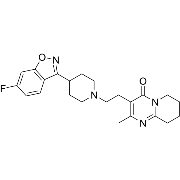
HY-11018
Risperidone5-HT2 Receptor Blocker -
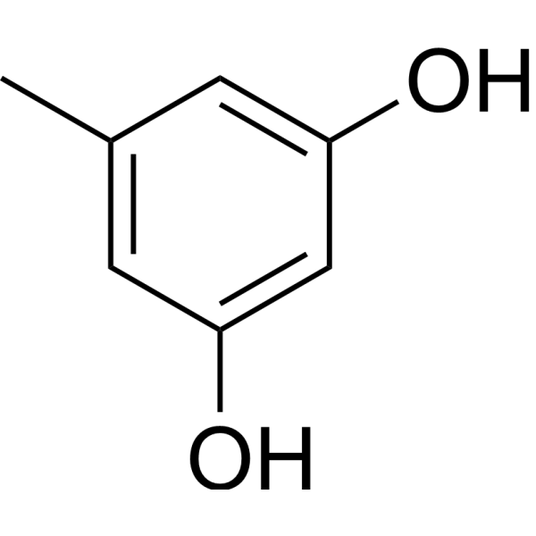
HY-D0168
OrcinolMelanogenesis Inhibitor -
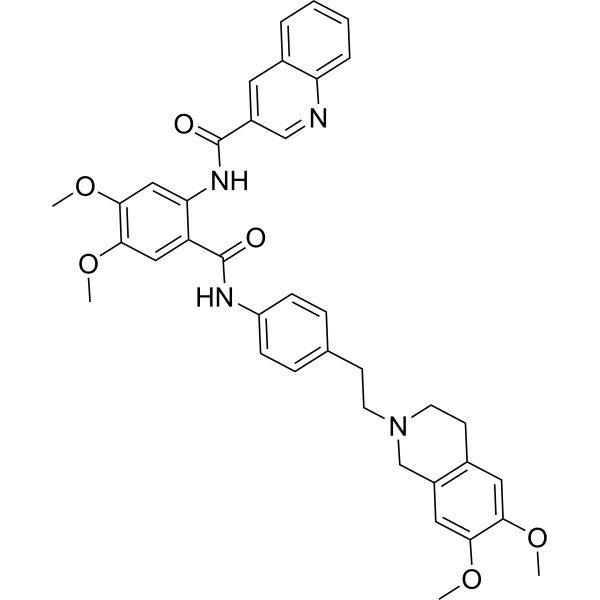
HY-10550
TariquidarP-gp Inhibitor -

HY-14275R
Verapamil (Standard)Calcium Channel Blocker -
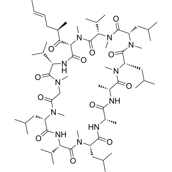
HY-17384
ValspodarP-glycoprotein Inhibitor -
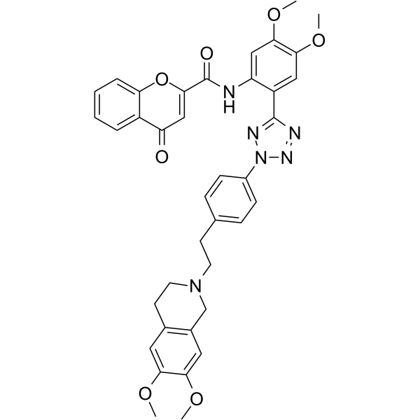
HY-13646
EncequidarP-glycoprotein Inhibitor -
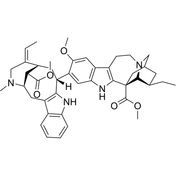
HY-N6932
VoacamineCB1/P-gp Inhibitor
Publications Citing Use of MedChemExpress (MCE) Coniferyl ferulate
MoreAll iGluR Isoforms
More

Biological Activity
Description
IC50 & Target
[2]|
CaMK II |
NMDA Receptor |
Cellular Effect
|
Cell Line

|
Type | Value | Description | References |
|---|---|---|---|---|
| MDA-MB-231 | IC50 |
19.8 μM
Compound: 40; coniferyl ferulate
|
Antimigratory activity against human MDA-MB-231 cells expressing c-MET assessed as inhibition of HGF-induced cell migration incubated for 24 hrs by Giemsa staining based wound healing assay
Antimigratory activity against human MDA-MB-231 cells expressing c-MET assessed as inhibition of HGF-induced cell migration incubated for 24 hrs by Giemsa staining based wound healing assay
|
[PMID: 27258622] |
| MDA-MB-231 | IC50 |
22.3 μM
Compound: 40; coniferyl ferulate
|
Antiproliferative activity against human MDA-MB-231 cells expressing c-MET assessed as inhibition of HGF-induced cell growth incubated for 72 hrs by MTT assay
Antiproliferative activity against human MDA-MB-231 cells expressing c-MET assessed as inhibition of HGF-induced cell growth incubated for 72 hrs by MTT assay
|
[PMID: 27258622] |
| MDA-MB-468 | IC50 |
25.6 μM
Compound: 40; coniferyl ferulate
|
Antiproliferative activity against human MDA-MB-468 cells expressing c-MET assessed as inhibition of HGF-induced cell growth incubated for 72 hrs by MTT assay
Antiproliferative activity against human MDA-MB-468 cells expressing c-MET assessed as inhibition of HGF-induced cell growth incubated for 72 hrs by MTT assay
|
[PMID: 27258622] |
In Vitro
Coniferyl ferulate (5-20 μM, 48 h) induces apoptosis and markedly inhibits cells in G1-phase in B-MD-C1 (ADR+/+) cells[1].
Coniferyl ferulate (5-20 μM, 48 h) inhibits the upregulation of MDR1/P-gp caused by Doxorubicin (HY-15142A), and bring the expression of P-gp close to the basal level in B-MD-C1 (ADR+/+) cells[1].
Coniferyl ferulate (0.2-20 μM, 28 h) protects against the decline in cell viability and apoptosis induced by Glutamate (HY-14608), and alleviates cell membrane damage by blocking the NMDA receptor-CaMKII-MAPKs signaling cascade, reducing the level of ROS and enhancing SOD activity in PC12 cells[2].
MedChemExpress (MCE) has not independently confirmed the accuracy of these methods. They are for reference only.
-
Cell Line:B-MD-C1(ADR+/+) cells
-
Concentration:5, 10 and 20 μM
-
Incubation Time:48 h
-
Result:Induced apoptosis.
-
Cell Line:B-MD-C1(ADR+/+) cells
-
Concentration:5, 10 and 20 μM
-
Incubation Time:48 h
-
Result:Markedly increased the proportion of cells in G1-phase, decreased the proportion of cells in S-phase.
-
Cell Line:B-MD-C1(ADR+/+) cells
-
Concentration:5, 10 and 20 μM
-
Incubation Time:48 h
-
Result:Dose-dependently inhibited Doxorubicin-induced MDR1 overexpression.
Brought the expression of P-gp close to the basal level.
-
Cell Line:Glutamate-damaged PC12 cells
-
Concentration:0.2, 2, 20 μM
-
Incubation Time:After 4 hours of pre-treatment, with Glutamate for another 24 hours
-
Result:Concentration-dependently protected the decline in cell viability induced by Glutamate.
Significantly alleviated the damage to the cell membrane caused by Glutamate.
-
Cell Line:Glutamate-damaged PC12 cells
-
Concentration:0.2, 2, 20 μM
-
Incubation Time:After 4 hours of pre-treatment, with Glutamate for another 24 hours
-
Result:Reduced nuclear shrinkage and PI-positive cells.
-
Cell Line:Glutamate-damaged PC12 cells
-
Concentration:0.2, 2, 20 μM
-
Incubation Time:After 4 hours of pre-treatment, with Glutamate for another 24 hours
-
Result:Decreased the level of p-NR2B, p-CaMKII, p-JNK and p-p38.
Increased the Bcl-2/Bax ratio and decreased the release of cytochrome C and activation of Caspase-3.
In Vivo
Coniferyl ferulate (50 mg/kg, i.g., once daily, for 4 weeks) attenuates depression-like and anxiety-like behaviors induced by chronic unpredicted mild stress (CUMS), and significantly ameliorates colonic inflammation in mice[3].
Coniferyl ferulate (50 mg/kg, i.g., once daily, for 25 days) alleviates xylene-caused hematopoietic stem and progenitor cell toxicity by inhibiting Mgst2 in mice[4].
MedChemExpress (MCE) has not independently confirmed the accuracy of these methods. They are for reference only.
-
Animal Model:Behavioral test (tail suspension and forced-swimming test) established in male ICR mice (18-22 g)[2]
-
Dosage:25, 50 and 100 mg/kg
-
Administration:Intragastric administration (i.g.), once daily, for 13 and 14 days
-
Result:Obviously decreased the immobility time in tail suspension test (TST) and forced swimming test (FST).
-
Animal Model:CUMS model established in in 8-week-old male C57BL/6 SPF mice (20 g)[3]
-
Dosage:50 mg/kg
-
Administration:Intragastric administration (i.g.), once daily, for 4 weeks
-
Result:Improved depressive in sucrose preference test (SPT), open field test (OFT), FTS and TST and anxiety-like behaviors in elevated plus-maze test (EPM).
Restored the reduction of intestinal microvilli and alleviated mitochondrial swelling.
Lowered the levels of IL-6, IL-1β, and TNF-α.
Restructured the gut microbiome, and microbial metabolism.
-
Animal Model:Xylene-induced hematotoxicity model established in male C57BL/6 SPF mice (8 weeks old, 20 g)
-
Dosage:50 mg/kg
-
Administration:Intragastric administration (i.g.), once daily, for 25 days
-
Result:Significantly reversed the White blood cell count (WBC) reduction induced by xylene.
Restored the proportion of bone marrow LSK cells to the normal level.
Reduced the level of ROS in mitochondria and restored the mitochondrial membrane potential inhibited by xylene.
Acted directly on the Mgst2 target site (KD = 8.0861 kcal/mol).
- RNA extraction experimental
- Research Protocol for Infectious Diseases
- Detection of Apoptosis
- TUNEL staining for apoptotic DNA fragmentation
- Annexin V plus membrane-impermeant dye apoptosis staining
- ROS/oxidative-stress fluorescent staining
- Apoptosis Solutions
- Research Protocol for Microbiome Analysis
- Research Protocol for Inflammation-related Diseases
- Gram Staining of Tissue Sections
- Bacterial live/dead nucleic-acid viability staining


Chemical Information
-
CAS No. 63644-62-2
-
Appearance Solid
-
Molecular Weight 356.37
-
Formula C20H20O6
-
Color White to off-white
-
SMILES
O=C(OC/C=C/C1=CC=C(O)C(OC)=C1)/C=C/C2=CC=C(O)C(OC)=C2
-
Structure Classification
-
Shipping
Room temperature in continental US; may vary elsewhere.
-
Storage
-20°C, protect from light
* In solvent : -80°C, 6 months; -20°C, 1 month (protect from light)


Publications (1)
-
Journal Impact Factor
-
Most Recent
-
Am J Respir Cell Mol Biol
2026 Mar 25:aanag065. PMID: 42089332


Solvent & Solubility
In Vitro:
DMSO : 100 mg/mL (280.61 mM; Need ultrasonic; Hygroscopic DMSO has a significant impact on the solubility of product, please use newly opened DMSO)
Please refer to the solubility information to select the appropriate solvent. Once prepared, please aliquot and store the solution to prevent product inactivation from repeated freeze-thaw cycles.
Storage method and period of stock solution: -80°C, 6 months; -20°C, 1 month (protect from light). When stored at -80°C, please use it within 6 months. When stored at -20°C, please use it within 1 month.
Please refer to the solubility information to select the appropriate solvent. Once prepared, please aliquot and store the solution to prevent product inactivation from repeated freeze-thaw cycles.
Storage method and period of stock solution: -80°C, 6 months; -20°C, 1 month (protect from light). When stored at -80°C, please use it within 6 months. When stored at -20°C, please use it within 1 month.
Concentration (start) × Volume (start) = Concentration (final) × Volume (final)
In Vivo:
Select the appropriate dissolution method based on your experimental animal and administration route.
- For the following dissolution methods, please ensure to first prepare a clear stock solution using an In Vitro approach and then sequentially add co-solvents:
- To ensure reliable experimental results, the clarified stock solution can be appropriately stored based on storage conditions. As for the working solution for In Vivo experiments, it is recommended to prepare freshly and use it on the same day.
- The percentages shown for the solvents indicate their volumetric ratio in the final prepared solution. If precipitation or phase separation occurs during preparation, heat and/or sonication can be used to aid dissolution.
Add each solvent one by one: 10% DMSO 90% (20% SBE-β-CD in Saline)
Solubility: 2.5 mg/mL (7.02 mM); Suspended solution; Need ultrasonic
This protocol yields a suspended solution of 2.5 mg/mL. Suspended solution can be used for oral and intraperitoneal injection.
Taking 1 mL working solution as an example, add 100 μL DMSO stock solution (25.0 mg/mL) to 900 μL 20% SBE-β-CD in Saline, and mix evenly.
Preparation of 20% SBE-β-CD in Saline (4°C, storage for one week): 2 g SBE-β-CD powder is dissolved in 10 mL Saline, completely dissolve until clear.
In Vivo Dissolution Calculator
Please enter the basic information of animal experiments:
-
-
-
-
Recommended: Prepare an additional quantity of animals to account for potential losses during experiments.
Please enter your animal formula composition:
-
%DMSO +
Recommended: Keep the proportion of DMSO in working solution below 2% if your animal is weak.
-
%+
-
+%Tween-80 + +
-
%Saline +
The co-solvents required include: DMSO, . All of co-solvents are available by MedChemExpress (MCE). , Tween 80. All of co-solvents are available by MedChemExpress (MCE).
Working solution concentration: 0.22 mg/mL
Method for preparing stock solution: mg drug dissolved in μL DMSO. Stock solution concentration: mg/mL. * In solvent : -80°C, 6 months; -20°C, 1 month (protect from light)
1. Take μL DMSO stock solution;
2. Add μL .
μL , mix evenly;
3. Then add μL Tween 80, mix evenly;
4. Then add μL
Please ensure that the stock solution in the first step is dissolved to a clear state, and add co-solvents in sequence. You can use ultrasonic heating (ultrasonic cleaner, recommended frequency 20-40 kHz), vortexing, etc. to assist dissolution.


Purity & Documentation
-
Data Sheet (282 KB)
-
SDS (252 KB)
- English - EN (252 KB)
- Français - FR (252 KB)
- Deutsch - DE (252 KB)
- Norwegian - NO (252 KB)
- Español - ES (252 KB)
- Swedish - SV (252 KB)
- Italian - IT (252 KB)
- Korean - KR (252 KB)
- Portuguese - PT (252 KB)
-
Handling Instructions (2659 KB)
References
[1]. Chen C, et al. Coniferyl Ferulate, a Strong Inhibitor of Glutathione S-Transferase Isolated from Radix Angelicae sinensis, Reverses Multidrug Resistance and Downregulates P-Glycoprotein. Evid Based Complement Alternat Med. 2013;2013:639083. [Content Brief]
[2]. Gong W, et al. Coniferyl ferulate exerts antidepressant effect via inhibiting the activation of NMDAR-CaMKII-MAPKs and mitochondrial apoptotic pathways. J Ethnopharmacol. 2020 Apr 6;251:112533. [Content Brief]
[3]. Hao WZ, et al. Oral coniferyl ferulate attenuated depression symptoms in mice via reshaping gut microbiota and microbial metabolism. Food Funct. 2021 Dec 13;12(24):12550-12564. [Content Brief]
[4]. Yin Z, et al. Coniferyl ferulate alleviate xylene-caused hematopoietic stem and progenitor cell toxicity by Mgst2. Front Pharmacol. 2024 Mar 8;15:1334445. [Content Brief]
[5]. Chou SC, et al. Antibacterial activity of components from Lomatium californicum. Phytother Res. 2006 Feb;20(2):153-6. [Content Brief]


Complete Stock Solution Preparation Table
Please refer to the solubility information to select the appropriate solvent. Once prepared, please aliquot and store the solution to prevent product inactivation from repeated freeze-thaw cycles.
Storage method and period of stock solution: -80°C, 6 months; -20°C, 1 month (protect from light). When stored at -80°C, please use it within 6 months. When stored at -20°C, please use it within 1 month.
| Optional Solvent | Concentration Solvent Mass | 1 mg | 5 mg | 10 mg | 25 mg |
|---|---|---|---|---|---|
| DMSO | 1 mM | 2.8061 mL | 14.0304 mL | 28.0607 mL | 70.1518 mL |
| 5 mM | 0.5612 mL | 2.8061 mL | 5.6121 mL | 14.0304 mL | |
| 10 mM | 0.2806 mL | 1.4030 mL | 2.8061 mL | 7.0152 mL | |
| 15 mM | 0.1871 mL | 0.9354 mL | 1.8707 mL | 4.6768 mL | |
| 20 mM | 0.1403 mL | 0.7015 mL | 1.4030 mL | 3.5076 mL | |
| 25 mM | 0.1122 mL | 0.5612 mL | 1.1224 mL | 2.8061 mL | |
| 30 mM | 0.0935 mL | 0.4677 mL | 0.9354 mL | 2.3384 mL | |
| 40 mM | 0.0702 mL | 0.3508 mL | 0.7015 mL | 1.7538 mL | |
| 50 mM | 0.0561 mL | 0.2806 mL | 0.5612 mL | 1.4030 mL | |
| 60 mM | 0.0468 mL | 0.2338 mL | 0.4677 mL | 1.1692 mL | |
| 80 mM | 0.0351 mL | 0.1754 mL | 0.3508 mL | 0.8769 mL | |
| 100 mM | 0.0281 mL | 0.1403 mL | 0.2806 mL | 0.7015 mL |


Coniferyl ferulate Related Classifications
HY-N1916 Related Classifications
 Powered by Bioz