


Erlotinib
Based on 138 publication(s) in Google Scholar
Erlotinib (CP-358774) is a selective, orally active EGFR tyrosine kinase inhibitor. Erlotinib also acts as a substrate and inhibitor of OATP2B1, with an IC50 of approximately 0.079 μM for inhibiting OATP2B1-mediated uptake of estrone 3-sulfate. Erlotinib blocks EGFR phosphorylation, downstream signal transduction, as well as the growth and proliferation of cancer cells. Erlotinib inhibits MMP-10-mediated renal injury, fibrotic lesions, the ERK1/2, GSK-3β and β-catenin signaling pathways, fibronectin, α-SMA, collagen deposition, and renal injury markers. Erlotinib is metabolized via CYP3A to produce the active metabolite OSI-420. Erlotinib can be used in research related to non-small cell lung cancer, gastric cancer, papillary renal cell carcinoma, pancreatic cancer, renal fibrosis, and other conditions.
For research use only. We do not sell to patients.
- Purity: 99.96%
- CAS No.: 183321-74-6
- Formula: C22H23N3O4
- Molecular Weight:393.44
-
Storage:Powder -20°C, 3 years , 4°C, 2 years ; In solvent -80°C, 1 year , -20°C, 6 months


-
 Biological Activity
-
Chemical Information
-
Solvent & Solubility
- Purity & Documentation
- References
-
 Help & FAQs
-


Immunology/Inflammation Compound Library
HY-L007
-
JAK/STAT Compound Library
HY-L008
-
Kinase Inhibitor Library
HY-L009
-
Protein Tyrosine Kinase Compound Library
HY-L016
-
FDA-Approved Drug Library
HY-L022
-
Anti-Cancer Compound Library
HY-L025
-
CNS-Penetrant Compound Library
HY-L028
-
Autophagy Compound Library
HY-L029
-
Drug Repurposing Compound Library
HY-L035
-
Differentiation Inducing Compound Library
HY-L038
-
Orally Active Compound Library
HY-L061
-
FDA Approved & Pharmacopeial Drug Library
HY-L066
-
Anti-Hepatitis C Virus Compound Library
HY-L073
-
Anti-Lung Cancer Compound Library
HY-L075
-
Anti-Pancreatic Cancer Compound Library
HY-L077
-
Targeted Therapy Drug Library
HY-L080
-
Anti-Obesity Compound Library
HY-L087
-
Angiogenesis-Related Compound Library
HY-L088
-
Anti-Liver Cancer Compound Library
HY-L101
-
Rare Diseases Drug Library
HY-L102
-
Anti-Colorectal Cancer Compound Library
HY-L103
-
Protein-protein Interaction Inhibitor Library
HY-L109
-
Chemotherapy Drug Library
HY-L112
-
EMA-Approved Drug Library
HY-L116
-
FDA-Approved Anticancer Drug Library
HY-L122
-
Human Metabolite Library
HY-L123
-
Anti-Prostate Cancer Compound Library
HY-L124
-
Cancer Stem Cells Compound Library
HY-L135
-
Heterocyclic Compound Library
HY-L138
-
Off-patent Drug Library
HY-L141
-
Membrane Protein-targeted Compound Library
HY-L149
-
Membrane Receptor-targeted Compound Library
HY-L150
-
Mitochondrial Toxicity Compound Library
HY-L155
-
Highly Selective Inhibitors Library
HY-L158
-
Highly Selective Activators Library
HY-L159
-
Cytokine Inhibitors Library
HY-L161
-
Cell Death Library
HY-L162
-
Anti-Ovarian Cancer Compound Library
HY-L173
-
Radiosensitizer Library
HY-L179
-
Bioactive Compound Library Max
HY-L181
-
Kinase Inhibitor Library Mini
HY-L009M
-
MCE Bioactive Compound Library
HY-L001V
-
Drug Repurposing Compound Library Plus
HY-L035P
-
FDA-Approved Drug Library Plus
HY-L022P
-
FDA-Approved Drug Library Mini
HY-L022M
-
Bioactive Compound Library
HY-L001
-
Anti-Brain Cancer Compound Library
HY-L188
-
Protein Kinase Compound Library
HY-L196
-
Cell Proliferation Compound Library
HY-L201
-
High-Throughput Bioactive Compound Library
HY-L205
-
Anti-Cancer Approved Drug Library
HY-L213
-
Mass Spectrometry Human Metabolite Library
HY-L215
-
Posttranslational Modification Library
HY-L226
-
Nephrotoxicity Compound Library
HY-L229
-
FDA Kinase Inhibitor Library
HY-L230
-
RNA Binding Bioactive Compound Library
HY-L248
-
Alkyne Compound Library
HY-L258


Publications Citing Use of MedChemExpress (MCE) Erlotinib
More- Signal Transduct Target Ther. 2025 Dec 15;10(1):406. [Abstract]
- Cancer Cell. 2025 Jul 25:S1535-6108(25)00310-1. [Abstract]
- Nat Immunol. 2022 Feb;23(2):251-261. [Abstract]
- Cell Res. 2020 Oct;30(10):833-853. [Abstract]
- Bioact Mater. 2024 May 11:38:384-398. [Abstract]
- Bioact Mater. 2021 Nov 4;13:312-323. [Abstract]
- Nat Cell Biol. 2026 Mar 27. [Abstract]
- Mol Cell. 2023 Dec 7;83(23):4334-4351.e7. [Abstract]
- Cancer Res. 2024 Feb 1;84(3):479-492. [Abstract]
- Mol Cell. 2022 Jul 7;82(13):2443-2457.e7. [Abstract]
- Nat Commun. 2026 Jan 21;17(1):1902. [Abstract]
- Nat Commun. 2023 Apr 24;14(1):2342. [Abstract]
- Nat Commun. 2023 Mar 28;14(1):1726. [Abstract]
- Nat Commun. 2019 Apr 18;10(1):1812 [Abstract]
- Cell Death Differ. 2024 Sep;31(9):1140-1156. [Abstract]
- Acta Pharm Sin B. 2025 Dec 29.
- Sci Transl Med. 2018 Jul 18;10(450):eaaq1093. [Abstract]
- J Clin Invest. 2020 Mar 2;130(3):1417-1430. [Abstract]
- Biomaterials. 2022 Oct:289:121800. [Abstract]
- J Exp Clin Cancer Res. 2026 Jan 6;45(1):32. [Abstract]
- J Exp Clin Cancer Res. 2024 Nov 20;43(1):308. [Abstract]
- J Exp Clin Cancer Res. 2021 Jan 9;40(1):25. [Abstract]
- Allergy. 2025 May;80(5):1335-1347. [Abstract]
- J Control Release. 2024 Dec:376:829-841. [Abstract]
- Genome Med. 2021 Sep 1;13(1):142. [Abstract]
- Hum Reprod Open. 2025 May 14;2025(3):hoaf027. [Abstract]
- Cell Rep Med. 2026 Apr 21;7(4):102691. [Abstract]
- Cell Rep Med. 2025 Apr 2:102053. [Abstract]
- Cell Rep Med. 2023 Feb 21;4(2):100911. [Abstract]
- Cancer Lett. 2025 Apr 10:217715. [Abstract]
- Cell Death Dis. 2022 Dec 27;13(12):1075. [Abstract]
- Cell Death Dis. 2022 Jan 13;13(1):55. [Abstract]
- Proc Natl Acad Sci U S A. 2024 May 21;121(21):e2403685121. [Abstract]
- J Pharm Anal. 2021 Dec;11(6):799-807. [Abstract]
- Dev Cell. 2025 Sep 29:S1534-5807(25)00539-8. [Abstract]
- Int J Biol Macromol. 2023 Oct 1:250:126147. [Abstract]
- Acta Pharmacol Sin. 2021 Jan;42(1):108-114. [Abstract]
- EMBO J. 2025 Mar;44(5):1464-1487. [Abstract]
- NPJ Precis Oncol. 2026 Apr 8;10(1):211. [Abstract]
- Cell Syst. 2020 Nov 18;11(5):478-494.e9. [Abstract]
- Biomed Pharmacother. 2024 Jun:175:116644. [Abstract]
- Oncogene. 2023 Sep;42(39):2869-2877. [Abstract]
- Stem Cell Res Ther. 2022 Jun 21;13(1):269. [Abstract]
- Oncogene. 2022 May;41(22):3064-3078. [Abstract]
- Oncogene. 2021 Jul;40(30):4884-4893. [Abstract]
- Oncogene. 2021 Apr;40(14):2596-2609. [Abstract]
- Oncogene. 2017 May 11;36(19):2643-2654. [Abstract]
- Cell Death Discov. 2026 Jan 9;12(1):11. [Abstract]
- Cell Death Discov. 2025 May 17;11(1):241. [Abstract]
- Cell Rep. 2024 Mar 27;43(4):114002. [Abstract]
- Cell Rep. 2023 May 29;42(6):112570. [Abstract]
- Br J Cancer. 2025 Dec 13. [Abstract]
- Br J Cancer. 2023 Feb;128(4):678-690. [Abstract]
- Ecotoxicol Environ Saf. 2024 Oct 15:285:117031. [Abstract]
- Ecotoxicol Environ Saf. 2023 Jan 1:249:114386. [Abstract]
- Biomater Adv. 2026 Feb 18:183:214782. [Abstract]
- Anal Chim Acta. 2018 Nov 22:1032:138-146. [Abstract]
- Cell Biol Toxicol. 2021 Apr;37(2):313-330. [Abstract]
- J Invest Dermatol. 2019 Jan;139(1):224-234. [Abstract]
- Biochem Pharmacol. 2025 Jul 18:241:117183. [Abstract]
- Neurobiol Dis. 2020 Aug;142:104961. [Abstract]
- J Enzyme Inhib Med Chem. 2025 Dec;40(1):2501743. [Abstract]
- Mar Drugs. 2024 Sep 28;22(10):444. [Abstract]
- Virulence. 2022 Dec;13(1):1849-1867. [Abstract]
- Commun Biol. 2021 Dec 13;4(1):1391. [Abstract]
- CNS Neurosci Ther. 2022 Jan;28(1):64-76. [Abstract]
- Int J Mol Sci. 2026 Mar 28;27(7):3092. [Abstract]
- PLoS Pathog. 2024 Aug 5;20(8):e1012437. [Abstract]
- J Nutr Biochem. 2023 Sep:119:109399. [Abstract]
- Pharmaceuticals (Basel). 2024 Dec 16;17(12):1696. [Abstract]
- Front Pharmacol. 2022 Apr 8;13:879751. [Abstract]
- Front Pharmacol. 2018 Jun 21:9:660. [Abstract]
- Bioorg Chem. 2025 Aug 9:164:108864. [Abstract]
- Mol Cancer Res. 2019 Nov;17(11):2233-2243. [Abstract]
- Cell Rep Methods. 2023 Oct 23;3(10):100599. [Abstract]
- Mol Oncol. 2023 Dec;17(12):2618-2636. [Abstract]
- Cell Rep Methods. 2023 Feb 21;3(2):100411. [Abstract]
- Mol Pharm. 2021 Jul 5;18(7):2495-2506. [Abstract]
- Mol Oncol. 2018 Mar;12(3):305-321. [Abstract]
- Lung Cancer. 2024 May 9:15:55-67. [Abstract]
- Cancers (Basel). 2022 Jun 5;14(11):2807. [Abstract]
- ACS Omega. 2025 Oct 29;10(44):52562-52575. [Abstract]
- J Cell Mol Med. 2020 Jan;24(2):1969-1979. [Abstract]
- Adv Ther. 2024 Jul 18.
- Oncol Rep. 2021 Mar;45(3):1306-1314. [Abstract]
- Oncol Rep. 2016 Dec;36(6):3588-3596. [Abstract]
- Mol Divers. 2025 Jun;29(3):2391-2405. [Abstract]
- Cancer Res Treat. 2023 Oct;55(4):1077-1086. [Abstract]
- Cancer Res Treat. 2023 Jul;55(3):778-803. [Abstract]
- Bioengineering (Basel). 2025 Oct 19;12(10):1121. [Abstract]
- Proteomes. 2023 Jun 2;11(2):20. [Abstract]
- Stem Cells. 2022 May 27;40(5):508-522. [Abstract]
- Mol Med Rep. 2026 Feb;33(2):77. [Abstract]
- Exp Cell Res. 2021 Feb 1;399(1):112424. [Abstract]
- Viruses. 2020 Jun 10;12(6):628. [Abstract]
- Exp Cell Res. 2020 Aug 1;393(1):112054. [Abstract]
- Saudi Pharm J. 2023 Oct;31(10):101756. [Abstract]
- J Cancer. 2024 Mar 25;15(10):2891-2899. [Abstract]
- Mol Carcinog. 2022 Jul;61(7):664-676. [Abstract]
- Cancer Med. 2024 May;13(10):e7083. [Abstract]
- Cancer Med. 2019 Dec;8(18):7793-7808. [Abstract]
- Breast Cancer Res Treat. 2025 Jun;211(2):467-478. [Abstract]
- PLoS One. 2026 Apr 24;21(4):e0347843.
- PLoS One. 2024 Nov 1;19(11):e0308647. [Abstract]
- Cancer Manag Res. 2024 May 28:16:507-525. [Abstract]
- PLoS One. 2018 Jun 14;13(6):e0199208. [Abstract]
- PeerJ. 2023 Oct 16:11:e16121. [Abstract]
- Thorac Cancer. 2020 Oct;11(10):2858-2866. [Abstract]
- Biomed Res Int. 2019 Nov 28;2019:2410845. [Abstract]
- Biochem Biophys Res Commun. 2026 Feb 12:800:153165.
- Biochem Biophys Res Commun. 2024 Sep 19:733:150711. [Abstract]
- Biochem Biophys Res Commun. 2022 Dec 25;636(Pt 1):125-131. [Abstract]
- J Med Food. 2018 Jul;21(7):701-708. [Abstract]
- Biomed Chromatogr. 2024 Nov 7:e6039. [Abstract]
- Biomed Chromatogr. 2016 Jul;30(7):1150-4. [Abstract]
- Am J Transl Res. 2020 May 15;12(5):2295-2304. [Abstract]
- Biol Methods Protoc. 2025 Feb 13;10(1):bpaf012. [Abstract]
- bioRxiv. 2026 Apr 15.
- Res Sq. 2026 Apr 28.
- bioRxiv. 2026 Mar 25.
- bioRxiv. 2026 Jan 27.
- bioRxiv. 2026 Jan 17.
- bioRxiv. 2026 Jan 9.
- bioRxiv. 2025 Sep 21.
- Res Sq. 2025 Jun 24:rs.3.rs-6456987. [Abstract]
- Complutense University of Madrid. 2025.
- Patent. US20240344020A1
- bioRxiv. 2024 Dec 20:2024.12.19.629301. [Abstract]
- Patent. US20240344020A1.
- Research Square Preprint. 2024 Nov 26.
- bioRxiv. 2023 Oct 20:2023.10.19.563158. [Abstract]
- Research Square Preprint. 2022 Jun.
- Cell Insight. 2022 Jun 16;1(4):100045. [Abstract]
- bioRxiv. October 28, 2021.
- SSRN. 31 Aug 2021.
- Research Square Preprint. 2021 May.
- bioRxiv. 2019 Sep.
- Oncotarget. 2017 Dec 2;8(68):112313-112329. [Abstract]
 Customer Validation & Images

-
Flow Cytometry
-
WB
-
IF
-
WB
-
WB
All EGFR Isoforms
More

Biological Activity
|
EGFR |
OATP2B1 |
β-catenin |
ERK1 |
ERK2 |
GSK-3β |
|
Cell Line
|
Type | Value | Description | References |
|---|---|---|---|---|
| 16HBE14o- | CC50 |
1.32 μM
Compound: Erlotinib
|
Cytotoxicity against human 16HBE14o- cells assessed as inhibition of cell growth by MTT assay
Cytotoxicity against human 16HBE14o- cells assessed as inhibition of cell growth by MTT assay
|
[PMID: 36509365] |
| A-375 | IC50 |
5.12 μM
Compound: 1
|
Antiproliferative activity against human A375 cells after 48 hrs by propidium iodide fluorescence assay
Antiproliferative activity against human A375 cells after 48 hrs by propidium iodide fluorescence assay
|
[PMID: 30744932] |
| A-375 | IC50 |
27.26 μM
Compound: Erlotinib
|
Antiproliferative activity aganist human A375 cells incubated for 72 hrs by MTT assay
Antiproliferative activity aganist human A375 cells incubated for 72 hrs by MTT assay
|
[PMID: 31493743] |
| A-375 | IC50 |
17.86 μM
Compound: Erlotinib
|
Cytotoxicity against human A-375 cells assessed as inhibition of cell growth incubated for 48 hrs by MTT assay
Cytotoxicity against human A-375 cells assessed as inhibition of cell growth incubated for 48 hrs by MTT assay
|
[PMID: 35685617] |
| A-375 | IC50 |
25.85 μM
Compound: Erlotinib
|
Antiproliferative activity against human A-375 cells incubated for 24 hrs by MTT assay
Antiproliferative activity against human A-375 cells incubated for 24 hrs by MTT assay
|
[PMID: 36513215] |
| A-431 | IC50 |
0.608 μg/mL
Compound: 2 (Tarceva)
|
Inhibition of A431 cell proliferation
Inhibition of A431 cell proliferation
|
[PMID: 12270171] |
| A-431 | GI50 |
0.47 μM
Compound: Tarceva
|
Growth inhibition of human A431 cells overexpressing EGFR after 72 hrs by MTT assay
Growth inhibition of human A431 cells overexpressing EGFR after 72 hrs by MTT assay
|
[PMID: 17983745] |
| A-431 | IC50 |
96 nM
Compound: 2
|
Cytotoxicity against human A431 cells after 72 hrs by SRB assay
Cytotoxicity against human A431 cells after 72 hrs by SRB assay
|
[PMID: 19888761] |
| A-431 | GI50 |
0.915 μM
Compound: Tar
|
Growth inhibition of human A431 cells after 72 hrs by MTT assay
Growth inhibition of human A431 cells after 72 hrs by MTT assay
|
[PMID: 19969465] |
| A-431 | IC50 |
34 nM
Compound: Erlotinib
|
Inhibition of EGFR intracellular phosphorylation in human A431 cells by ELISA
Inhibition of EGFR intracellular phosphorylation in human A431 cells by ELISA
|
[PMID: 20346655] |
| A-431 | IC50 |
>10000 nM
Compound: Erlotinib
|
Inhibition of IGF1R intracellular phosphorylation in human A431 cells by ELISA
Inhibition of IGF1R intracellular phosphorylation in human A431 cells by ELISA
|
[PMID: 20346655] |
| A-431 | IC50 |
1.2 μM
Compound: 1
|
Inhibition of EGFR phosphorylation in human A431 cells after 10 mins by FLISA
Inhibition of EGFR phosphorylation in human A431 cells after 10 mins by FLISA
|
[PMID: 20403700] |
| A-431 | IC50 |
1.2 μM
Compound: Erlotinib
|
Inhibition of EGFR expressed in human A431 cells
Inhibition of EGFR expressed in human A431 cells
|
[PMID: 20558072] |
| A-431 | IC50 |
124.7 μM
Compound: Erlotinib
|
Inhibition of VEGFR2 expressed in human A431 cells
Inhibition of VEGFR2 expressed in human A431 cells
|
[PMID: 20558072] |
| A-431 | IC50 |
83.1 μM
Compound: Erlotinib
|
Inhibition of PDGFRbeta expressed in human A431 cells
Inhibition of PDGFRbeta expressed in human A431 cells
|
[PMID: 20558072] |
| A-431 | IC50 |
1.9 μM
Compound: Chemical Probe: Erlotinib
|
Antiproliferative activity against human A-431 cells assessed as reduction in cell proliferation measured after 72 hrs by celltiter-glo assay
Antiproliferative activity against human A-431 cells assessed as reduction in cell proliferation measured after 72 hrs by celltiter-glo assay
|
[PMID: 20571069] |
| A-431 | IC50 |
1.2 μM
Compound: 1
|
Inhibition of EGFR tyrosine kinase activity in EGF-stimulated human A431 cells after 60 mins by ELISA
Inhibition of EGFR tyrosine kinase activity in EGF-stimulated human A431 cells after 60 mins by ELISA
|
[PMID: 22204741] |
| A-431 | IC50 |
55 nM
Compound: Erlotinib
|
Cytotoxicity against human A431 cells over expressing EGFR after 48 hrs by MTT assay
Cytotoxicity against human A431 cells over expressing EGFR after 48 hrs by MTT assay
|
[PMID: 22414612] |
| A-431 | IC50 |
1.2 μM
Compound: Erlotinib
|
Inhibition of EGFR in human A431 cells pretreated for 60 mins measured after 1 hr by ELISA
Inhibition of EGFR in human A431 cells pretreated for 60 mins measured after 1 hr by ELISA
|
[PMID: 22739090] |
| A-431 | IC50 |
1.87 μg/mL
Compound: Erlotinib
|
Antiproliferative activity against human A431 cells after 24 to 48 hrs by WST1 assay
Antiproliferative activity against human A431 cells after 24 to 48 hrs by WST1 assay
|
[PMID: 22901387] |
| A-431 | IC50 |
1.2 μM
Compound: 2
|
Inhibition of EGF-induced EGFR phosphorylation in human A431 cells overexpressing EGFR pretreated for 60 mins prior to EGF addition measured after 10 mins by phosphotyrosine ELISA cytoblot analysis
Inhibition of EGF-induced EGFR phosphorylation in human A431 cells overexpressing EGFR pretreated for 60 mins prior to EGF addition measured after 10 mins by phosphotyrosine ELISA cytoblot analysis
|
[PMID: 23375090] |
| A-431 | IC50 |
1.2 μM
Compound: Erlotinib
|
Inhibition of EGFR in human A431 cells assessed as inhibition of EGF-induced tyrosine phosphorylation incubated for 60 mins prior to EGF-activation measured 10 mins by ELISA
Inhibition of EGFR in human A431 cells assessed as inhibition of EGF-induced tyrosine phosphorylation incubated for 60 mins prior to EGF-activation measured 10 mins by ELISA
|
[PMID: 23434139] |
| A-431 | IC50 |
0.409 μM
Compound: 2
|
Antiproliferative activity against human A431 cells after 72 hrs by SRB assay
Antiproliferative activity against human A431 cells after 72 hrs by SRB assay
|
[PMID: 24183742] |
| A-431 | IC50 |
0.75 μM
Compound: Erlotinib, Tarceva
|
Cytotoxicity against human A431 cells expressing EGFR after 72 hrs by MTT assay
Cytotoxicity against human A431 cells expressing EGFR after 72 hrs by MTT assay
|
[PMID: 24411123] |
| A-431 | IC50 |
0.56 μM
Compound: 2
|
Antiproliferative activity against human A431 cells assessed as growth inhibition after 72 hrs by SRB assay
Antiproliferative activity against human A431 cells assessed as growth inhibition after 72 hrs by SRB assay
|
[PMID: 24565969] |
| A-431 | IC50 |
1.2 nM
Compound: Erlotinib
|
Inhibition of EGFR in human A431 cells by phosphotyrosine ELISA assay
Inhibition of EGFR in human A431 cells by phosphotyrosine ELISA assay
|
[PMID: 24890652] |
| A-431 | IC50 |
1.2 nM
Compound: Erlotinib
|
Inhibition of EGFR in human A431 cells compound pretreated for 60 min before EGF stimulation for 10 mins by phosphotyrosine ELISA cytoblot method
Inhibition of EGFR in human A431 cells compound pretreated for 60 min before EGF stimulation for 10 mins by phosphotyrosine ELISA cytoblot method
|
[PMID: 25882519] |
| A-431 | IC50 |
0.844 μM
Compound: Erlotinib
|
Antiproliferative activity against human A431 cells after 72 hrs by CCK-8 assay
Antiproliferative activity against human A431 cells after 72 hrs by CCK-8 assay
|
[PMID: 26487917] |
| A-431 | IC50 |
0.75 μM
Compound: Tarceva
|
Cytotoxicity against human A431 cells overexpressing EGFR assessed as growth inhibition after 72 hrs by MTT assay
Cytotoxicity against human A431 cells overexpressing EGFR assessed as growth inhibition after 72 hrs by MTT assay
|
[PMID: 27524310] |
| A-431 | IC50 |
0.4 μM
Compound: Erlotinib
|
Antiproliferative activity against human A431 cells
Antiproliferative activity against human A431 cells
|
[PMID: 27614407] |
| A-431 | IC50 |
5.14 μM
Compound: Erlotinib
|
Antiproliferative activity against human A431 cells assessed as reduction in cell viability after 24 hrs by MTT assay
Antiproliferative activity against human A431 cells assessed as reduction in cell viability after 24 hrs by MTT assay
|
[PMID: 31078412] |
| A-431 | IC50 |
2.2 μM
Compound: 2
|
Antiproliferative activity against human A-431 cells harboring wild type EGFR assessed as inhibition of cell growth by MTT assay
Antiproliferative activity against human A-431 cells harboring wild type EGFR assessed as inhibition of cell growth by MTT assay
|
[PMID: 31869655] |
| A-431 | IC50 |
0.75 μM
Compound: Erlotinib
|
Antiproliferative activity against human A431 cells overexpressing EGFR assessed as cell growth inhibition measured after 72 hrs by MTT assay
Antiproliferative activity against human A431 cells overexpressing EGFR assessed as cell growth inhibition measured after 72 hrs by MTT assay
|
[PMID: 32139324] |
| A-431 | IC50 |
1.41 μM
Compound: Erlotinib
|
Cytotoxicity against human A-431 cells assessed as reduction in cell viability by MTT assay
Cytotoxicity against human A-431 cells assessed as reduction in cell viability by MTT assay
|
[PMID: 32320239] |
| A-431 | IC50 |
11.85 μM
Compound: Erlotinib
|
Antiproliferative activity against human A-431 cells assessed as reduction in cell viability incubated for 72 hrs by MTT assay
Antiproliferative activity against human A-431 cells assessed as reduction in cell viability incubated for 72 hrs by MTT assay
|
[PMID: 33640672] |
| A-431 | IC50 |
28.7 μM
Compound: Erlotinib
|
Cytotoxicity against human A-431 cells expressing EGFR assessed as reduction in cell viability incubated for 72 hrs by MTT assay
Cytotoxicity against human A-431 cells expressing EGFR assessed as reduction in cell viability incubated for 72 hrs by MTT assay
|
[PMID: 34046625] |
| A-431 | IC50 |
31.94 μM
Compound: Erlotinib
|
Antiproliferative activity against human A-431 cells overexpressing EGFR assessed as cell growth inhibition by MTT assay
Antiproliferative activity against human A-431 cells overexpressing EGFR assessed as cell growth inhibition by MTT assay
|
[PMID: 34995690] |
| A-431 | EC50 |
2805 nM
Compound: 16
|
Cytotoxicity against human A-431 cells expressing wild type EGFR assessed as reduction in cell viability incubated for 96 hrs by CellTiter-Glo assay
Cytotoxicity against human A-431 cells expressing wild type EGFR assessed as reduction in cell viability incubated for 96 hrs by CellTiter-Glo assay
|
[PMID: 37197473] |
| A-431 | GI50 |
222 nM
Compound: 1
|
Antiproliferative activity against human A-431 cells harboring EGFR assessed as cell growth inhibition incubated for 72 hrs by Celltiter Glo assay
Antiproliferative activity against human A-431 cells harboring EGFR assessed as cell growth inhibition incubated for 72 hrs by Celltiter Glo assay
|
[PMID: 37499291] |
| A-431 | IC50 |
2.5 μM
Compound: 6
|
Cytotoxicity against human A431 cells by Alamar Blue assay
Cytotoxicity against human A431 cells by Alamar Blue assay
|
10.1039/C2MD00317A |
| A498 | IC50 |
25.6 μM
Compound: 2
|
Antiproliferative activity against human A498 cells expressing EGFR and Src after 48 hrs by SRB assay
Antiproliferative activity against human A498 cells expressing EGFR and Src after 48 hrs by SRB assay
|
[PMID: 26188620] |
| A498 | IC50 |
12500 nM
Compound: Erlotinib
|
Antiproliferative activity against human A498 cells after 48 hrs by MTT assay
Antiproliferative activity against human A498 cells after 48 hrs by MTT assay
|
[PMID: 29057042] |
| A549 | IC50 |
33.03 μM
Compound: Erlotinib
|
Cytotoxicity against human A549 cells after 4 to 5 days by MTT assay
Cytotoxicity against human A549 cells after 4 to 5 days by MTT assay
|
[PMID: 20466555] |
| A549 | IC50 |
0.13 μg/mL
Compound: Erlotinib, ZD-1839
|
Antiproliferative activity against human A549 cells after 48 hrs by MTT assay
Antiproliferative activity against human A549 cells after 48 hrs by MTT assay
|
[PMID: 21763148] |
| A549 | IC50 |
0.27 μM
Compound: Erlotinib, ZD-1839
|
Antiproliferative activity against human A549 cells expressing EGFR assessed as inhibition of basal cell growth after 48 hrs by MTT assay
Antiproliferative activity against human A549 cells expressing EGFR assessed as inhibition of basal cell growth after 48 hrs by MTT assay
|
[PMID: 21763148] |
| A549 | IC50 |
0.13 μg/mL
Compound: Erlotinib
|
Antiproliferative activity against human A549 cells after 48 hrs by MTT assay
Antiproliferative activity against human A549 cells after 48 hrs by MTT assay
|
[PMID: 21920766] |
| A549 | IC50 |
9.48 μg/mL
Compound: Erlotinib
|
Antiproliferative activity against human A549 cells after 24 to 48 hrs by WST1 assay
Antiproliferative activity against human A549 cells after 24 to 48 hrs by WST1 assay
|
[PMID: 22901387] |
| A549 | GI50 |
0.03 μM
Compound: Erlotinib
|
Antiproliferative activity against human A549 cells after 24 hrs by MTT assay
Antiproliferative activity against human A549 cells after 24 hrs by MTT assay
|
[PMID: 23391364] |
| A549 | IC50 |
0.13 μM
Compound: Erlotinib
|
Antiproliferative activity against human A549 cells after 48 hrs by MTT assay
Antiproliferative activity against human A549 cells after 48 hrs by MTT assay
|
[PMID: 23962660] |
| A549 | IC50 |
0.13 μM
Compound: Erlotinib
|
Cytotoxicity against human A549 cells assessed as growth inhibition after 48 hrs by MTT assay
Cytotoxicity against human A549 cells assessed as growth inhibition after 48 hrs by MTT assay
|
[PMID: 24144854] |
| A549 | IC50 |
0.13 μM
Compound: Erlotinib
|
Antiproliferative activity against human A549 cells after 48 hrs by MTT assay
Antiproliferative activity against human A549 cells after 48 hrs by MTT assay
|
[PMID: 24607998] |
| A549 | IC50 |
0.13 μM
Compound: Erlotinib
|
Antiproliferative activity against human A549 cells after 48 hrs by MTT assay
Antiproliferative activity against human A549 cells after 48 hrs by MTT assay
|
[PMID: 24630412] |
| A549 | IC50 |
7.099 μM
Compound: Erlotinib
|
Cytotoxicity against human A549 cells after 48 hrs by MTT assay
Cytotoxicity against human A549 cells after 48 hrs by MTT assay
|
[PMID: 24900830] |
| A549 | IC50 |
0.1 μM
Compound: Erlotinib
|
Antiproliferative activity against human A549 cells after 48 hrs by MTT assay
Antiproliferative activity against human A549 cells after 48 hrs by MTT assay
|
[PMID: 25172421] |
| A549 | IC50 |
7.59 μM
Compound: 4
|
Antiproliferative activity against human A549 cells assessed as inhibition of cell proliferation under normoxia conditions after 72 hrs by SRB assay
Antiproliferative activity against human A549 cells assessed as inhibition of cell proliferation under normoxia conditions after 72 hrs by SRB assay
|
[PMID: 25468044] |
| A549 | IC50 |
9.1 μM
Compound: 4
|
Antiproliferative activity against human A549 cells assessed as inhibition of cell proliferation under hypoxia conditions after 72 hrs by SRB assay
Antiproliferative activity against human A549 cells assessed as inhibition of cell proliferation under hypoxia conditions after 72 hrs by SRB assay
|
[PMID: 25468044] |
| A549 | IC50 |
11 nM
Compound: 3
|
Inhibition of EGF-stimulated wild type EGFR autophosphorylation expressed in human A549 cells by sandwich ELISA
Inhibition of EGF-stimulated wild type EGFR autophosphorylation expressed in human A549 cells by sandwich ELISA
|
[PMID: 26756222] |
| A549 | IC50 |
6.26 μM
Compound: Erlotinib
|
Antiproliferative activity against human A549 cells assessed as viable cells after 48 hrs by CCK8 assay
Antiproliferative activity against human A549 cells assessed as viable cells after 48 hrs by CCK8 assay
|
[PMID: 26879314] |
| A549 | IC50 |
0.02 μM
Compound: Erlotinib
|
Antiproliferative activity against human A549 cells assessed as inhibition of cell growth incubated for 48 hrs by propidium iodide staining based fluorometer assay
Antiproliferative activity against human A549 cells assessed as inhibition of cell growth incubated for 48 hrs by propidium iodide staining based fluorometer assay
|
[PMID: 27010345] |
| A549 | EC50 |
>1000 nM
Compound: Erlotinib
|
Antiproliferative activity against human A549 cells after 72 hrs by MTT assay
Antiproliferative activity against human A549 cells after 72 hrs by MTT assay
|
[PMID: 27010810] |
| A549 | IC50 |
7.35 μM
Compound: Erlotinib
|
Antiproliferative activity against human A549 cells assessed as reduction in cell viability after 48 hrs by cck8 assay
Antiproliferative activity against human A549 cells assessed as reduction in cell viability after 48 hrs by cck8 assay
|
[PMID: 27234887] |
| A549 | IC50 |
>100 μM
Compound: Erlotinib
|
Antiproliferative activity against human A549 cells assessed as inhibition of cell growth incubated for 72 hrs by Cell Titer Glo luminescent cell viability assay
Antiproliferative activity against human A549 cells assessed as inhibition of cell growth incubated for 72 hrs by Cell Titer Glo luminescent cell viability assay
|
[PMID: 27614407] |
| A549 | IC50 |
21.3 μM
Compound: Erlotinib
|
Antiproliferative activity against human A549 cells after 72 hrs by MTT assay
Antiproliferative activity against human A549 cells after 72 hrs by MTT assay
|
[PMID: 28886509] |
| A549 | IC50 |
0.73 μM
Compound: Erlotinib
|
Cytotoxicity against human A549 cells after 24 hrs by MTT assay
Cytotoxicity against human A549 cells after 24 hrs by MTT assay
|
[PMID: 29028534] |
| A549 | IC50 |
0.04 μM
Compound: Erlotinib
|
Antiproliferative activity human A549 cells after 48 hrs by propidium iodide staining-based fluorescence assay
Antiproliferative activity human A549 cells after 48 hrs by propidium iodide staining-based fluorescence assay
|
[PMID: 29407956] |
| A549 | IC50 |
17.32 μM
Compound: Erlotinib
|
Anticancer activity against human A549 cells assessed as reduction in cell viability after 96 hrs by MTT assay
Anticancer activity against human A549 cells assessed as reduction in cell viability after 96 hrs by MTT assay
|
[PMID: 29549841] |
| A549 | IC50 |
10.39 μM
Compound: 1; Erlo
|
Antiproliferative activity against human A549 cells after 24 hrs by MTT assay
Antiproliferative activity against human A549 cells after 24 hrs by MTT assay
|
[PMID: 30108891] |
| A549 | IC50 |
4.28 μM
Compound: Erlotinib
|
Antiproliferative activity against human A549 cells after 48 hrs by MTT assay
Antiproliferative activity against human A549 cells after 48 hrs by MTT assay
|
[PMID: 30503938] |
| A549 | IC50 |
10.26 μM
Compound: Erlotinib
|
Cytotoxicity against human A549 cells after 24 hrs by MTT assay
Cytotoxicity against human A549 cells after 24 hrs by MTT assay
|
[PMID: 30562699] |
| A549 | IC50 |
0.064 μM
Compound: Erlotinib
|
Inhibition of human HER2 in human A549 cells by ELISA method
Inhibition of human HER2 in human A549 cells by ELISA method
|
[PMID: 30826508] |
| A549 | IC50 |
27.55 μM
Compound: Erlotinib
|
Antiproliferative activity against human A549 cells assessed as reduction in cell viability after 24 hrs by MTT assay
Antiproliferative activity against human A549 cells assessed as reduction in cell viability after 24 hrs by MTT assay
|
[PMID: 31078412] |
| A549 | IC50 |
0.03 μM
Compound: ERL
|
Cytotoxicity against human A549 Cells by MTT assay
Cytotoxicity against human A549 Cells by MTT assay
|
[PMID: 31129455] |
| A549 | IC50 |
20 μM
Compound: Erlotinib
|
Antiproliferative activity against wild type human A549 cells assessed as reduction in cell viability incubated for 72 hrs by MTT assay
Antiproliferative activity against wild type human A549 cells assessed as reduction in cell viability incubated for 72 hrs by MTT assay
|
[PMID: 31446247] |
| A549 | IC50 |
22.35 μM
Compound: Erlotinib
|
Antiproliferative activity aganist human A549 cells incubated for 72 hrs by MTT assay
Antiproliferative activity aganist human A549 cells incubated for 72 hrs by MTT assay
|
[PMID: 31493743] |
| A549 | IC50 |
3.8 μM
Compound: Erlotinib
|
Antiproliferative activity against human A549 cells incubated for 48 hrs by MTT assay
Antiproliferative activity against human A549 cells incubated for 48 hrs by MTT assay
|
[PMID: 31671308] |
| A549 | IC50 |
11.8 μM
Compound: Erlotinib
|
Cytotoxicity against human A549 cells assessed as reduction in cell viability by MTT assay
Cytotoxicity against human A549 cells assessed as reduction in cell viability by MTT assay
|
[PMID: 32320239] |
| A549 | IC50 |
0.008 μM
Compound: Erlotinib
|
Antiproliferative activity against human A549 cells harboring wild type EGFR assessed as reduction in cell viability measured after 72 hrs by MTT assay
Antiproliferative activity against human A549 cells harboring wild type EGFR assessed as reduction in cell viability measured after 72 hrs by MTT assay
|
[PMID: 32402937] |
| A549 | IC50 |
>50 μM
Compound: Erlotinib
|
Antiproliferative activity against human A549 cells assessed as cell growth inhibition incubated for 24 hrs by MTT assay
Antiproliferative activity against human A549 cells assessed as cell growth inhibition incubated for 24 hrs by MTT assay
|
[PMID: 32739648] |
| A549 | IC50 |
0.22 μM
Compound: Erlotinib
|
Anticancer activity against human A549 cells assessed as cell growth inhibition incubated for 24 hrs by MTT assay
Anticancer activity against human A549 cells assessed as cell growth inhibition incubated for 24 hrs by MTT assay
|
[PMID: 32739648] |
| A549 | IC50 |
2.98 μM
Compound: Erlotinib
|
Antiproliferative activity against human A549 cells assessed as cell growth inhibition incubated for 48 hrs by MTT assay
Antiproliferative activity against human A549 cells assessed as cell growth inhibition incubated for 48 hrs by MTT assay
|
[PMID: 32739648] |
| A549 | IC50 |
2.63 μM
Compound: Erlotinib
|
Antiproliferative activity against human A549 cells assessed as reduction in cell growth by MTT assay
Antiproliferative activity against human A549 cells assessed as reduction in cell growth by MTT assay
|
[PMID: 32949719] |
| A549 | IC50 |
16.83 μM
Compound: Erlotinib
|
Antiproliferative activity against human A549 cells
Antiproliferative activity against human A549 cells
|
[PMID: 32961382] |
| A549 | IC50 |
20.86 μM
Compound: Erlotinib
|
Antiproliferative activity against human A549 cells assessed as reduction in cell viability incubated for 72 hrs by CCK8 assay
Antiproliferative activity against human A549 cells assessed as reduction in cell viability incubated for 72 hrs by CCK8 assay
|
[PMID: 33423463] |
| A549 | IC50 |
1.04 μM
Compound: Erlotinib
|
Antiproliferative activity against human A549 cells assessed as reduction in cell viability incubated for 48 hrs by MTT assay
Antiproliferative activity against human A549 cells assessed as reduction in cell viability incubated for 48 hrs by MTT assay
|
[PMID: 33479688] |
| A549 | IC50 |
16.43 μM
Compound: Erlotinib
|
Antiproliferative activity against human A549 cells assessed as reduction in cell viability incubated for 72 hrs by MTT assay
Antiproliferative activity against human A549 cells assessed as reduction in cell viability incubated for 72 hrs by MTT assay
|
[PMID: 33640672] |
| A549 | IC50 |
10 μM
Compound: Erlotinib
|
Cytotoxicity against human A549 cells incubated for 2 to 4 hrs by MTT assay
Cytotoxicity against human A549 cells incubated for 2 to 4 hrs by MTT assay
|
[PMID: 33771586] |
| A549 | IC50 |
47.74 μM
Compound: Erlotinib
|
Cytotoxicity against human A549 cells expressing EGFR assessed as reduction in cell viability incubated for 72 hrs by MTT assay
Cytotoxicity against human A549 cells expressing EGFR assessed as reduction in cell viability incubated for 72 hrs by MTT assay
|
[PMID: 34046625] |
| A549 | IC50 |
11.68 μM
Compound: Erlotinib
|
Anticancer activity against human A549 cells assessed as inhibition of cell proliferation measured after 48 hrs by MTT assay based microtiter plate analysis
Anticancer activity against human A549 cells assessed as inhibition of cell proliferation measured after 48 hrs by MTT assay based microtiter plate analysis
|
[PMID: 34438126] |
| A549 | EC50 |
0.29 μM
Compound: Erlotinib
|
Cytotoxicity against human A549 cells measured after 48 hrs by MTT assay
Cytotoxicity against human A549 cells measured after 48 hrs by MTT assay
|
[PMID: 35569690] |
| A549 | IC50 |
17.86 μM
Compound: Erlotinib
|
Cytotoxicity against human A549 cells assessed as inhibition of cell growth incubated for 48 hrs by MTT assay
Cytotoxicity against human A549 cells assessed as inhibition of cell growth incubated for 48 hrs by MTT assay
|
[PMID: 35685617] |
| A549 | IC50 |
34.16 μM
Compound: Erlotinib
|
Antiproliferative activity against human A549 cells incubated for 24 hrs by MTT assay
Antiproliferative activity against human A549 cells incubated for 24 hrs by MTT assay
|
[PMID: 36513215] |
| A549 | IC50 |
10.2 μM
Compound: Erlotinib
|
Antiproliferative activity against human A549 cells assessed as inhibition of cell growth by MTT assay
Antiproliferative activity against human A549 cells assessed as inhibition of cell growth by MTT assay
|
[PMID: 36893627] |
| A549 | IC50 |
1.23 μM
Compound: Erlotinib
|
Antiproliferative activity against human A549 cells assessed as inhibition of cell growth incubated for 48 hrs by MTT assay
Antiproliferative activity against human A549 cells assessed as inhibition of cell growth incubated for 48 hrs by MTT assay
|
[PMID: 37122544] |
| A549 | IC50 |
30 nM
Compound: Erlotinib
|
Antiproliferative activity against human A549 cells by MTT assay
Antiproliferative activity against human A549 cells by MTT assay
|
[PMID: 37122549] |
| A549 | IC50 |
19.75 μM
Compound: Erlotinib
|
Antiproliferative activity against human A549 cells assessed as cell growth inhibition incubated for 48 hrs by MTT assay
Antiproliferative activity against human A549 cells assessed as cell growth inhibition incubated for 48 hrs by MTT assay
|
[PMID: 38107169] |
| A549 | IC50 |
23.53 μM
Compound: Erlotinib
|
Antiproliferative activity against human A549 cells assessed as inhibition of cell growth incubated for 48 hrs by MTT assay
Antiproliferative activity against human A549 cells assessed as inhibition of cell growth incubated for 48 hrs by MTT assay
|
[PMID: 38244939] |
| A549 | IC50 |
7.74 μM
Compound: Erlotinib
|
Antiproliferative activity against human A549 cells assessed as inhibition of cell growth incubated for 48 hrs in presence of 10-hydroxyl-evodiamine by MTT assay
Antiproliferative activity against human A549 cells assessed as inhibition of cell growth incubated for 48 hrs in presence of 10-hydroxyl-evodiamine by MTT assay
|
[PMID: 38244939] |
| A549 | IC50 |
9.19 μM
Compound: Erlotinib
|
Antiproliferative activity against human A549 cells assessed as inhibition of cell growth incubated for 48 hrs in presence of evodiamine by MTT assay
Antiproliferative activity against human A549 cells assessed as inhibition of cell growth incubated for 48 hrs in presence of evodiamine by MTT assay
|
[PMID: 38244939] |
| A549 | IC50 |
30 nM
Compound: Erlotinib
|
Antiproliferative activity against human A549 cells incubated for 48 hrs by MTT assay
Antiproliferative activity against human A549 cells incubated for 48 hrs by MTT assay
|
[PMID: 39026636] |
| A549 | IC50 |
4.68 μM
Compound: Erlotinib
|
Antiproliferative activity against human A549 cells incubated for 48 hrs by MTT assay
Antiproliferative activity against human A549 cells incubated for 48 hrs by MTT assay
|
[PMID: 39026650] |
| A549 | IC50 |
52.17 μM
Compound: Erlotinib
|
Cytotoxicity against human A549 cells incubated for 24 hrs by MTT assay
Cytotoxicity against human A549 cells incubated for 24 hrs by MTT assay
|
[PMID: 39047611] |
| A549 | IC50 |
>50 μM
Compound: 6
|
Cytotoxicity against human A549 cells by Alamar Blue assay
Cytotoxicity against human A549 cells by Alamar Blue assay
|
10.1039/C2MD00317A |
| AU565 | IC50 |
3.3 μM
Compound: Erlotinib
|
Antiproliferative activity against human AU565 cells incubated for 72 hrs by ATPlite luminescence assay
Antiproliferative activity against human AU565 cells incubated for 72 hrs by ATPlite luminescence assay
|
[PMID: 27614407] |
| B16-F10 | IC50 |
0.02 μg/mL
Compound: Erlotinib, ZD-1839
|
Antiproliferative activity against mouse B16F10 cells assessed as inhibition of serum-stimulated cell growth after 48 hrs by MTT assay
Antiproliferative activity against mouse B16F10 cells assessed as inhibition of serum-stimulated cell growth after 48 hrs by MTT assay
|
[PMID: 21763148] |
| B16-F10 | IC50 |
0.022 μg/mL
Compound: Erlotinib
|
Antiproliferative activity against mouse B16-F10 cells after 48 hrs by MTT assay
Antiproliferative activity against mouse B16-F10 cells after 48 hrs by MTT assay
|
[PMID: 21920766] |
| B16-F10 | IC50 |
0.1 μM
Compound: Tarceva
|
Antiproliferative activity against mouse B16F10 cells after 48 hrs by MTT assay
Antiproliferative activity against mouse B16F10 cells after 48 hrs by MTT assay
|
[PMID: 22361272] |
| B16-F10 | IC50 |
0.94 μg/mL
Compound: Erlotinib
|
Antiproliferative activity against mouse B16F10 cells after 24 to 48 hrs by WST1 assay
Antiproliferative activity against mouse B16F10 cells after 24 to 48 hrs by WST1 assay
|
[PMID: 22901387] |
| B16-F10 | IC50 |
0.05 μM
Compound: Erlotinib
|
Antiproliferative activity against mouse B16/F10 cells after 48 hrs by MTT assay
Antiproliferative activity against mouse B16/F10 cells after 48 hrs by MTT assay
|
[PMID: 23245802] |
| B16-F10 | IC50 |
0.17 μM
Compound: Erlotinib
|
Antiproliferative activity against mouse B16F10 cells after 12 hrs by MTT assay
Antiproliferative activity against mouse B16F10 cells after 12 hrs by MTT assay
|
[PMID: 26652482] |
| B16-F10 | IC50 |
0.12 μM
Compound: Erlotinib
|
Antiproliferative activity against mouse B16F10 cells after 48 hrs by MTT assay
Antiproliferative activity against mouse B16F10 cells after 48 hrs by MTT assay
|
[PMID: 28988624] |
| BaF3 | IC50 |
12.5 nM
Compound: erlotinib
|
Cytotoxicity against mouse BA/F3 cells expressing EGFR L858R mutant
Cytotoxicity against mouse BA/F3 cells expressing EGFR L858R mutant
|
[PMID: 19239229] |
| BaF3 | GI50 |
<0.5 μM
Compound: Erlotinib
|
Growth inhibition of mouse BA/F3 cells expressing EGFR Del1 mutant after 96 hrs by cell titer-glo assay
Growth inhibition of mouse BA/F3 cells expressing EGFR Del1 mutant after 96 hrs by cell titer-glo assay
|
[PMID: 20222733] |
| BaF3 | GI50 |
<0.5 μM
Compound: Erlotinib
|
Growth inhibition of mouse BA/F3 cells expressing EGFR Del3 mutant after 96 hrs by cell titer-glo assay
Growth inhibition of mouse BA/F3 cells expressing EGFR Del3 mutant after 96 hrs by cell titer-glo assay
|
[PMID: 20222733] |
| BaF3 | GI50 |
<0.5 μM
Compound: Erlotinib
|
Growth inhibition of mouse BA/F3 cells expressing EGFR Del5 mutant after 96 hrs by cell titer-glo assay
Growth inhibition of mouse BA/F3 cells expressing EGFR Del5 mutant after 96 hrs by cell titer-glo assay
|
[PMID: 20222733] |
| BaF3 | GI50 |
<0.5 μM
Compound: Erlotinib
|
Growth inhibition of mouse BA/F3 cells expressing EGFR L858R mutant after 96 hrs by cell titer-glo assay
Growth inhibition of mouse BA/F3 cells expressing EGFR L858R mutant after 96 hrs by cell titer-glo assay
|
[PMID: 20222733] |
| BaF3 | GI50 |
>5 μM
Compound: Erlotinib
|
Growth inhibition of mouse BA/F3 cells expressing EGFR Del1 and Thr790Met mutant after 96 hrs by cell titer-glo assay
Growth inhibition of mouse BA/F3 cells expressing EGFR Del1 and Thr790Met mutant after 96 hrs by cell titer-glo assay
|
[PMID: 20222733] |
| BaF3 | GI50 |
>5 μM
Compound: Erlotinib
|
Growth inhibition of mouse BA/F3 cells expressing EGFR Del3 and Thr790Met mutant after 96 hrs by cell titer-glo assay
Growth inhibition of mouse BA/F3 cells expressing EGFR Del3 and Thr790Met mutant after 96 hrs by cell titer-glo assay
|
[PMID: 20222733] |
| BaF3 | GI50 |
>5 μM
Compound: Erlotinib
|
Growth inhibition of mouse BA/F3 cells expressing EGFR Del5 and Thr790Met mutant after 96 hrs by cell titer-glo assay
Growth inhibition of mouse BA/F3 cells expressing EGFR Del5 and Thr790Met mutant after 96 hrs by cell titer-glo assay
|
[PMID: 20222733] |
| BaF3 | GI50 |
>5 μM
Compound: Erlotinib
|
Growth inhibition of mouse BA/F3 cells expressing EGFR L858R and Thr790Met mutant after 96 hrs by cell titer-glo assay
Growth inhibition of mouse BA/F3 cells expressing EGFR L858R and Thr790Met mutant after 96 hrs by cell titer-glo assay
|
[PMID: 20222733] |
| BaF3 | IC50 |
87 nM
Compound: Erlotinib
|
Inhibition of EGFR L858R mutant (unknown origin) expressed in mouse BA/F3 cells assessed as reduction in cell viability after 48 hrs by XTT assay
Inhibition of EGFR L858R mutant (unknown origin) expressed in mouse BA/F3 cells assessed as reduction in cell viability after 48 hrs by XTT assay
|
[PMID: 27235841] |
| BaF3 | IC50 |
87 nM
Compound: Erlotinib
|
Antiproliferative activity against mouse Ba/F3 cells expressing EGFR-L858R mutant assessed as inhibition of cell growth incubated for 48 hrs by XTT assay
Antiproliferative activity against mouse Ba/F3 cells expressing EGFR-L858R mutant assessed as inhibition of cell growth incubated for 48 hrs by XTT assay
|
[PMID: 27614407] |
| BaF3 | EC50 |
16 nM
Compound: 16
|
Cytotoxicity against mouse BaF3 cells expressing EGFR L858R/C797S mutant assessed as reduction in cell viability incubated for 96 hrs by CellTiter-Glo assay
Cytotoxicity against mouse BaF3 cells expressing EGFR L858R/C797S mutant assessed as reduction in cell viability incubated for 96 hrs by CellTiter-Glo assay
|
[PMID: 37197473] |
| BaF3 | GI50 |
112.7 nM
Compound: 1
|
Antiproliferative activity against mouse BaF3 cells harboring TEL-EGFR-C797S mutant assessed as cell growth inhibition incubated for 72 hrs by Celltiter Glo assay
Antiproliferative activity against mouse BaF3 cells harboring TEL-EGFR-C797S mutant assessed as cell growth inhibition incubated for 72 hrs by Celltiter Glo assay
|
[PMID: 37499291] |
| BaF3 | IC50 |
2320 nM
Compound: Erlotinib
|
Antiproliferative activity against mouse BaF3 cells harboring EGFR-D770-N771insNPG mutant incubated for 3 days by Cell Titer-Glo assay
Antiproliferative activity against mouse BaF3 cells harboring EGFR-D770-N771insNPG mutant incubated for 3 days by Cell Titer-Glo assay
|
[PMID: 37669428] |
| BaF3 | IC50 |
71 nM
Compound: Erlotinib
|
Antiproliferative activity against mouse BaF3 cells harboring wild type EGFR incubated for 3 days by Cell Titer-Glo assay
Antiproliferative activity against mouse BaF3 cells harboring wild type EGFR incubated for 3 days by Cell Titer-Glo assay
|
[PMID: 37669428] |
| BGC-823 | IC50 |
11.79 μM
Compound: 3a; Erlotinib
|
Antiproliferative activity against human BGC-823 cells assessed as cell growth inhibition measured after 72 hrs by MTT assay
Antiproliferative activity against human BGC-823 cells assessed as cell growth inhibition measured after 72 hrs by MTT assay
|
[PMID: 34416665] |
| BT-474 | IC50 |
6730 nM
Compound: Erlotinib
|
Inhibition of ERBb2 phosphorylation in human BT474 cells by ELISA
Inhibition of ERBb2 phosphorylation in human BT474 cells by ELISA
|
[PMID: 20817523] |
| BT-474 | IC50 |
>10 μM
Compound: Erlotinib, Tarceva
|
Cytotoxicity against human BT474 cells expressing HER2 after 72 hrs by MTT assay
Cytotoxicity against human BT474 cells expressing HER2 after 72 hrs by MTT assay
|
[PMID: 24411123] |
| BT-474 | IC50 |
13 μM
Compound: Erlotinib
|
Antiproliferative activity against human BT474 cells after 72 hrs by CellTiter blue assay
Antiproliferative activity against human BT474 cells after 72 hrs by CellTiter blue assay
|
[PMID: 26599532] |
| BT-474 | IC50 |
>10 μM
Compound: Tarceva
|
Cytotoxicity against human BT474 cells overexpressing HER2 assessed as growth inhibition after 72 hrs by MTT assay
Cytotoxicity against human BT474 cells overexpressing HER2 assessed as growth inhibition after 72 hrs by MTT assay
|
[PMID: 27524310] |
| BT-474 | IC50 |
>10 μM
Compound: Erlotinib
|
Antiproliferative activity against human BT474 cells overexpressing HER2 assessed as cell growth inhibition measured after 72 hrs by MTT assay
Antiproliferative activity against human BT474 cells overexpressing HER2 assessed as cell growth inhibition measured after 72 hrs by MTT assay
|
[PMID: 32139324] |
| BXPC-3 | IC50 |
7.6 μM
Compound: erlotinib
|
Antiproliferative activity against human BxPC3 cells after hrs by ATP content assay
Antiproliferative activity against human BxPC3 cells after hrs by ATP content assay
|
[PMID: 20143778] |
| BXPC-3 | EC50 |
>1000 nM
Compound: Erlotinib
|
Antiproliferative activity against human BxPC3 cells after 72 hrs by MTT assay
Antiproliferative activity against human BxPC3 cells after 72 hrs by MTT assay
|
[PMID: 27010810] |
| BXPC-3 | IC50 |
1.9 μM
Compound: Erlotinib
|
Antiproliferative activity against human BXPC-3 cells measured for 96 hrs by sytox green dye staining based proliferation assay
Antiproliferative activity against human BXPC-3 cells measured for 96 hrs by sytox green dye staining based proliferation assay
|
[PMID: 27614407] |
| C-33-A | IC50 |
0.9 μM
Compound: Erlotinib
|
Antiproliferative activity against human C-33A cells incubated for 72 hrs by ATPlite luminescence assay
Antiproliferative activity against human C-33A cells incubated for 72 hrs by ATPlite luminescence assay
|
[PMID: 27614407] |
| CAKI-2 | EC50 |
>1000 nM
Compound: Erlotinib
|
Antiproliferative activity against human Caki2 cells after 72 hrs by MTT assay
Antiproliferative activity against human Caki2 cells after 72 hrs by MTT assay
|
[PMID: 27010810] |
| CAL-27 | IC50 |
10.15 μM
Compound: erlotinib
|
Antiproliferative activity against human CAL27 cells
Antiproliferative activity against human CAL27 cells
|
[PMID: 20143778] |
| CAL-27 | IC50 |
0.239 μM
Compound: 3, Tarceva
|
Cytotoxicity against human CAL27 cells after 4 days by propidium iodide staining-based fluorometric analysis
Cytotoxicity against human CAL27 cells after 4 days by propidium iodide staining-based fluorometric analysis
|
[PMID: 22169601] |
| CAL-27 | IC50 |
1.3 μM
Compound: Erlotinib
|
Antiproliferative activity against human CAL27 cells
Antiproliferative activity against human CAL27 cells
|
[PMID: 27614407] |
| CAL-27 | IC50 |
0.074 μM
Compound: 6
|
Cytotoxicity against human CAL27 cells by Alamar Blue assay
Cytotoxicity against human CAL27 cells by Alamar Blue assay
|
10.1039/C2MD00317A |
| Calu-3 | IC50 |
0.93 μM
Compound: Erlotinib, Tarceva
|
Cytotoxicity against human Calu3 cells expressing HER2 after 72 hrs by MTT assay
Cytotoxicity against human Calu3 cells expressing HER2 after 72 hrs by MTT assay
|
[PMID: 24411123] |
| Calu-3 | IC50 |
4.7 μM
Compound: Erlotinib
|
Antiproliferative activity against human Calu3 cells after 72 hrs by CellTiter blue assay
Antiproliferative activity against human Calu3 cells after 72 hrs by CellTiter blue assay
|
[PMID: 26599532] |
| Calu-3 | IC50 |
0.926 μM
Compound: Erlotinib
|
Antiproliferative activity against human Calu3 cells overexpressing HER2 assessed as cell growth inhibition measured after 72 hrs by MTT assay
Antiproliferative activity against human Calu3 cells overexpressing HER2 assessed as cell growth inhibition measured after 72 hrs by MTT assay
|
[PMID: 32139324] |
| Calu-6 | IC50 |
35.6 μM
Compound: Chemical Probe: Erlotinib
|
Antiproliferative activity against human Calu-6 cells assessed as reduction in cell proliferation measured after 72 hrs by celltiter-glo assay
Antiproliferative activity against human Calu-6 cells assessed as reduction in cell proliferation measured after 72 hrs by celltiter-glo assay
|
[PMID: 20571069] |
| CAPAN-1 | IC50 |
>20 μM
Compound: erlotinib
|
Antiproliferative activity against human Capan1 cells after hrs by ATP content assay
Antiproliferative activity against human Capan1 cells after hrs by ATP content assay
|
[PMID: 20143778] |
| CL97 | IC50 |
>20 μM
Compound: Erlotinib
|
Antiproliferative activity against human CL97 cells harbouring EGFR G719A/T790M mutant assessed as reduction in cell viability incubated for 72 hrs by MTT assay
Antiproliferative activity against human CL97 cells harbouring EGFR G719A/T790M mutant assessed as reduction in cell viability incubated for 72 hrs by MTT assay
|
[PMID: 31446247] |
| COS-7 | IC50 |
>20 μM
Compound: erlotinib
|
Inhibition of human JAK2 expressed in COS7 cells
Inhibition of human JAK2 expressed in COS7 cells
|
[PMID: 17178722] |
| COS-7 | IC50 |
4 μM
Compound: erlotinib
|
Inhibition of human JAK2 V617F mutant expressed in COS7 cells
Inhibition of human JAK2 V617F mutant expressed in COS7 cells
|
[PMID: 17178722] |
| CWR22R | IC50 |
100 μM
Compound: 3, Tarceva
|
Cytotoxicity against human 22Rv1 cells after 4 days by propidium iodide staining-based fluorometric analysis
Cytotoxicity against human 22Rv1 cells after 4 days by propidium iodide staining-based fluorometric analysis
|
[PMID: 22169601] |
| DU-145 | IC50 |
19.4 μM
Compound: Erlotinib
|
Cytotoxicity against human DU145 cells after 4 to 5 days by MTT assay
Cytotoxicity against human DU145 cells after 4 to 5 days by MTT assay
|
[PMID: 20466555] |
| DU-145 | IC50 |
0.966 μM
Compound: 3, Tarceva
|
Cytotoxicity against human DU145 cells after 4 days by propidium iodide staining-based fluorometric analysis
Cytotoxicity against human DU145 cells after 4 days by propidium iodide staining-based fluorometric analysis
|
[PMID: 22169601] |
| DU-145 | IC50 |
18.4 μM
Compound: 1; Erlo
|
Antiproliferative activity against human DU145 cells after 24 hrs by MTT assay
Antiproliferative activity against human DU145 cells after 24 hrs by MTT assay
|
[PMID: 30108891] |
| ECa-109 cell line | IC50 |
19.22 μM
Compound: 3a; Erlotinib
|
Antiproliferative activity against human EC109 cells assessed as cell growth inhibition measured after 72 hrs by MTT assay
Antiproliferative activity against human EC109 cells assessed as cell growth inhibition measured after 72 hrs by MTT assay
|
[PMID: 34416665] |
| Erythrocyte | IC50 |
16.23 μM
Compound: Erlotinib
|
Cytotoxicity against human RBC cells assessed as reduction in cell viability after 24 hrs by MTT assay
Cytotoxicity against human RBC cells assessed as reduction in cell viability after 24 hrs by MTT assay
|
[PMID: 31546197] |
| FaDu | IC50 |
18 μM
Compound: Erlotinib
|
Antiproliferative activity against human FADU cells
Antiproliferative activity against human FADU cells
|
[PMID: 26599532] |
| FaDu | IC50 |
>11 μM
Compound: Erlotinib
|
Antiproliferative activity against human FaDU cells assessed as inhibition of cell growth incubated for 72 hrs by Cell Titer Glo luminescent cell viability assay
Antiproliferative activity against human FaDU cells assessed as inhibition of cell growth incubated for 72 hrs by Cell Titer Glo luminescent cell viability assay
|
[PMID: 27614407] |
| GES1 | IC50 |
17.99 μM
Compound: 3a; Erlotinib
|
Cytotoxicity against human GES1 cells assessed as cell growth inhibition measured after 72 hrs by MTT assay
Cytotoxicity against human GES1 cells assessed as cell growth inhibition measured after 72 hrs by MTT assay
|
[PMID: 34416665] |
| H9 | IC50 |
1.3 μM
Compound: Erlotinib
|
Antiproliferative activity against human H9 cells after 72 hrs by CellTiter-Glo assay
Antiproliferative activity against human H9 cells after 72 hrs by CellTiter-Glo assay
|
[PMID: 26599532] |
| HCC827 | IC50 |
7.5 μM
Compound: erlotinib
|
Antiproliferative activity against human HCC827 cells after hrs by ATP content assay
Antiproliferative activity against human HCC827 cells after hrs by ATP content assay
|
[PMID: 20143778] |
| HCC827 | IC50 |
0.02 μM
Compound: Chemical Probe: Erlotinib
|
Antiproliferative activity against human HCC827 cells assessed as reduction in cell proliferation measured after 72 hrs by celltiter-glo assay
Antiproliferative activity against human HCC827 cells assessed as reduction in cell proliferation measured after 72 hrs by celltiter-glo assay
|
[PMID: 20571069] |
| HCC827 | IC50 |
0.002 μM
Compound: Erlotinib
|
Cytotoxicity against human HCC827 cells after 48 hrs by MTT assay
Cytotoxicity against human HCC827 cells after 48 hrs by MTT assay
|
[PMID: 24900830] |
| HCC827 | IC50 |
0.029 μM
Compound: Erlotinib
|
Antiproliferative activity against human HCC827 cells after 72 hrs by CCK-8 assay
Antiproliferative activity against human HCC827 cells after 72 hrs by CCK-8 assay
|
[PMID: 26487917] |
| HCC827 | GI50 |
10 nM
Compound: 1
|
Antiproliferative activity against human HCC827 cells harboring EGFR del19 mutant assessed as cell growth inhibition incubated for 72 hrs by Celltiter Glo assay
Antiproliferative activity against human HCC827 cells harboring EGFR del19 mutant assessed as cell growth inhibition incubated for 72 hrs by Celltiter Glo assay
|
[PMID: 37499291] |
| HCT-116 | IC50 |
65.8 μM
Compound: 3, Tarceva
|
Cytotoxicity against human HCT116 cells after 4 days by propidium iodide staining-based fluorometric analysis
Cytotoxicity against human HCT116 cells after 4 days by propidium iodide staining-based fluorometric analysis
|
[PMID: 22169601] |
| HCT-116 | IC50 |
0.14 μM
Compound: Tarceva
|
Antiproliferative activity against human HCT116 cells after 48 hrs by MTT assay
Antiproliferative activity against human HCT116 cells after 48 hrs by MTT assay
|
[PMID: 22361272] |
| HCT-116 | IC50 |
7.31 μM
Compound: Erlotinib
|
Cytotoxicity against human HCT116 cells after 24 hrs by MTT assay
Cytotoxicity against human HCT116 cells after 24 hrs by MTT assay
|
[PMID: 30562699] |
| HCT-116 | IC50 |
11.57 μM
Compound: Erlotinib
|
Antiproliferative activity against human HCT116 cells assessed as reduction in cell viability after 24 hrs by MTT assay
Antiproliferative activity against human HCT116 cells assessed as reduction in cell viability after 24 hrs by MTT assay
|
[PMID: 31546197] |
| HCT-116 | IC50 |
23.29 μM
Compound: Erlotinib
|
Antiproliferative activity against human HCT116 cells incubated for 48 hrs by MTT assay
Antiproliferative activity against human HCT116 cells incubated for 48 hrs by MTT assay
|
[PMID: 31671308] |
| HCT-116 | IC50 |
13.91 μM
Compound: Erlotinib
|
Cytotoxicity against human HCT-116 cells assessed as reduction in cell viability by SRB assay
Cytotoxicity against human HCT-116 cells assessed as reduction in cell viability by SRB assay
|
[PMID: 32361329] |
| HCT-116 | IC50 |
11.21 μM
Compound: Erlotinib
|
Antitumor activity against human HCT-116 cells assessed as inhibition of cell viability by MTT assay
Antitumor activity against human HCT-116 cells assessed as inhibition of cell viability by MTT assay
|
[PMID: 33065442] |
| HCT-116 | IC50 |
20.46 μM
Compound: Erlotinib
|
Anticancer activity against human HCT-116 cells assessed as cell growth inhibition measured by MTT assay
Anticancer activity against human HCT-116 cells assessed as cell growth inhibition measured by MTT assay
|
[PMID: 34015505] |
| HCT-116 | IC50 |
17.86 μM
Compound: Erlotinib
|
Cytotoxicity against human HCT-116 cells assessed as inhibition of cell growth incubated for 48 hrs by MTT assay
Cytotoxicity against human HCT-116 cells assessed as inhibition of cell growth incubated for 48 hrs by MTT assay
|
[PMID: 35685617] |
| HCT-116 | IC50 |
1.11 μM
Compound: Erlotinib
|
Antiproliferative activity against human HCT-116 cells assessed as inhibition of cell growth incubated for 48 hrs by MTT assay
Antiproliferative activity against human HCT-116 cells assessed as inhibition of cell growth incubated for 48 hrs by MTT assay
|
[PMID: 37122544] |
| HCT-116 | IC50 |
18.73 μM
Compound: Erlotinib
|
Antiproliferative activity against human HCT-116 cells assessed as cell growth inhibition incubated for 48 hrs by MTT assay
Antiproliferative activity against human HCT-116 cells assessed as cell growth inhibition incubated for 48 hrs by MTT assay
|
[PMID: 38107169] |
| HEK293 | IC50 |
0.55 μM
Compound: Erlotinib
|
Inhibition of human liver OATP2B1 expressed in HEK293 Flp-In cells assessed as reduction in [3H]E3S uptake incubated for 5 mins by scintillation counting
Inhibition of human liver OATP2B1 expressed in HEK293 Flp-In cells assessed as reduction in [3H]E3S uptake incubated for 5 mins by scintillation counting
|
[PMID: 22541068] |
| HEK293 | IC50 |
21 μM
Compound: Erlotinib
|
Inhibition of human liver OATP1B1 expressed in HEK293 Flp-In cells assessed as reduction in E17-betaG uptake by scintillation counting
Inhibition of human liver OATP1B1 expressed in HEK293 Flp-In cells assessed as reduction in E17-betaG uptake by scintillation counting
|
[PMID: 22541068] |
| HEK293 | IC50 |
44 μM
Compound: Erlotinib
|
Inhibition of human liver OATP1B3 expressed in HEK293 Flp-In cells assessed as reduction in [3H]E17-betaG uptake incubated for 5 mins by scintillation counting
Inhibition of human liver OATP1B3 expressed in HEK293 Flp-In cells assessed as reduction in [3H]E17-betaG uptake incubated for 5 mins by scintillation counting
|
[PMID: 22541068] |
| HEK293 | IC50 |
16.2 μM
Compound: Erlotinib
|
Inhibition of human OCT1 expressed in HEK293 cells assessed as decrease in uptake of ASP+ after 2 mins by fluorescence assay
Inhibition of human OCT1 expressed in HEK293 cells assessed as decrease in uptake of ASP+ after 2 mins by fluorescence assay
|
[PMID: 28230985] |
| HEK293 | IC50 |
30.3 μM
Compound: 1; Erlo
|
Antiproliferative activity against HEK293 cells after 24 hrs by MTT assay
Antiproliferative activity against HEK293 cells after 24 hrs by MTT assay
|
[PMID: 30108891] |
| HEK293 | IC50 |
910 nM
Compound: Erlotinib
|
Inhibition of tracer 5 binding to human N-terminal nano luciferase-fused GAK expressed in HEK293 cells measured after 2 hrs by nanoBRET assay
Inhibition of tracer 5 binding to human N-terminal nano luciferase-fused GAK expressed in HEK293 cells measured after 2 hrs by nanoBRET assay
|
[PMID: 30973735] |
| HEK293 | IC50 |
>32 μM
Compound: 6
|
Inhibition of human recombinant HDAC1 expressed in human HEK293 cells using Ac-NH-GGK(Ac)-AMC as substrate after 180 mins by fluorescence assay
Inhibition of human recombinant HDAC1 expressed in human HEK293 cells using Ac-NH-GGK(Ac)-AMC as substrate after 180 mins by fluorescence assay
|
10.1039/C2MD00317A |
| HEK293 | IC50 |
>32 μM
Compound: 6
|
Inhibition of human recombinant HDAC6 expressed in human HEK293 cells using Ac-NH-GGK(Ac)-AMC as substrate after 180 mins by fluorescence assay
Inhibition of human recombinant HDAC6 expressed in human HEK293 cells using Ac-NH-GGK(Ac)-AMC as substrate after 180 mins by fluorescence assay
|
10.1039/C2MD00317A |
| HEK-293T | CC50 |
104.76 μM
Compound: Erlotinib
|
Cytotoxicity against human HEK293T cells for 24 hrs by MTT assay
Cytotoxicity against human HEK293T cells for 24 hrs by MTT assay
|
[PMID: 26652482] |
| HEK-293T | CC50 |
28.22 μM
Compound: Erlotinib
|
Cytotoxicity against HEK293T assessed as reduction in cell viability by MTT assay
Cytotoxicity against HEK293T assessed as reduction in cell viability by MTT assay
|
[PMID: 27234887] |
| HEK-293T | IC50 |
73 μM
Compound: Erlotinib
|
Cytotoxicity against human HEK293T cells after 24 hrs by MTT assay
Cytotoxicity against human HEK293T cells after 24 hrs by MTT assay
|
[PMID: 30562699] |
| HeLa | IC50 |
0.2 μM
Compound: Erlotinib
|
Antiproliferative activity against human HeLa cells after 12 hrs by MTT assay
Antiproliferative activity against human HeLa cells after 12 hrs by MTT assay
|
[PMID: 26652482] |
| HeLa | IC50 |
2.72 μM
Compound: Erlotinib
|
Inhibition of EGFR-TK in human HeLa cells overexpressing EGFR incubated for 18 to 24 hrs by kinase assay
Inhibition of EGFR-TK in human HeLa cells overexpressing EGFR incubated for 18 to 24 hrs by kinase assay
|
[PMID: 31416740] |
| HeLa | IC50 |
7.48 μM
Compound: Erlotinib
|
Antiproliferative activity against human HeLa cells incubated for 48 hrs by MTT assay
Antiproliferative activity against human HeLa cells incubated for 48 hrs by MTT assay
|
[PMID: 31671308] |
| HeLa | IC50 |
14.93 μM
Compound: Erlotinib
|
Antiproliferative activity against human HeLa cells assessed as cell growth inhibition by MTT assay
Antiproliferative activity against human HeLa cells assessed as cell growth inhibition by MTT assay
|
[PMID: 34995690] |
| HeLa | IC50 |
>15.8 μM
Compound: 6
|
Inhibition of HDAC in human HeLa cells using Boc-K(Ac)-AMC as substrate preincubated for 4 hrs before substrate addition measured after 3 hrs by fluorescence assay
Inhibition of HDAC in human HeLa cells using Boc-K(Ac)-AMC as substrate preincubated for 4 hrs before substrate addition measured after 3 hrs by fluorescence assay
|
10.1039/C2MD00317A |
| HeLa | IC50 |
>32 μM
Compound: 6
|
Inhibition of HDAC in human HeLa cell nuclear extracts using Ac-NH-GGK(Ac)-AMC as substrate after 180 mins by fluorescence assay
Inhibition of HDAC in human HeLa cell nuclear extracts using Ac-NH-GGK(Ac)-AMC as substrate after 180 mins by fluorescence assay
|
10.1039/C2MD00317A |
| HeLa | IC50 |
8.3 μM
Compound: 6
|
Cytotoxicity against human HeLa cells by Alamar Blue assay
Cytotoxicity against human HeLa cells by Alamar Blue assay
|
10.1039/C2MD00317A |
| Hep 3B2 | IC50 |
>20 μM
Compound: erlotinib
|
Antiproliferative activity against human Hep3B2 cells after hrs by ATP content assay
Antiproliferative activity against human Hep3B2 cells after hrs by ATP content assay
|
[PMID: 20143778] |
| HepG2 | IC50 |
>20 μM
Compound: erlotinib
|
Antiproliferative activity against human HepG2 cells after hrs by ATP content assay
Antiproliferative activity against human HepG2 cells after hrs by ATP content assay
|
[PMID: 20143778] |
| HepG2 | IC50 |
0.08 μM
Compound: Erlotinib
|
Antiproliferative activity against human HepG2 cells after 48 hrs by MTT assay
Antiproliferative activity against human HepG2 cells after 48 hrs by MTT assay
|
[PMID: 21816517] |
| HepG2 | IC50 |
0.12 μM
Compound: Erlotinib
|
Antiproliferative activity against human HepG2 cells after 48 hrs by MTT assay
Antiproliferative activity against human HepG2 cells after 48 hrs by MTT assay
|
[PMID: 22112541] |
| HepG2 | IC50 |
0.12 μM
Compound: ZD 1839
|
Antiproliferative activity against human HepG2 cells after 48 hrs by MTT assay
Antiproliferative activity against human HepG2 cells after 48 hrs by MTT assay
|
[PMID: 23245570] |
| HepG2 | IC50 |
0.12 μM
Compound: Erlotinib
|
Antiproliferative activity against human HepG2 cells after 48 hrs by MTT assay
Antiproliferative activity against human HepG2 cells after 48 hrs by MTT assay
|
[PMID: 23962660] |
| HepG2 | IC50 |
0.12 μM
Compound: Erlotinib
|
Cytotoxicity against human HepG2 cells assessed as growth inhibition after 48 hrs by MTT assay
Cytotoxicity against human HepG2 cells assessed as growth inhibition after 48 hrs by MTT assay
|
[PMID: 24144854] |
| HepG2 | IC50 |
0.12 μM
Compound: Erlotinib
|
Antiproliferative activity against human HepG2 cells after 48 hrs by MTT assay
Antiproliferative activity against human HepG2 cells after 48 hrs by MTT assay
|
[PMID: 24607998] |
| HepG2 | IC50 |
0.12 μM
Compound: Erlotinib
|
Antiproliferative activity against human HepG2 cells after 48 hrs by MTT assay
Antiproliferative activity against human HepG2 cells after 48 hrs by MTT assay
|
[PMID: 24630412] |
| HepG2 | IC50 |
0.1 μM
Compound: Erlotinib
|
Antiproliferative activity against human HepG2 cells after 48 hrs by MTT assay
Antiproliferative activity against human HepG2 cells after 48 hrs by MTT assay
|
[PMID: 25172421] |
| HepG2 | IC50 |
0.12 μM
Compound: Erlotinib
|
Antiproliferative activity against human HepG2 cells after 12 hrs by MTT assay
Antiproliferative activity against human HepG2 cells after 12 hrs by MTT assay
|
[PMID: 26652482] |
| HepG2 | IC50 |
5.8 μM
Compound: Erlotinib
|
Antiproliferative activity against human HepG2 cells after 48 hrs by MTT assay
Antiproliferative activity against human HepG2 cells after 48 hrs by MTT assay
|
[PMID: 26896708] |
| HepG2 | IC50 |
25 μM
Compound: Erlotinib
|
Cytotoxicity against human HepG2 cells assessed as decrease in cell viability after 24 hrs by spectrophotometry
Cytotoxicity against human HepG2 cells assessed as decrease in cell viability after 24 hrs by spectrophotometry
|
[PMID: 28487127] |
| HepG2 | IC50 |
25 μM
Compound: Erlotinib
|
Antiproliferative activity against human HepG2 cells after 48 hrs by MTT assay
Antiproliferative activity against human HepG2 cells after 48 hrs by MTT assay
|
[PMID: 30047410] |
| HepG2 | IC50 |
25.01 μM
Compound: Erlotinib
|
Cytotoxicity against human HepG2 cells after 3 days by MTT assay
Cytotoxicity against human HepG2 cells after 3 days by MTT assay
|
[PMID: 30337229] |
| HepG2 | IC50 |
25 μM
Compound: Erlotinib
|
Antiproliferative activity against human HepG2 cells assessed as reduction in cell growth incubated for 48 hrs by MTT assay
Antiproliferative activity against human HepG2 cells assessed as reduction in cell growth incubated for 48 hrs by MTT assay
|
[PMID: 31022583] |
| HepG2 | IC50 |
11.57 μM
Compound: Erlotinib
|
Antiproliferative activity against human HepG2 cells assessed as reduction in cell viability after 24 hrs by MTT assay
Antiproliferative activity against human HepG2 cells assessed as reduction in cell viability after 24 hrs by MTT assay
|
[PMID: 31546197] |
| HepG2 | IC50 |
28.8 μM
Compound: Erlotinib
|
Antiproliferative activity against human HepG2 cells incubated for 48 hrs by MTT assay
Antiproliferative activity against human HepG2 cells incubated for 48 hrs by MTT assay
|
[PMID: 31671308] |
| HepG2 | IC50 |
6.73 μM
Compound: Erlotinib
|
Cytotoxicity against human HepG2 cells assessed as reduction in cell viability by SRB assay
Cytotoxicity against human HepG2 cells assessed as reduction in cell viability by SRB assay
|
[PMID: 32361329] |
| HepG2 | IC50 |
8.19 μM
Compound: Erlotinib
|
Antitumor activity against human HepG2 cells assessed as inhibition of cell viability by MTT assay
Antitumor activity against human HepG2 cells assessed as inhibition of cell viability by MTT assay
|
[PMID: 33065442] |
| HepG2 | IC50 |
10.23 μM
Compound: Erlotinib
|
Antiproliferative activity against human HepG2 cells assessed as reduction in cell viability incubated for 72 hrs by CCK8 assay
Antiproliferative activity against human HepG2 cells assessed as reduction in cell viability incubated for 72 hrs by CCK8 assay
|
[PMID: 33423463] |
| HepG2 | IC50 |
17.68 μM
Compound: Erlotinib
|
Anticancer activity against human HepG2 cells assessed as cell growth inhibition measured by MTT assay
Anticancer activity against human HepG2 cells assessed as cell growth inhibition measured by MTT assay
|
[PMID: 34015505] |
| HepG2 | IC50 |
10.17 μM
Compound: Erlotinib
|
Cytotoxicity against human HepG2 cells measured after 24 hrs by MTT assay
Cytotoxicity against human HepG2 cells measured after 24 hrs by MTT assay
|
[PMID: 34363937] |
| HepG2 | IC50 |
9.76 μM
Compound: 3a; Erlotinib
|
Antiproliferative activity against human HepG2 cells assessed as cell growth inhibition measured after 72 hrs by MTT assay
Antiproliferative activity against human HepG2 cells assessed as cell growth inhibition measured after 72 hrs by MTT assay
|
[PMID: 34416665] |
| HS27 | IC50 |
>10000 nM
Compound: 2
|
Cytotoxicity against human HS27 cells after 72 hrs by SRB assay
Cytotoxicity against human HS27 cells after 72 hrs by SRB assay
|
[PMID: 19888761] |
| Hs-578T | IC50 |
>100 μM
Compound: Erlotinib
|
Antiproliferative activity against human Hs578T cells after 72 hrs by CellTiter-Glo assay
Antiproliferative activity against human Hs578T cells after 72 hrs by CellTiter-Glo assay
|
[PMID: 26599532] |
| HT-29 | IC50 |
100 μM
Compound: 3, Tarceva
|
Cytotoxicity against human HT-29 cells after 4 days by propidium iodide staining-based fluorometric analysis
Cytotoxicity against human HT-29 cells after 4 days by propidium iodide staining-based fluorometric analysis
|
[PMID: 22169601] |
| HT-29 | IC50 |
2.98 μM
Compound: 4
|
Antiproliferative activity against human HT-29 cells assessed as inhibition of cell proliferation under normoxia conditions after 72 hrs by SRB assay
Antiproliferative activity against human HT-29 cells assessed as inhibition of cell proliferation under normoxia conditions after 72 hrs by SRB assay
|
[PMID: 25468044] |
| HT-29 | IC50 |
4.37 μM
Compound: 4
|
Antiproliferative activity against human HT-29 cells assessed as inhibition of cell proliferation under hypoxia conditions after 72 hrs by SRB assay
Antiproliferative activity against human HT-29 cells assessed as inhibition of cell proliferation under hypoxia conditions after 72 hrs by SRB assay
|
[PMID: 25468044] |
| HT-29 | IC50 |
0.03 μM
Compound: Erlotinib
|
Antiproliferative activity against human HT-29 cells assessed as inhibition of cell growth incubated for 48 hrs by propidium iodide staining based fluorometer assay
Antiproliferative activity against human HT-29 cells assessed as inhibition of cell growth incubated for 48 hrs by propidium iodide staining based fluorometer assay
|
[PMID: 27010345] |
| HT-29 | EC50 |
>1000 nM
Compound: Erlotinib
|
Antiproliferative activity against human HT-29 cells after 72 hrs by MTT assay
Antiproliferative activity against human HT-29 cells after 72 hrs by MTT assay
|
[PMID: 27010810] |
| HT-29 | IC50 |
0.02 μM
Compound: Erlotinib
|
Antiproliferative activity human HT-29 cells after 48 hrs by propidium iodide staining-based fluorescence assay
Antiproliferative activity human HT-29 cells after 48 hrs by propidium iodide staining-based fluorescence assay
|
[PMID: 29407956] |
| HT-29 | IC50 |
>20 μM
Compound: Erlotinib
|
Anticancer activity against human HT-29 cells assessed as reduction in cell viability after 96 hrs by MTT assay
Anticancer activity against human HT-29 cells assessed as reduction in cell viability after 96 hrs by MTT assay
|
[PMID: 29549841] |
| HT-29 | IC50 |
0.05 μM
Compound: 1
|
Antiproliferative activity against human HT-29 cells after 48 hrs by propidium iodide fluorescence assay
Antiproliferative activity against human HT-29 cells after 48 hrs by propidium iodide fluorescence assay
|
[PMID: 30744932] |
| HT-29 | IC50 |
0.02 μM
Compound: ERL
|
Cytotoxicity against human HT-29 cells by MTT assay
Cytotoxicity against human HT-29 cells by MTT assay
|
[PMID: 31129455] |
| HT-29 | IC50 |
4.63 μM
Compound: Erlotinib
|
Antiproliferative activity against human HT-29 cells assessed as reduction in cell viability incubated for 48 hrs by MTT assay
Antiproliferative activity against human HT-29 cells assessed as reduction in cell viability incubated for 48 hrs by MTT assay
|
[PMID: 33479688] |
| HT-29 | IC50 |
30 nM
Compound: Erlotinib
|
Antiproliferative activity against human HT-29 cells by MTT assay
Antiproliferative activity against human HT-29 cells by MTT assay
|
[PMID: 37122549] |
| HT-29 | IC50 |
30 nM
Compound: Erlotinib
|
Antiproliferative activity against human HT-29 cells incubated for 48 hrs by MTT assay
Antiproliferative activity against human HT-29 cells incubated for 48 hrs by MTT assay
|
[PMID: 39026636] |
| Huh-7 | EC50 |
0.5 μM
Compound: Erlotinib
|
Antiviral activity against Hepatitis C virus J6/JFH electroporated in human Huh7.5 cells assessed as inhibition of intracellular infectivity drug treated every 24 hrs for 72 hrs measured 72 hrs post electroporation by luciferase reporter gene assay
Antiviral activity against Hepatitis C virus J6/JFH electroporated in human Huh7.5 cells assessed as inhibition of intracellular infectivity drug treated every 24 hrs for 72 hrs measured 72 hrs post electroporation by luciferase reporter gene assay
|
[PMID: 25822739] |
| Huh-7 | EC50 |
0.5 μM
Compound: Erlotinib
|
Antiviral activity against Hepatitis C virus J6/JFH infected in human Huh7.5 cells assessed as inhibition of extracellular infectivity drug treated every 24 hrs for 72 hrs measured after 72 hrs by luciferase reporter gene assay
Antiviral activity against Hepatitis C virus J6/JFH infected in human Huh7.5 cells assessed as inhibition of extracellular infectivity drug treated every 24 hrs for 72 hrs measured after 72 hrs by luciferase reporter gene assay
|
[PMID: 25822739] |
| Huh-7 | EC50 |
0.5 μM
Compound: Erlotinib
|
Antiviral activity against Hepatitis C virus J6/JFH infected in human Huh7.5 cells assessed as inhibition of viral replication measured after 72 hrs by luciferase reporter gene assay
Antiviral activity against Hepatitis C virus J6/JFH infected in human Huh7.5 cells assessed as inhibition of viral replication measured after 72 hrs by luciferase reporter gene assay
|
[PMID: 25822739] |
| Huh-7 | IC50 |
25 μM
Compound: Erlotinib
|
Antiproliferative activity against human HuH7 cells after 48 hrs by MTT assay
Antiproliferative activity against human HuH7 cells after 48 hrs by MTT assay
|
[PMID: 29057042] |
| Huh-7 | CC50 |
>10 μM
Compound: Erlotinib
|
Cytotoxicity against human HuH7 cells assessed as reduction in cell viability incubated for 48 hrs by alamar blue reagent based cell viability assay
Cytotoxicity against human HuH7 cells assessed as reduction in cell viability incubated for 48 hrs by alamar blue reagent based cell viability assay
|
[PMID: 31757682] |
| Huh-7 | EC50 |
1.22 μM
Compound: Erlotinib
|
Antiviral activity against Dengue virus 2 infected in human HuH7 cells incubated for 48 hrs by luciferase reporter gene assay
Antiviral activity against Dengue virus 2 infected in human HuH7 cells incubated for 48 hrs by luciferase reporter gene assay
|
[PMID: 31757682] |
| Huh-7 | CC50 |
>20 μM
Compound: Erlotinib
|
Cytotoxicity against DENV-2 New Guinea C infected human Huh-7 cells assessed as reduction in cell viability preincubated for 1 hr followed by viral infection and measured after 48 hrs by Alamar blue assay
Cytotoxicity against DENV-2 New Guinea C infected human Huh-7 cells assessed as reduction in cell viability preincubated for 1 hr followed by viral infection and measured after 48 hrs by Alamar blue assay
|
[PMID: 33497888] |
| Huh-7.5 | CC50 |
>5 μM
Compound: 23
|
Cytotoxicity against human Huh7.5 cells
Cytotoxicity against human Huh7.5 cells
|
[PMID: 33539089] |
| HUVEC | IC50 |
165.2 nM
Compound: Erlotinib
|
Cytotoxicity against HUVEC
Cytotoxicity against HUVEC
|
[PMID: 24890652] |
| Jurkat | IC50 |
2 μM
Compound: erlotinib
|
Growth inhibition of human Jurkat cells after 24 hrs
Growth inhibition of human Jurkat cells after 24 hrs
|
[PMID: 17178722] |
| Jurkat | IC50 |
12.07 μM
Compound: Erlotinib
|
Antiproliferative activity aganist human Jurkat cells incubated for 72 hrs by MTT assay
Antiproliferative activity aganist human Jurkat cells incubated for 72 hrs by MTT assay
|
[PMID: 31493743] |
| K562 | IC50 |
55 μM
Compound: Erlotinib
|
Antiproliferative activity against human K562 cells assessed as inhibition of cell growth incubated for 72 hrs by Cell Titer Glo luminescent cell viability assay
Antiproliferative activity against human K562 cells assessed as inhibition of cell growth incubated for 72 hrs by Cell Titer Glo luminescent cell viability assay
|
[PMID: 27614407] |
| K562 | IC50 |
3.29 μM
Compound: Erlotinib
|
Antiproliferative activity against human K562 cells assessed as cell growth inhibition at 10 uM relative to control
Antiproliferative activity against human K562 cells assessed as cell growth inhibition at 10 uM relative to control
|
[PMID: 33271454] |
| KB | IC50 |
0.019 μM
Compound: Erlotinib, Tarceva
|
Inhibition of EGF-stimulated autophosphorylation of EGFR in human KB cells incubated for 1 hr prior to EGF challenge measured after 6 mins by ELISA
Inhibition of EGF-stimulated autophosphorylation of EGFR in human KB cells incubated for 1 hr prior to EGF challenge measured after 6 mins by ELISA
|
[PMID: 24411123] |
| KB | IC50 |
5 μM
Compound: 2
|
Antiproliferative activity against human KB cells expressing EGFR and Src after 48 hrs by SRB assay
Antiproliferative activity against human KB cells expressing EGFR and Src after 48 hrs by SRB assay
|
[PMID: 26188620] |
| KB | IC50 |
13.01 μM
Compound: Erlotinib
|
Cytotoxicity against human KB cells after 3 days by MTT assay
Cytotoxicity against human KB cells after 3 days by MTT assay
|
[PMID: 30337229] |
| KB | IC50 |
0.019 μM
Compound: Erlotinib
|
Inhibition of EGFR in human KB cells assessed as reduction in EGF-stimulated EGFR phosphorylation preincubated for 1 hr followed by EGF stimulation and measured after 6 mins by ELISA
Inhibition of EGFR in human KB cells assessed as reduction in EGF-stimulated EGFR phosphorylation preincubated for 1 hr followed by EGF stimulation and measured after 6 mins by ELISA
|
[PMID: 32139324] |
| L02 | IC50 |
18.5 μM
Compound: Erlotinib
|
Cytotoxicity against human LO2 cells after 48 hrs by MTT assay
Cytotoxicity against human LO2 cells after 48 hrs by MTT assay
|
[PMID: 26896708] |
| L02 | IC50 |
7.25 μM
Compound: Erlotinib
|
Cytotoxicity against human L0-2 cells assessed as reduction in cell viability measured after 72 hrs by MTT assay
Cytotoxicity against human L0-2 cells assessed as reduction in cell viability measured after 72 hrs by MTT assay
|
[PMID: 32402937] |
| L3.6pl | IC50 |
4 μM
Compound: Erlotinib
|
Growth inhibition of human L3.6PL cells after 72 hrs by CCK-8 assay
Growth inhibition of human L3.6PL cells after 72 hrs by CCK-8 assay
|
[PMID: 26547690] |
| Leukemia cell | IC50 |
2 μM
Compound: erlotinib
|
Growth inhibition of JAK2 V617F mutant expressing human erythroleukemia cells after 24 hrs
Growth inhibition of JAK2 V617F mutant expressing human erythroleukemia cells after 24 hrs
|
[PMID: 17178722] |
| Leukemia cell | IC50 |
2 μM
Compound: erlotinib
|
Growth inhibition of mouse erythroleukemia cells after 24 hrs
Growth inhibition of mouse erythroleukemia cells after 24 hrs
|
[PMID: 17178722] |
| LNCaP | IC50 |
18.35 μM
Compound: erlotinib
|
Antiproliferative activity against human LNCAP cells
Antiproliferative activity against human LNCAP cells
|
[PMID: 20143778] |
| LNCaP | IC50 |
6.63 μM
Compound: 3, Tarceva
|
Cytotoxicity against human LNCAP cells after 4 days by propidium iodide staining-based fluorometric analysis
Cytotoxicity against human LNCAP cells after 4 days by propidium iodide staining-based fluorometric analysis
|
[PMID: 22169601] |
| LoVo | IC50 |
0.077 μM
Compound: 2
|
Inhibition of wild type EGFR phosphorylation in human LoVo cells after 2 hrs by fluorescence assay
Inhibition of wild type EGFR phosphorylation in human LoVo cells after 2 hrs by fluorescence assay
|
[PMID: 23930994] |
| LoVo | IC50 |
8.61 μM
Compound: Erlotinib
|
Antiproliferative activity against human LoVo cells assessed as reduction in cell viability incubated for 72 hrs by CCK8 assay
Antiproliferative activity against human LoVo cells assessed as reduction in cell viability incubated for 72 hrs by CCK8 assay
|
[PMID: 33423463] |
| MCF7 | IC50 |
0.02 μM
Compound: Erlotinib
|
Antiproliferative activity against human MCF7 cells after 48 hrs by MTT assay
Antiproliferative activity against human MCF7 cells after 48 hrs by MTT assay
|
[PMID: 19914835] |
| MCF7 | IC50 |
0.01 μM
Compound: Erlotinib
|
Antiproliferative activity against human MCF7 cells after 48 hrs by MTT assay
Antiproliferative activity against human MCF7 cells after 48 hrs by MTT assay
|
[PMID: 19914837] |
| MCF7 | IC50 |
0.02 μM
Compound: Erlotinib
|
Antiproliferative activity against human MCF7 cells after 48 hrs by MTT assay
Antiproliferative activity against human MCF7 cells after 48 hrs by MTT assay
|
[PMID: 20005116] |
| MCF7 | IC50 |
>20 μM
Compound: erlotinib
|
Antiproliferative activity against human MCF7 cells after hrs by ATP content assay
Antiproliferative activity against human MCF7 cells after hrs by ATP content assay
|
[PMID: 20143778] |
| MCF7 | IC50 |
0.02 μM
Compound: Erlotinib
|
Antiproliferative activity against human MCF7 cells after 48 hrs by MTT assay
Antiproliferative activity against human MCF7 cells after 48 hrs by MTT assay
|
[PMID: 20594859] |
| MCF7 | IC50 |
0.02 μM
Compound: Erlotinib
|
Cytotoxicity against human MCF7 cells after 48 hrs by MTT assay
Cytotoxicity against human MCF7 cells after 48 hrs by MTT assay
|
[PMID: 20627597] |
| MCF7 | IC50 |
0.02 μM
Compound: Erlotinib
|
Cytotoxicity against human MCF7 cells after 48 hrs by MTT assay
Cytotoxicity against human MCF7 cells after 48 hrs by MTT assay
|
[PMID: 21802290] |
| MCF7 | IC50 |
100 μM
Compound: 3, Tarceva
|
Cytotoxicity against human MCF7 cells after 4 days by propidium iodide staining-based fluorometric analysis
Cytotoxicity against human MCF7 cells after 4 days by propidium iodide staining-based fluorometric analysis
|
[PMID: 22169601] |
| MCF7 | IC50 |
0.02 μM
Compound: Erlotinib
|
Antiproliferative activity against human MCF7 cells after 48 hrs by MTT assay
Antiproliferative activity against human MCF7 cells after 48 hrs by MTT assay
|
[PMID: 22277588] |
| MCF7 | IC50 |
0.07 μM
Compound: Tarceva
|
Antiproliferative activity against human MCF7 cells after 48 hrs by MTT assay
Antiproliferative activity against human MCF7 cells after 48 hrs by MTT assay
|
[PMID: 22361272] |
| MCF7 | IC50 |
4.03 μg/mL
Compound: Erlotinib
|
Antiproliferative activity against human MCF7 cells after 24 to 48 hrs by WST1 assay
Antiproliferative activity against human MCF7 cells after 24 to 48 hrs by WST1 assay
|
[PMID: 22901387] |
| MCF7 | IC50 |
8.97 μM
Compound: Erlotinib
|
Antiproliferative activity against human MCF7 cells after 24 hrs by MTT assay
Antiproliferative activity against human MCF7 cells after 24 hrs by MTT assay
|
[PMID: 22985957] |
| MCF7 | IC50 |
0.02 μM
Compound: Erlotinib
|
Antiproliferative activity against human MCF7 cells after 48 hrs by MTT assay
Antiproliferative activity against human MCF7 cells after 48 hrs by MTT assay
|
[PMID: 23245802] |
| MCF7 | GI50 |
0.08 μM
Compound: Erlotinib
|
Antiproliferative activity against human MCF7 cells after 24 hrs by MTT assay
Antiproliferative activity against human MCF7 cells after 24 hrs by MTT assay
|
[PMID: 23391364] |
| MCF7 | IC50 |
1.2 nM
Compound: Erlotinib
|
Cytotoxicity against human ER-positive MCF7 cells
Cytotoxicity against human ER-positive MCF7 cells
|
[PMID: 24890652] |
| MCF7 | IC50 |
0.1 μM
Compound: Erlotinib
|
Antiproliferative activity against human MCF7 cells after 12 hrs by MTT assay
Antiproliferative activity against human MCF7 cells after 12 hrs by MTT assay
|
[PMID: 26652482] |
| MCF7 | IC50 |
0.02 μM
Compound: Erlotinib
|
Antiproliferative activity against human MCF7 cells assessed as inhibition of cell growth incubated for 48 hrs by propidium iodide staining based fluorometer assay
Antiproliferative activity against human MCF7 cells assessed as inhibition of cell growth incubated for 48 hrs by propidium iodide staining based fluorometer assay
|
[PMID: 27010345] |
| MCF7 | IC50 |
0.08 μM
Compound: Erlotinib
|
Antiproliferative activity against human MCF7 cells after 48 hrs by MTT assay
Antiproliferative activity against human MCF7 cells after 48 hrs by MTT assay
|
[PMID: 28988624] |
| MCF7 | IC50 |
0.03 μM
Compound: Erlotinib
|
Antiproliferative activity human MCF7 cells after 48 hrs by propidium iodide staining-based fluorescence assay
Antiproliferative activity human MCF7 cells after 48 hrs by propidium iodide staining-based fluorescence assay
|
[PMID: 29407956] |
| MCF7 | IC50 |
>20 μM
Compound: Erlotinib
|
Anticancer activity against human MCF7 cells assessed as reduction in cell viability after 96 hrs by MTT assay
Anticancer activity against human MCF7 cells assessed as reduction in cell viability after 96 hrs by MTT assay
|
[PMID: 29549841] |
| MCF7 | IC50 |
20 μM
Compound: Erlotinib
|
Antiproliferative activity against human MCF7 cells after 48 hrs by MTT assay
Antiproliferative activity against human MCF7 cells after 48 hrs by MTT assay
|
[PMID: 30047410] |
| MCF7 | IC50 |
2.32 μM
Compound: Erlotinib
|
Antiproliferative activity against human MCF7 cells after 48 hrs by MTT assay
Antiproliferative activity against human MCF7 cells after 48 hrs by MTT assay
|
[PMID: 30503938] |
| MCF7 | IC50 |
12.42 μM
Compound: Erlotinib
|
Cytotoxicity against human MCF7 cells after 24 hrs by MTT assay
Cytotoxicity against human MCF7 cells after 24 hrs by MTT assay
|
[PMID: 30562699] |
| MCF7 | IC50 |
0.03 μM
Compound: ERL
|
Cytotoxicity against human MCF7 cells by MTT assay
Cytotoxicity against human MCF7 cells by MTT assay
|
[PMID: 31129455] |
| MCF7 | IC50 |
1.88 μM
Compound: Erlotinib
|
Inhibition of EGFR-TK in human MCF7 cells overexpressing EGFR incubated for 18 to 24 hrs by kinase assay
Inhibition of EGFR-TK in human MCF7 cells overexpressing EGFR incubated for 18 to 24 hrs by kinase assay
|
[PMID: 31416740] |
| MCF7 | IC50 |
8.24 μM
Compound: Erlotinib
|
Antiproliferative activity aganist human MCF7 cells incubated for 72 hrs by MTT assay
Antiproliferative activity aganist human MCF7 cells incubated for 72 hrs by MTT assay
|
[PMID: 31493743] |
| MCF7 | IC50 |
11.57 μM
Compound: Erlotinib
|
Antiproliferative activity against human MCF7 cells assessed as reduction in cell viability after 24 hrs by MTT assay
Antiproliferative activity against human MCF7 cells assessed as reduction in cell viability after 24 hrs by MTT assay
|
[PMID: 31546197] |
| MCF7 | IC50 |
11.7 μM
Compound: Erlotinib
|
Antiproliferative activity against human MCF7 cells incubated for 48 hrs by MTT assay
Antiproliferative activity against human MCF7 cells incubated for 48 hrs by MTT assay
|
[PMID: 31671308] |
| MCF7 | IC50 |
2.7 μM
Compound: Erlotinib
|
Cytotoxicity against human MCF7 cells assessed as reduction in cell viability in presence of SAHA by MTT assay
Cytotoxicity against human MCF7 cells assessed as reduction in cell viability in presence of SAHA by MTT assay
|
[PMID: 32320239] |
| MCF7 | IC50 |
3.11 μM
Compound: Erlotinib
|
Cytotoxicity against human MCF7 cells assessed as reduction in cell viability by SRB assay
Cytotoxicity against human MCF7 cells assessed as reduction in cell viability by SRB assay
|
[PMID: 32361329] |
| MCF7 | IC50 |
4.15 μM
Compound: Erlotinib
|
Antiproliferative activity against human MCF7 cells assessed as reduction in cell viability measured after 3 days by MTT assay
Antiproliferative activity against human MCF7 cells assessed as reduction in cell viability measured after 3 days by MTT assay
|
[PMID: 32828424] |
| MCF7 | IC50 |
4.16 μM
Compound: Erlotinib
|
Antitumor activity against human MCF7 cells assessed as inhibition of cell viability by MTT assay
Antitumor activity against human MCF7 cells assessed as inhibition of cell viability by MTT assay
|
[PMID: 33065442] |
| MCF7 | IC50 |
>30 μM
Compound: Erlotinib
|
Antiproliferative activity against human MCF-7 cells assessed as reduction in cell viability incubated for 48 hrs by MTT assay
Antiproliferative activity against human MCF-7 cells assessed as reduction in cell viability incubated for 48 hrs by MTT assay
|
[PMID: 33479688] |
| MCF7 | IC50 |
11.18 μM
Compound: Erlotinib
|
Cytotoxicity against human MCF7 cells incubated for 2 to 4 hrs by MTT assay
Cytotoxicity against human MCF7 cells incubated for 2 to 4 hrs by MTT assay
|
[PMID: 33771586] |
| MCF7 | IC50 |
4.1 μM
Compound: Erlotinib
|
Cytotoxicity against human MCF7 cells assessed as cell growth inhibition and measured after 72 hrs by SRB assay
Cytotoxicity against human MCF7 cells assessed as cell growth inhibition and measured after 72 hrs by SRB assay
|
[PMID: 34000484] |
| MCF7 | IC50 |
11.57 μM
Compound: Erlotinib
|
Anticancer activity against human MCF7 cells assessed as cell growth inhibition measured by MTT assay
Anticancer activity against human MCF7 cells assessed as cell growth inhibition measured by MTT assay
|
[PMID: 34015505] |
| MCF7 | IC50 |
12.4 μM
Compound: Erlotinib
|
Cytotoxicity against human MCF7 cells measured after 24 hrs by MTT assay
Cytotoxicity against human MCF7 cells measured after 24 hrs by MTT assay
|
[PMID: 34363937] |
| MCF7 | IC50 |
>50 μM
Compound: 3a; Erlotinib
|
Antiproliferative activity against human MCF7 cells assessed as cell growth inhibition measured after 72 hrs by MTT assay
Antiproliferative activity against human MCF7 cells assessed as cell growth inhibition measured after 72 hrs by MTT assay
|
[PMID: 34416665] |
| MCF7 | EC50 |
2.83 μM
Compound: Erlotinib
|
Cytotoxicity against human MCF7 cells measured after 48 hrs by MTT assay
Cytotoxicity against human MCF7 cells measured after 48 hrs by MTT assay
|
[PMID: 35569690] |
| MCF7 | IC50 |
40 nM
Compound: Erlotinib
|
Antiproliferative activity against human MCF7 cells by MTT assay
Antiproliferative activity against human MCF7 cells by MTT assay
|
[PMID: 37122549] |
| MCF7 | IC50 |
32.29 μM
Compound: Erlotinib
|
Antiproliferative activity against human MCF7 cells assessed as cell growth inhibition incubated for 48 hrs by MTT assay
Antiproliferative activity against human MCF7 cells assessed as cell growth inhibition incubated for 48 hrs by MTT assay
|
[PMID: 38107169] |
| MCF7 | IC50 |
40 nM
Compound: Erlotinib
|
Antiproliferative activity against human MCF7 cells incubated for 48 hrs by MTT assay
Antiproliferative activity against human MCF7 cells incubated for 48 hrs by MTT assay
|
[PMID: 39026636] |
| MCF7 | IC50 |
>10 μM
Compound: Erlotinib
|
Antiproliferative activity against human MCF7 cells incubated for 48 hrs by MTT assay
Antiproliferative activity against human MCF7 cells incubated for 48 hrs by MTT assay
|
[PMID: 39026650] |
| MCF7 | IC50 |
0.22 μM
Compound: Erlotinib
|
Antiproliferative activity against Homo sapiens (human) MCF-7 cells overexpressing EGFR assessed as inhibition of cell survival after 48 hr by MTT assay
Antiproliferative activity against Homo sapiens (human) MCF-7 cells overexpressing EGFR assessed as inhibition of cell survival after 48 hr by MTT assay
|
10.1007/s00044-012-0093-z |
| MDA-MB-231 | IC50 |
>20 μM
Compound: erlotinib
|
Antiproliferative activity against human MDA-MB-231 cells after hrs by ATP content assay
Antiproliferative activity against human MDA-MB-231 cells after hrs by ATP content assay
|
[PMID: 20143778] |
| MDA-MB-231 | IC50 |
34.5 μM
Compound: 3, Tarceva
|
Cytotoxicity against human MDA231 cells after 4 days by propidium iodide staining-based fluorometric analysis
Cytotoxicity against human MDA231 cells after 4 days by propidium iodide staining-based fluorometric analysis
|
[PMID: 22169601] |
| MDA-MB-231 | IC50 |
438.5 nM
Compound: Erlotinib
|
Cytotoxicity against human triple negative MDA-MB-231 cells
Cytotoxicity against human triple negative MDA-MB-231 cells
|
[PMID: 24890652] |
| MDA-MB-231 | EC50 |
>1000 nM
Compound: Erlotinib
|
Antiproliferative activity against human MDA-MB-231 cells after 72 hrs by MTT assay
Antiproliferative activity against human MDA-MB-231 cells after 72 hrs by MTT assay
|
[PMID: 27010810] |
| MDA-MB-231 | IC50 |
30.12 μM
Compound: Erlotinib
|
Antiproliferative activity against human MDA-MB-231 cells incubated for 72 hrs by MTT assay
Antiproliferative activity against human MDA-MB-231 cells incubated for 72 hrs by MTT assay
|
[PMID: 28923383] |
| MDA-MB-231 | IC50 |
33.46 μM
Compound: Erlotinib
|
Antiproliferative activity against human MDA-MB-231 cells incubated for 48 hrs by MTT assay
Antiproliferative activity against human MDA-MB-231 cells incubated for 48 hrs by MTT assay
|
[PMID: 28923383] |
| MDA-MB-231 | IC50 |
55.51 μM
Compound: Erlotinib
|
Antiproliferative activity against human MDA-MB-231 cells incubated for 24 hrs by MTT assay
Antiproliferative activity against human MDA-MB-231 cells incubated for 24 hrs by MTT assay
|
[PMID: 28923383] |
| MDA-MB-231 | IC50 |
14.74 μM
Compound: 1; Erlo
|
Antiproliferative activity against human MDA-MB-231 cells after 24 hrs by MTT assay
Antiproliferative activity against human MDA-MB-231 cells after 24 hrs by MTT assay
|
[PMID: 30108891] |
| MDA-MB-231 | IC50 |
0.05 μM
Compound: Erlotinib
|
Cytotoxicity against human MDA-MB-231 cells after 24 hrs by MTT assay
Cytotoxicity against human MDA-MB-231 cells after 24 hrs by MTT assay
|
[PMID: 30562699] |
| MDA-MB-231 | IC50 |
8.3 μM
Compound: Erlotinib
|
Antiproliferative activity against human MDA-MB-231 cells incubated for 48 hrs by MTT assay
Antiproliferative activity against human MDA-MB-231 cells incubated for 48 hrs by MTT assay
|
[PMID: 31671308] |
| MDA-MB-231 | IC50 |
0.07182 μM
Compound: Erlotinib
|
Effect on Bcl2 expression in human MDA-MB-231 cells
Effect on Bcl2 expression in human MDA-MB-231 cells
|
[PMID: 32739648] |
| MDA-MB-231 | IC50 |
0.48 μM
Compound: Erlotinib
|
Anticancer activity against human MDA-MB-231 cells assessed as cell growth inhibition incubated for 24 hrs by MTT assay
Anticancer activity against human MDA-MB-231 cells assessed as cell growth inhibition incubated for 24 hrs by MTT assay
|
[PMID: 32739648] |
| MDA-MB-231 | IC50 |
0.48 μM
Compound: Erlotinib
|
Antiproliferative activity against human MDA-MB-231 cells assessed as cell growth inhibition incubated for 24 hrs by MTT assay
Antiproliferative activity against human MDA-MB-231 cells assessed as cell growth inhibition incubated for 24 hrs by MTT assay
|
[PMID: 32739648] |
| MDA-MB-231 | IC50 |
11.0418 μM
Compound: Erlotinib
|
Effect on Bax expression in human MDA-MB-231 cells
Effect on Bax expression in human MDA-MB-231 cells
|
[PMID: 32739648] |
| MDA-MB-231 | IC50 |
4.56 μM
Compound: Erlotinib
|
Antiproliferative activity against human MDA-MB-231 cells assessed as cell growth inhibition incubated for 48 hrs by MTT assay
Antiproliferative activity against human MDA-MB-231 cells assessed as cell growth inhibition incubated for 48 hrs by MTT assay
|
[PMID: 32739648] |
| MDA-MB-231 | IC50 |
15.51 μM
Compound: Erlotinib
|
Antiproliferative activity against human MDA-MB-231 cells assessed as reduction in cell growth by MTT assay
Antiproliferative activity against human MDA-MB-231 cells assessed as reduction in cell growth by MTT assay
|
[PMID: 32949719] |
| MDA-MB-231 | IC50 |
5.46 μM
Compound: Erlotinib
|
Antiproliferative activity against human MDA-MB-231 cells assessed as reduction in cell viability incubated for 48 hrs by MTT assay
Antiproliferative activity against human MDA-MB-231 cells assessed as reduction in cell viability incubated for 48 hrs by MTT assay
|
[PMID: 33479688] |
| MDA-MB-231 | IC50 |
2.54 μM
Compound: Erlotinib
|
Antiproliferative activity against human MDA-MB-231 cells assessed as inhibition of cell growth incubated for 48 hrs by MTT assay
Antiproliferative activity against human MDA-MB-231 cells assessed as inhibition of cell growth incubated for 48 hrs by MTT assay
|
[PMID: 37122544] |
| MDA-MB-231 | IC50 |
>10 μM
Compound: Erlotinib
|
Antiproliferative activity against human MDA-MB-231 cells incubated for 48 hrs by MTT assay
Antiproliferative activity against human MDA-MB-231 cells incubated for 48 hrs by MTT assay
|
[PMID: 39026650] |
| MDA-MB-435 | IC50 |
622.1 nM
Compound: Erlotinib
|
Cytotoxicity against human MDA-MB-435 cells
Cytotoxicity against human MDA-MB-435 cells
|
[PMID: 24890652] |
| MDA-MB-435 | IC50 |
7.05 μM
Compound: Erlotinib
|
Antiproliferative activity against human MDA-MB-435 cells assessed as cell growth inhibition at 10 uM relative to control
Antiproliferative activity against human MDA-MB-435 cells assessed as cell growth inhibition at 10 uM relative to control
|
[PMID: 33271454] |
| MDA-MB-468 | IC50 |
11.4 μM
Compound: erlotinib
|
Antiproliferative activity against human MDA-MB-468 cells
Antiproliferative activity against human MDA-MB-468 cells
|
[PMID: 20143778] |
| MDA-MB-468 | IC50 |
235.2 nM
Compound: Erlotinib
|
Cytotoxicity against human triple negative MDA-MB-468 cells
Cytotoxicity against human triple negative MDA-MB-468 cells
|
[PMID: 24890652] |
| MEXF276L | IC50 |
18.6 μM
Compound: 3, Tarceva
|
Cytotoxicity against human MEXF 276L cells after 4 days by propidium iodide staining-based fluorometric analysis
Cytotoxicity against human MEXF 276L cells after 4 days by propidium iodide staining-based fluorometric analysis
|
[PMID: 22169601] |
| MGC-803 | IC50 |
10.78 μM
Compound: 3a; Erlotinib
|
Antiproliferative activity against human MGC-803 cells assessed as cell growth inhibition measured after 72 hrs by MTT assay
Antiproliferative activity against human MGC-803 cells assessed as cell growth inhibition measured after 72 hrs by MTT assay
|
[PMID: 34416665] |
| MIA PaCa-2 | IC50 |
82.47 μM
Compound: Erlotinib
|
Cytotoxicity against human MIAPaCa2 cells after 4 to 5 days by MTT assay
Cytotoxicity against human MIAPaCa2 cells after 4 to 5 days by MTT assay
|
[PMID: 20466555] |
| MIA PaCa-2 | IC50 |
>10 μM
Compound: Erlotinib
|
Growth inhibition of human MIAPaCa2 cells after 72 hrs by CCK-8 assay
Growth inhibition of human MIAPaCa2 cells after 72 hrs by CCK-8 assay
|
[PMID: 26547690] |
| MIA PaCa-2 | IC50 |
0.03 μM
Compound: Erlotinib
|
Antiproliferative activity against human MIA PaCa-2 cells assessed as inhibition of cell growth incubated for 48 hrs by propidium iodide staining based fluorometer assay
Antiproliferative activity against human MIA PaCa-2 cells assessed as inhibition of cell growth incubated for 48 hrs by propidium iodide staining based fluorometer assay
|
[PMID: 27010345] |
| MIA PaCa-2 | EC50 |
27 μM
Compound: Erlotinib
|
Antiproliferative activity against human MIA PaCa-2 cells assessed as cell viability measured after 72 hrs by MTT assay
Antiproliferative activity against human MIA PaCa-2 cells assessed as cell viability measured after 72 hrs by MTT assay
|
[PMID: 28105281] |
| MIA PaCa-2 | IC50 |
0.03 μM
Compound: Erlotinib
|
Antiproliferative activity human MIAPaCa2 cells after 48 hrs by propidium iodide staining-based fluorescence assay
Antiproliferative activity human MIAPaCa2 cells after 48 hrs by propidium iodide staining-based fluorescence assay
|
[PMID: 29407956] |
| MIA PaCa-2 | IC50 |
0.04 μM
Compound: 1
|
Antiproliferative activity against human MIAPaCa2 cells after 48 hrs by propidium iodide fluorescence assay
Antiproliferative activity against human MIAPaCa2 cells after 48 hrs by propidium iodide fluorescence assay
|
[PMID: 30744932] |
| MIA PaCa-2 | IC50 |
0.04 μM
Compound: ERL
|
Cytotoxicity against human MIAPaCa2 cells by MTT assay
Cytotoxicity against human MIAPaCa2 cells by MTT assay
|
[PMID: 31129455] |
| MKN-45 | IC50 |
76.7 μM
Compound: 3, Tarceva
|
Cytotoxicity against human MKN45 cells after 4 days by propidium iodide staining-based fluorometric analysis
Cytotoxicity against human MKN45 cells after 4 days by propidium iodide staining-based fluorometric analysis
|
[PMID: 22169601] |
| MRC5 | IC50 |
5.8 μM
Compound: Erlotinib
|
Antiproliferative activity against human MRC5 cells after 72 hrs by CellTiter-Glo assay
Antiproliferative activity against human MRC5 cells after 72 hrs by CellTiter-Glo assay
|
[PMID: 26599532] |
| NCI-H1299 | IC50 |
>=21.1 μM
Compound: Chemical Probe: Erlotinib
|
Antiproliferative activity against human NCI-H1299 cells assessed as reduction in cell proliferation measured after 72 hrs by celltiter-glo assay
Antiproliferative activity against human NCI-H1299 cells assessed as reduction in cell proliferation measured after 72 hrs by celltiter-glo assay
|
[PMID: 20571069] |
| NCI-H1299 | IC50 |
3.33 μM
Compound: Erlotinib
|
Antiproliferative activity against human NCI-H1299 cells assessed as cell growth inhibition incubated for 48 hrs by MTT assay
Antiproliferative activity against human NCI-H1299 cells assessed as cell growth inhibition incubated for 48 hrs by MTT assay
|
[PMID: 32739648] |
| NCI-H1299 | IC50 |
28.92 μM
Compound: Erlotinib
|
Antiproliferative activity against human NCI-H1299 cells assessed as reduction in cell growth by MTT assay
Antiproliferative activity against human NCI-H1299 cells assessed as reduction in cell growth by MTT assay
|
[PMID: 32949719] |
| NCI-H1299 | IC50 |
3.05 μM
Compound: Erlotinib
|
Antiproliferative activity against human H1299 cells assessed as reduction in cell viability incubated for 48 hrs by MTT assay
Antiproliferative activity against human H1299 cells assessed as reduction in cell viability incubated for 48 hrs by MTT assay
|
[PMID: 33479688] |
| NCI-H1563 | IC50 |
8.17 μM
Compound: Erlotinib
|
Antiproliferative activity against human NCI-H1563 cells assessed as inhibition of cell growth
Antiproliferative activity against human NCI-H1563 cells assessed as inhibition of cell growth
|
[PMID: 36509365] |
| NCI-H1650 | IC50 |
>50 μM
Compound: 3a; Erlotinib
|
Antiproliferative activity against human NCI-H1650 cells assessed as cell growth inhibition measured after 72 hrs by MTT assay
Antiproliferative activity against human NCI-H1650 cells assessed as cell growth inhibition measured after 72 hrs by MTT assay
|
[PMID: 34416665] |
| NCI-H1650 | IC50 |
40 μM
Compound: Erlotinib
|
Antiproliferative activity against human NCI-H1650 cells assessed as reduction in cell growth incubated for 72 hrs by cell-titer glo assay
Antiproliferative activity against human NCI-H1650 cells assessed as reduction in cell growth incubated for 72 hrs by cell-titer glo assay
|
[PMID: 38889607] |
| NCI-H1975 | IC50 |
>1000 nM
Compound: 2
|
Cytotoxicity against human NCI-H1975 cells after 24 hrs by MTT assay
Cytotoxicity against human NCI-H1975 cells after 24 hrs by MTT assay
|
[PMID: 19888761] |
| NCI-H1975 | IC50 |
7.6 μM
Compound: 2
|
Inhibition of EGFR L858R/T970M double mutant phosphorylation in human NCI-H1975 cells after 2 hrs by fluorescence assay
Inhibition of EGFR L858R/T970M double mutant phosphorylation in human NCI-H1975 cells after 2 hrs by fluorescence assay
|
[PMID: 23930994] |
| NCI-H1975 | IC50 |
7.6 μM
Compound: 2
|
Inhibition of EGFR L858R/T790M double mutant phosphorylation in human NCI-H1975 cells after 2 hrs by fluorescence assay
Inhibition of EGFR L858R/T790M double mutant phosphorylation in human NCI-H1975 cells after 2 hrs by fluorescence assay
|
[PMID: 23930994] |
| NCI-H1975 | IC50 |
6.9 μM
Compound: 2
|
Antiproliferative activity against human NCI-H1975 cells expressing EGFR T790M mutant after 72 hrs by SRB assay
Antiproliferative activity against human NCI-H1975 cells expressing EGFR T790M mutant after 72 hrs by SRB assay
|
[PMID: 24183742] |
| NCI-H1975 | IC50 |
5.51 μM
Compound: Erlotinib, Tarceva
|
Cytotoxicity against human NCI-H1975 cells expressing EGFR L858R/T790M mutant after 72 hrs by MTT assay
Cytotoxicity against human NCI-H1975 cells expressing EGFR L858R/T790M mutant after 72 hrs by MTT assay
|
[PMID: 24411123] |
| NCI-H1975 | IC50 |
7.06 μM
Compound: 2
|
Antiproliferative activity against gefitinib-resistant human NCI-H1975 cells assessed as growth inhibition after 72 hrs by SRB assay
Antiproliferative activity against gefitinib-resistant human NCI-H1975 cells assessed as growth inhibition after 72 hrs by SRB assay
|
[PMID: 24565969] |
| NCI-H1975 | IC50 |
7.498 μM
Compound: Erlotinib
|
Cytotoxicity against human NCI-H1975 cells after 48 hrs by MTT assay
Cytotoxicity against human NCI-H1975 cells after 48 hrs by MTT assay
|
[PMID: 24900830] |
| NCI-H1975 | IC50 |
9 μM
Compound: 3
|
Inhibition of EGFR L858R/T790M double mutant autophosphorylation in human NCI-H1975 cells after 2 hrs by sandwich ELISA
Inhibition of EGFR L858R/T790M double mutant autophosphorylation in human NCI-H1975 cells after 2 hrs by sandwich ELISA
|
[PMID: 26756222] |
| NCI-H1975 | EC50 |
>1000 nM
Compound: Erlotinib
|
Antiproliferative activity against human NCI-H1975 cells harboring EGFR L858R/T790M double mutant after 72 hrs by MTT assay
Antiproliferative activity against human NCI-H1975 cells harboring EGFR L858R/T790M double mutant after 72 hrs by MTT assay
|
[PMID: 27010810] |
| NCI-H1975 | IC50 |
5.51 μM
Compound: Tarceva
|
Cytotoxicity against human NCI-H1975 cells harboring EGFR L858R/T790M double mutant assessed as growth inhibition after 72 hrs by MTT assay
Cytotoxicity against human NCI-H1975 cells harboring EGFR L858R/T790M double mutant assessed as growth inhibition after 72 hrs by MTT assay
|
[PMID: 27524310] |
| NCI-H1975 | IC50 |
10 μM
Compound: Erlotinib
|
Antiproliferative activity against human NCI-H1975 cells harbouring EGFR L858R/T790M double mutant assessed as reduction in cell viability incubated for 72 hrs by MTT assay
Antiproliferative activity against human NCI-H1975 cells harbouring EGFR L858R/T790M double mutant assessed as reduction in cell viability incubated for 72 hrs by MTT assay
|
[PMID: 31446247] |
| NCI-H1975 | IC50 |
47.6 μM
Compound: 2
|
Antiproliferative activity against human NCI-H1975 cells expressing EGFR L858R/T790M mutant assessed as inhibition of cell growth by MTT assay
Antiproliferative activity against human NCI-H1975 cells expressing EGFR L858R/T790M mutant assessed as inhibition of cell growth by MTT assay
|
[PMID: 31869655] |
| NCI-H1975 | IC50 |
5.51 μM
Compound: Erlotinib
|
Antiproliferative activity against human NCI-H1975 cells harboring EGFR L858R/T790M mutant assessed as cell growth inhibition measured after 72 hrs by MTT assay
Antiproliferative activity against human NCI-H1975 cells harboring EGFR L858R/T790M mutant assessed as cell growth inhibition measured after 72 hrs by MTT assay
|
[PMID: 32139324] |
| NCI-H1975 | IC50 |
>20 μM
Compound: Erlotinib
|
Antiproliferative activity against human H1975 cells harboring EGFR L858R/T790M double mutant assessed as reduction in cell viability measured after 72 hrs by MTT assay
Antiproliferative activity against human H1975 cells harboring EGFR L858R/T790M double mutant assessed as reduction in cell viability measured after 72 hrs by MTT assay
|
[PMID: 32402937] |
| NCI-H1975 | IC50 |
13.12 μM
Compound: Erlotinib
|
Antiproliferative activity against human NCI-H1975 cells assessed as reduction in cell viability incubated for 72 hrs by MTT assay
Antiproliferative activity against human NCI-H1975 cells assessed as reduction in cell viability incubated for 72 hrs by MTT assay
|
[PMID: 33640672] |
| NCI-H1975 | IC50 |
25.63 μM
Compound: 3a; Erlotinib
|
Antiproliferative activity against human NCI-H1975 cells assessed as cell growth inhibition measured after 72 hrs by MTT assay
Antiproliferative activity against human NCI-H1975 cells assessed as cell growth inhibition measured after 72 hrs by MTT assay
|
[PMID: 34416665] |
| NCI-H1975 | IC50 |
4.67 μM
Compound: Erlotinib
|
Antiproliferative activity against human NCI-H1975 cells harboring T790M mutation assessed as inhibition of cell growth
Antiproliferative activity against human NCI-H1975 cells harboring T790M mutation assessed as inhibition of cell growth
|
[PMID: 36509365] |
| NCI-H1975 | IC50 |
10.2 μM
Compound: Erlotinib
|
Antiproliferative activity against human NCI-H1975 cells assessed as inhibition of cell growth by MTT assay
Antiproliferative activity against human NCI-H1975 cells assessed as inhibition of cell growth by MTT assay
|
[PMID: 36893627] |
| NCI-H1975 | GI50 |
>10000 nM
Compound: 1
|
Antiproliferative activity against human NCI-H1975 cells harboring EGFR L858R/T790M mutant assessed as cell growth inhibition incubated for 72 hrs by CCK8 assay
Antiproliferative activity against human NCI-H1975 cells harboring EGFR L858R/T790M mutant assessed as cell growth inhibition incubated for 72 hrs by CCK8 assay
|
[PMID: 37499291] |
| NCI-H1975 | IC50 |
37 μM
Compound: Erlotinib
|
Antiproliferative activity against human NCI-H1975 cells assessed as reduction in cell growth incubated for 72 hrs by cell-titer glo assay
Antiproliferative activity against human NCI-H1975 cells assessed as reduction in cell growth incubated for 72 hrs by cell-titer glo assay
|
[PMID: 38889607] |
| NCI-H1993 | IC50 |
2.043 μM
Compound: Erlotinib
|
Cytotoxicity against human NCI-H1993 cells after 48 hrs by MTT assay
Cytotoxicity against human NCI-H1993 cells after 48 hrs by MTT assay
|
[PMID: 24900830] |
| NCI-H2122 | IC50 |
1 μM
Compound: erlotinib
|
Antiproliferative activity against human NCI-H2122 cells
Antiproliferative activity against human NCI-H2122 cells
|
[PMID: 20143778] |
| NCI-H23 | IC50 |
10.6 μM
Compound: Chemical Probe: Erlotinib
|
Antiproliferative activity against human NCI-H23 cells assessed as reduction in cell proliferation measured after 72 hrs by celltiter-glo assay
Antiproliferative activity against human NCI-H23 cells assessed as reduction in cell proliferation measured after 72 hrs by celltiter-glo assay
|
[PMID: 20571069] |
| NCI-H23 | IC50 |
49.3 μM
Compound: Erlotinib
|
Cytotoxicity against human NCI-H23 cells assessed as reduction in cell viability after 46 hrs by MTT assay
Cytotoxicity against human NCI-H23 cells assessed as reduction in cell viability after 46 hrs by MTT assay
|
[PMID: 29138027] |
| NCI-H292 | IC50 |
0.5 μM
Compound: Chemical Probe: Erlotinib
|
Antiproliferative activity against human NCI-H292 cells assessed as reduction in cell proliferation measured after 72 hrs by celltiter-glo assay
Antiproliferative activity against human NCI-H292 cells assessed as reduction in cell proliferation measured after 72 hrs by celltiter-glo assay
|
[PMID: 20571069] |
| NCI-H292 | IC50 |
1 μM
Compound: Erlotinib
|
Cytotoxicity against human NCI-H292 cells after 72 hrs by SRB assay
Cytotoxicity against human NCI-H292 cells after 72 hrs by SRB assay
|
[PMID: 25215856] |
| NCI-H292 | EC50 |
31 nM
Compound: erlotinib
|
Antiproliferative activity against human NCI-H292 cells expressing wild type EGFR after 72 hrs by luminescence assay
Antiproliferative activity against human NCI-H292 cells expressing wild type EGFR after 72 hrs by luminescence assay
|
[PMID: 26455919] |
| NCI-H292 | IC50 |
6.3 nM
Compound: erlotinib
|
Inhibition of EGFR phosphorylation in human NCI-H292 cells preincubated for 1 hr followed by stimulation with EGF for 8 mins by electrochemiluminescent immunoassay
Inhibition of EGFR phosphorylation in human NCI-H292 cells preincubated for 1 hr followed by stimulation with EGF for 8 mins by electrochemiluminescent immunoassay
|
[PMID: 26455919] |
| NCI-H3255 | GI50 |
458 nM
Compound: 1
|
Antiproliferative activity against human NCI-H3255 cells harboring EGFR L858R mutant assessed as cell growth inhibition incubated for 72 hrs by CCK8 assay
Antiproliferative activity against human NCI-H3255 cells harboring EGFR L858R mutant assessed as cell growth inhibition incubated for 72 hrs by CCK8 assay
|
[PMID: 37499291] |
| NCI-H358 | IC50 |
6 μM
Compound: erlotinib
|
Antiproliferative activity against human NCI-H358 cells after hrs by ATP content assay
Antiproliferative activity against human NCI-H358 cells after hrs by ATP content assay
|
[PMID: 20143778] |
| NCI-H358 | IC50 |
4.4 μM
Compound: Chemical Probe: Erlotinib
|
Antiproliferative activity against human NCI-H358 cells assessed as reduction in cell proliferation measured after 72 hrs by celltiter-glo assay
Antiproliferative activity against human NCI-H358 cells assessed as reduction in cell proliferation measured after 72 hrs by celltiter-glo assay
|
[PMID: 20571069] |
| NCI-H358 | IC50 |
6.85 μM
Compound: Erlotinib
|
Antiproliferative activity against human NCI-H358 cells assessed as viable cells after 48 hrs by CCK8 assay
Antiproliferative activity against human NCI-H358 cells assessed as viable cells after 48 hrs by CCK8 assay
|
[PMID: 26879314] |
| NCI-H358 | IC50 |
3.91 μM
Compound: Erlotinib
|
Antiproliferative activity against human NCI-H358 cells assessed as reduction in cell viability after 48 hrs by cck8 assay
Antiproliferative activity against human NCI-H358 cells assessed as reduction in cell viability after 48 hrs by cck8 assay
|
[PMID: 27234887] |
| NCI-H460 | IC50 |
8.2 μM
Compound: erlotinib
|
Antiproliferative activity against human H460 cells after hrs by ATP content assay
Antiproliferative activity against human H460 cells after hrs by ATP content assay
|
[PMID: 20143778] |
| NCI-H460 | IC50 |
19.9 μM
Compound: Chemical Probe: Erlotinib
|
Antiproliferative activity against human NCI-H460 cells assessed as reduction in cell proliferation measured after 72 hrs by celltiter-glo assay
Antiproliferative activity against human NCI-H460 cells assessed as reduction in cell proliferation measured after 72 hrs by celltiter-glo assay
|
[PMID: 20571069] |
| NCI-H460 | IC50 |
74.3 μM
Compound: 3, Tarceva
|
Cytotoxicity against human H460 cells after 4 days by propidium iodide staining-based fluorometric analysis
Cytotoxicity against human H460 cells after 4 days by propidium iodide staining-based fluorometric analysis
|
[PMID: 22169601] |
| NCI-H460 | IC50 |
9.5 μM
Compound: Erlotinib
|
Cytotoxicity against human H460 cells after 72 hrs by SRB assay
Cytotoxicity against human H460 cells after 72 hrs by SRB assay
|
[PMID: 25215856] |
| NCI-H460 | IC50 |
0.04 μM
Compound: Erlotinib
|
Antiproliferative activity against human NCI-H460 cells assessed as inhibition of cell growth incubated for 48 hrs by propidium iodide staining based fluorometer assay
Antiproliferative activity against human NCI-H460 cells assessed as inhibition of cell growth incubated for 48 hrs by propidium iodide staining based fluorometer assay
|
[PMID: 27010345] |
| NCI-H460 | IC50 |
0.02 μM
Compound: 1
|
Antiproliferative activity against human NCI-H460 cells after 48 hrs by propidium iodide fluorescence assay
Antiproliferative activity against human NCI-H460 cells after 48 hrs by propidium iodide fluorescence assay
|
[PMID: 30744932] |
| NCI-H460 | IC50 |
24.6 μM
Compound: Erlotinib
|
Antiproliferative activity against human H460 cells assessed as reduction in cell viability after 24 hrs by MTT assay
Antiproliferative activity against human H460 cells assessed as reduction in cell viability after 24 hrs by MTT assay
|
[PMID: 31078412] |
| NCI-H460 | IC50 |
0.02 μM
Compound: ERL
|
Cytotoxicity against human H460 cells by MTT assay
Cytotoxicity against human H460 cells by MTT assay
|
[PMID: 31129455] |
| NCI-H460 | IC50 |
25.32 μM
Compound: Erlotinib
|
Antiproliferative activity against human NCI-H460 cells assessed as reduction in cell growth by MTT assay
Antiproliferative activity against human NCI-H460 cells assessed as reduction in cell growth by MTT assay
|
[PMID: 32949719] |
| NCI-H460 | IC50 |
12.86 μM
Compound: 3a; Erlotinib
|
Antiproliferative activity against human NCI-H460 cells assessed as cell growth inhibition measured after 72 hrs by MTT assay
Antiproliferative activity against human NCI-H460 cells assessed as cell growth inhibition measured after 72 hrs by MTT assay
|
[PMID: 34416665] |
| NCI-H460 | IC50 |
10.2 μM
Compound: Erlotinib
|
Antiproliferative activity against human NCI-H460 cells assessed as inhibition of cell growth by MTT assay
Antiproliferative activity against human NCI-H460 cells assessed as inhibition of cell growth by MTT assay
|
[PMID: 36893627] |
| NCI-H460 | IC50 |
12.84 μM
Compound: Erlotinib
|
Antiproliferative activity against human NCI-H460 cells assessed as inhibition of cell growth incubated for 48 hrs in presence of 10-hydroxyl-evodiamine by MTT assay
Antiproliferative activity against human NCI-H460 cells assessed as inhibition of cell growth incubated for 48 hrs in presence of 10-hydroxyl-evodiamine by MTT assay
|
[PMID: 38244939] |
| NCI-H460 | IC50 |
13.29 μM
Compound: Erlotinib
|
Antiproliferative activity against human NCI-H460 cells assessed as inhibition of cell growth incubated for 48 hrs by MTT assay
Antiproliferative activity against human NCI-H460 cells assessed as inhibition of cell growth incubated for 48 hrs by MTT assay
|
[PMID: 38244939] |
| NCI-H460 | IC50 |
13.44 μM
Compound: Erlotinib
|
Antiproliferative activity against human NCI-H460 cells assessed as inhibition of cell growth incubated for 48 hrs in presence of evodiamine by MTT assay
Antiproliferative activity against human NCI-H460 cells assessed as inhibition of cell growth incubated for 48 hrs in presence of evodiamine by MTT assay
|
[PMID: 38244939] |
| NCI-H661 | IC50 |
12.71 μM
Compound: Erlotinib
|
Cytotoxicity against human NCI-H661 cells after 4 to 5 days by MTT assay
Cytotoxicity against human NCI-H661 cells after 4 to 5 days by MTT assay
|
[PMID: 20466555] |
| NCI-H727 | IC50 |
>6240 nM
Compound: Erlotinib
|
Antiproliferative activity against human NCI-H727 cells after 48 hrs by MTT assay
Antiproliferative activity against human NCI-H727 cells after 48 hrs by MTT assay
|
[PMID: 29057042] |
| NCI-N87 | IC50 |
5200 nM
Compound: Erlotinib
|
Inhibition of ErbB2 intracellular phosphorylation in human N87 cells by ELISA
Inhibition of ErbB2 intracellular phosphorylation in human N87 cells by ELISA
|
[PMID: 20346655] |
| NCI-N87 | IC50 |
>10 μM
Compound: Erlotinib, Tarceva
|
Cytotoxicity against human NCI-N87 cells expressing HER2 after 72 hrs by MTT assay
Cytotoxicity against human NCI-N87 cells expressing HER2 after 72 hrs by MTT assay
|
[PMID: 24411123] |
| NCI-N87 | IC50 |
>10 μM
Compound: Tarceva
|
Cytotoxicity against human NCI-N87 cells overexpressing HER2 assessed as growth inhibition after 72 hrs by MTT assay
Cytotoxicity against human NCI-N87 cells overexpressing HER2 assessed as growth inhibition after 72 hrs by MTT assay
|
[PMID: 27524310] |
| NCI-N87 | IC50 |
>10 μM
Compound: Erlotinib
|
Antiproliferative activity against human NCI-N87 cells overexpressing HER2 assessed as cell growth inhibition measured after 72 hrs by MTT assay
Antiproliferative activity against human NCI-N87 cells overexpressing HER2 assessed as cell growth inhibition measured after 72 hrs by MTT assay
|
[PMID: 32139324] |
| NIH3T3 | IC50 |
19.3 nM
Compound: erlotinib
|
Inhibition of EGF stimulated human erbB1 autophosphorylation in NIH3T3 cells
Inhibition of EGF stimulated human erbB1 autophosphorylation in NIH3T3 cells
|
[PMID: 16480284] |
| NIH3T3 | IC50 |
>10 μM
Compound: Erlotinib
|
Cytotoxicity against mouse NIH/3T3 cells after 48 hrs by MTT assay
Cytotoxicity against mouse NIH/3T3 cells after 48 hrs by MTT assay
|
[PMID: 24900830] |
| NIH3T3 | IC50 |
7 μM
Compound: CP-358,774
|
Antiproliferative activity against raf-transformed mouse NIH3T3 cells assessed as inhibition of cell viability
Antiproliferative activity against raf-transformed mouse NIH3T3 cells assessed as inhibition of cell viability
|
[PMID: 9354447] |
| OVCAR-3 | IC50 |
100 μM
Compound: 3, Tarceva
|
Cytotoxicity against human OVCAR3 cells after 4 days by propidium iodide staining-based fluorometric analysis
Cytotoxicity against human OVCAR3 cells after 4 days by propidium iodide staining-based fluorometric analysis
|
[PMID: 22169601] |
| OVCAR-3 | IC50 |
4.6 μM
Compound: Erlotinib
|
Antiproliferative activity against human OVCAR3 cells after 72 hrs by CellTiter-Glo assay
Antiproliferative activity against human OVCAR3 cells after 72 hrs by CellTiter-Glo assay
|
[PMID: 26599532] |
| PANC-1 | IC50 |
>100 μM
Compound: Erlotinib
|
Cytotoxicity against human PANC1 cells after 4 to 5 days by MTT assay
Cytotoxicity against human PANC1 cells after 4 to 5 days by MTT assay
|
[PMID: 20466555] |
| PANC-1 | IC50 |
75.8 μM
Compound: 3, Tarceva
|
Cytotoxicity against human PANC1 cells after 4 days by propidium iodide staining-based fluorometric analysis
Cytotoxicity against human PANC1 cells after 4 days by propidium iodide staining-based fluorometric analysis
|
[PMID: 22169601] |
| PANC-1 | IC50 |
3.1 μM
Compound: Erlotinib
|
Growth inhibition of human PANC1 cells after 72 hrs by CCK-8 assay
Growth inhibition of human PANC1 cells after 72 hrs by CCK-8 assay
|
[PMID: 26547690] |
| PANC-1 | IC50 |
0.02 μM
Compound: Erlotinib
|
Antiproliferative activity against human PANC-1 cells assessed as inhibition of cell growth incubated for 48 hrs by propidium iodide staining based fluorometer assay
Antiproliferative activity against human PANC-1 cells assessed as inhibition of cell growth incubated for 48 hrs by propidium iodide staining based fluorometer assay
|
[PMID: 27010345] |
| PANC-1 | IC50 |
0.06 μM
Compound: 1
|
Antiproliferative activity against human PANC1 cells after 48 hrs by propidium iodide fluorescence assay
Antiproliferative activity against human PANC1 cells after 48 hrs by propidium iodide fluorescence assay
|
[PMID: 30744932] |
| PANC-1 | IC50 |
0.02 μM
Compound: ERL
|
Cytotoxicity against human PANC1 cells by MTT assay
Cytotoxicity against human PANC1 cells by MTT assay
|
[PMID: 31129455] |
| PANC-1 | IC50 |
20 μM
Compound: Erlotinib
|
Cytotoxicity against human PANC-1 cells assessed as reduction in cell viability measured after 72 hrs by SRB assay
Cytotoxicity against human PANC-1 cells assessed as reduction in cell viability measured after 72 hrs by SRB assay
|
[PMID: 32736230] |
| PANC-1 | IC50 |
30 nM
Compound: Erlotinib
|
Antiproliferative activity against human PANC-1 cells by MTT assay
Antiproliferative activity against human PANC-1 cells by MTT assay
|
[PMID: 37122549] |
| PANC-1 | IC50 |
30 nM
Compound: Erlotinib
|
Antiproliferative activity against human PANC-1 cells incubated for 48 hrs by MTT assay
Antiproliferative activity against human PANC-1 cells incubated for 48 hrs by MTT assay
|
[PMID: 39026636] |
| Panel NCI-60 (60 carcinoma cell lines) | GI50 |
7.29 μM
Compound: Erlotinib
|
Anticancer activity against human Panel NCI-60 (60 carcinoma cell lines) cells assessed as cell growth inhibition incubated for 48 hrs by sulforhodamine B assay
Anticancer activity against human Panel NCI-60 (60 carcinoma cell lines) cells assessed as cell growth inhibition incubated for 48 hrs by sulforhodamine B assay
|
[PMID: 31881454] |
| Panel NCI-60 cells | GI50 |
7.29 μM
Compound: Erlotinib
|
Anticancer activity against human Panel NCI-60 (60 carcinoma cell lines) cells assessed as cell growth inhibition incubated for 48 hrs by sulforhodamine B assay
Anticancer activity against human Panel NCI-60 (60 carcinoma cell lines) cells assessed as cell growth inhibition incubated for 48 hrs by sulforhodamine B assay
|
[PMID: 31881454] |
| PBMC | IC50 |
39.32 μM
Compound: Erlotinib
|
Cytotoxicity against human PBMC cells assessed as reduction in cell viability incubated for 72 hrs by MTT assay
Cytotoxicity against human PBMC cells assessed as reduction in cell viability incubated for 72 hrs by MTT assay
|
[PMID: 31493743] |
| PBMC | IC50 |
20.46 μM
Compound: Erlotinib
|
Cytotoxicity against human PBMC cells assessed as reduction in cell viability after 24 hrs by MTT assay
Cytotoxicity against human PBMC cells assessed as reduction in cell viability after 24 hrs by MTT assay
|
[PMID: 31546197] |
| PC-3 | IC50 |
36.76 μM
Compound: Erlotinib
|
Cytotoxicity against human PC3 cells after 4 to 5 days by MTT assay
Cytotoxicity against human PC3 cells after 4 to 5 days by MTT assay
|
[PMID: 20466555] |
| PC-3 | IC50 |
0.02 μM
Compound: Erlotinib
|
Antiproliferative activity against human PC-3 cells assessed as inhibition of cell growth incubated for 48 hrs by propidium iodide staining based fluorometer assay
Antiproliferative activity against human PC-3 cells assessed as inhibition of cell growth incubated for 48 hrs by propidium iodide staining based fluorometer assay
|
[PMID: 27010345] |
| PC-3 | EC50 |
>1000 nM
Compound: Erlotinib
|
Antiproliferative activity against human PC3 cells after 72 hrs by MTT assay
Antiproliferative activity against human PC3 cells after 72 hrs by MTT assay
|
[PMID: 27010810] |
| PC-3 | IC50 |
0.03 μM
Compound: Erlotinib
|
Antiproliferative activity human PC3 cells after 48 hrs by propidium iodide staining-based fluorescence assay
Antiproliferative activity human PC3 cells after 48 hrs by propidium iodide staining-based fluorescence assay
|
[PMID: 29407956] |
| PC-3 | IC50 |
0.02 μM
Compound: ERL
|
Cytotoxicity against human PC3 cells by MTT assay
Cytotoxicity against human PC3 cells by MTT assay
|
[PMID: 31129455] |
| PC-3 | IC50 |
11.57 μM
Compound: Erlotinib
|
Antiproliferative activity against human PC3 cells assessed as reduction in cell viability after 24 hrs by MTT assay
Antiproliferative activity against human PC3 cells assessed as reduction in cell viability after 24 hrs by MTT assay
|
[PMID: 31546197] |
| PC-3 | IC50 |
7 μM
Compound: Erlotinib
|
Antiproliferative activity against human PC3 cells incubated for 48 hrs by MTT assay
Antiproliferative activity against human PC3 cells incubated for 48 hrs by MTT assay
|
[PMID: 31671308] |
| PC-3 | IC50 |
26.85 μM
Compound: Erlotinib
|
Anticancer activity against human PC-3 cells assessed as cell growth inhibition measured by MTT assay
Anticancer activity against human PC-3 cells assessed as cell growth inhibition measured by MTT assay
|
[PMID: 34015505] |
| PC-3 | IC50 |
9.53 μM
Compound: 3a; Erlotinib
|
Antiproliferative activity against human PC-3 cells assessed as cell growth inhibition measured after 72 hrs by MTT assay
Antiproliferative activity against human PC-3 cells assessed as cell growth inhibition measured after 72 hrs by MTT assay
|
[PMID: 34416665] |
| PC-3M | IC50 |
17.2 μM
Compound: 3, Tarceva
|
Cytotoxicity against human PC3M cells after 4 days by propidium iodide staining-based fluorometric analysis
Cytotoxicity against human PC3M cells after 4 days by propidium iodide staining-based fluorometric analysis
|
[PMID: 22169601] |
| PC-9 | IC50 |
1.36 μM
Compound: Erlotinib
|
Inhibition of EGFR phosphorylation in human PC9 cells after 24 hrs by by Western blot analysis
Inhibition of EGFR phosphorylation in human PC9 cells after 24 hrs by by Western blot analysis
|
[PMID: 22980218] |
| PC-9 | IC50 |
0.0059 μM
Compound: 2
|
Inhibition of EGFR exon 19 deletion activating mutant phosphorylation in human PC9 cells after 2 hrs by fluorescence assay
Inhibition of EGFR exon 19 deletion activating mutant phosphorylation in human PC9 cells after 2 hrs by fluorescence assay
|
[PMID: 23930994] |
| PC-9 | IC50 |
0.07 μM
Compound: Erlotinib
|
Antiproliferative activity against human PC9 cells after 96 hrs by Sytox green marker based fluorescence analysis
Antiproliferative activity against human PC9 cells after 96 hrs by Sytox green marker based fluorescence analysis
|
[PMID: 26599532] |
| PC-9 | IC50 |
11 nM
Compound: 3
|
Inhibition of EGFR deletion mutant autophosphorylation in human PC9 cells after 2 hrs by sandwich ELISA
Inhibition of EGFR deletion mutant autophosphorylation in human PC9 cells after 2 hrs by sandwich ELISA
|
[PMID: 26756222] |
| PC-9 | IC50 |
<0.1 μM
Compound: Erlotinib
|
Antiproliferative activity against human PC9 cells measured for 96 hrs by sytox green dye staining based proliferation assay
Antiproliferative activity against human PC9 cells measured for 96 hrs by sytox green dye staining based proliferation assay
|
[PMID: 27614407] |
| PC-9 | IC50 |
0.1 μM
Compound: Erlotinib
|
Antiproliferative activity against human PC9 cells harbouring EGFR delE746-A750 mutant assessed as reduction in cell viability incubated for 72 hrs by MTT assay
Antiproliferative activity against human PC9 cells harbouring EGFR delE746-A750 mutant assessed as reduction in cell viability incubated for 72 hrs by MTT assay
|
[PMID: 31446247] |
| PC-9 | IC50 |
>50 μM
Compound: Erlotinib
|
Antiproliferative activity against human PC9 cells harboring EGFRdel19 mutant assessed as reduction in cell viability measured after 72 hrs by MTT assay
Antiproliferative activity against human PC9 cells harboring EGFRdel19 mutant assessed as reduction in cell viability measured after 72 hrs by MTT assay
|
[PMID: 32402937] |
| PC-9 | IC50 |
1.72 μM
Compound: Erlotinib
|
Antiproliferative activity against human PC-9 cells assessed as inhibition of cell growth incubated for 48 hrs in presence of 10-hydroxyl-evodiamine by MTT assay
Antiproliferative activity against human PC-9 cells assessed as inhibition of cell growth incubated for 48 hrs in presence of 10-hydroxyl-evodiamine by MTT assay
|
[PMID: 38244939] |
| PC-9 | IC50 |
1.8 μM
Compound: Erlotinib
|
Antiproliferative activity against human PC-9 cells assessed as inhibition of cell growth incubated for 48 hrs by MTT assay
Antiproliferative activity against human PC-9 cells assessed as inhibition of cell growth incubated for 48 hrs by MTT assay
|
[PMID: 38244939] |
| PC-9 | IC50 |
2.37 μM
Compound: Erlotinib
|
Antiproliferative activity against human PC-9 cells assessed as inhibition of cell growth incubated for 48 hrs in presence of evodiamine by MTT assay
Antiproliferative activity against human PC-9 cells assessed as inhibition of cell growth incubated for 48 hrs in presence of evodiamine by MTT assay
|
[PMID: 38244939] |
| RKO | IC50 |
100 μM
Compound: 3, Tarceva
|
Cytotoxicity against human RKO cells after 4 days by propidium iodide staining-based fluorometric analysis
Cytotoxicity against human RKO cells after 4 days by propidium iodide staining-based fluorometric analysis
|
[PMID: 22169601] |
| SAOS-2 | IC50 |
21 μM
Compound: 3, Tarceva
|
Cytotoxicity against human Saos2 cells after 4 days by propidium iodide staining-based fluorometric analysis
Cytotoxicity against human Saos2 cells after 4 days by propidium iodide staining-based fluorometric analysis
|
[PMID: 22169601] |
| SCC-15 | IC50 |
1.94 μM
Compound: erlotinib
|
Antiproliferative activity against human SCC15 cells
Antiproliferative activity against human SCC15 cells
|
[PMID: 20143778] |
| SCC-25 | IC50 |
3.34 μM
Compound: erlotinib
|
Antiproliferative activity against human SCC25 cells
Antiproliferative activity against human SCC25 cells
|
[PMID: 20143778] |
| SF-539 | IC50 |
83.1 μM
Compound: 1
|
Inhibition of PDGFRbeta phosphorylation in human SF539 cells after 10 mins by FLISA
Inhibition of PDGFRbeta phosphorylation in human SF539 cells after 10 mins by FLISA
|
[PMID: 20403700] |
| SF-539 | IC50 |
83.1 μM
Compound: 1
|
Inhibition of PDGFRbeta tyrosine kinase activity in PDGF-BB-stimulated human SF-539 cells after 60 mins by ELISA
Inhibition of PDGFRbeta tyrosine kinase activity in PDGF-BB-stimulated human SF-539 cells after 60 mins by ELISA
|
[PMID: 22204741] |
| SF-539 | IC50 |
12.2 μM
Compound: Erlotinib
|
Inhibition of PDGFRbeta in human SF539 cells pretreated for 60 mins measured after 1 hr by ELISA
Inhibition of PDGFRbeta in human SF539 cells pretreated for 60 mins measured after 1 hr by ELISA
|
[PMID: 22739090] |
| SF-539 | IC50 |
12.2 μM
Compound: 2
|
Inhibition of PDGF-BB-induced PDGFR beta phosphorylation in human SF539 cells overexpressing PDGFR beta pretreated for 60 mins prior to PDGF-BB addition measured after 10 mins by phosphotyrosine ELISA cytoblot analysis
Inhibition of PDGF-BB-induced PDGFR beta phosphorylation in human SF539 cells overexpressing PDGFR beta pretreated for 60 mins prior to PDGF-BB addition measured after 10 mins by phosphotyrosine ELISA cytoblot analysis
|
[PMID: 23375090] |
| SF-539 | IC50 |
12.2 μM
Compound: Erlotinib
|
Inhibition of PDGFRbeta in human SF539 cells assessed as inhibition of PDGFR-BB-induced tyrosine phosphorylation incubated for 60 mins prior to EGF-activation measured 10 mins by ELISA
Inhibition of PDGFRbeta in human SF539 cells assessed as inhibition of PDGFR-BB-induced tyrosine phosphorylation incubated for 60 mins prior to EGF-activation measured 10 mins by ELISA
|
[PMID: 23434139] |
| Sf9 | IC50 |
0.03 μM
Compound: Erlotinib
|
Inhibition of cloned EGFR autophosphorylation expressed in baculovirus-infected Sf9 cells by solid-phase ELISA
Inhibition of cloned EGFR autophosphorylation expressed in baculovirus-infected Sf9 cells by solid-phase ELISA
|
[PMID: 19914835] |
| Sf9 | IC50 |
0.16 μM
Compound: Erlotinib
|
Inhibition of human cloned HER2 autophosphorylation expressed in baculovirus-infected Sf9 cells by solid-phase ELISA
Inhibition of human cloned HER2 autophosphorylation expressed in baculovirus-infected Sf9 cells by solid-phase ELISA
|
[PMID: 19914835] |
| Sf9 | IC50 |
0.02 μM
Compound: Erlotinib
|
Inhibition of cloned EGFR autophosphorylation expressed in baculovirus-infected Sf9 cells by solid-phase ELISA
Inhibition of cloned EGFR autophosphorylation expressed in baculovirus-infected Sf9 cells by solid-phase ELISA
|
[PMID: 19914837] |
| Sf9 | IC50 |
0.12 μM
Compound: Erlotinib
|
Inhibition of human cloned HER2 autophosphorylation expressed in baculovirus-infected Sf9 cells by solid-phase ELISA
Inhibition of human cloned HER2 autophosphorylation expressed in baculovirus-infected Sf9 cells by solid-phase ELISA
|
[PMID: 19914837] |
| Sf9 | IC50 |
0.02 μM
Compound: Erlotinib
|
Inhibition of recombinant EGFR expressed in Sf9-baculovirus system by ELISA
Inhibition of recombinant EGFR expressed in Sf9-baculovirus system by ELISA
|
[PMID: 20005116] |
| Sf9 | IC50 |
0.16 μM
Compound: Erlotinib
|
Inhibition of human recombinant HER2 expressed in Sf9-baculovirus system by ELISA
Inhibition of human recombinant HER2 expressed in Sf9-baculovirus system by ELISA
|
[PMID: 20005116] |
| Sf9 | IC50 |
0.03 μM
Compound: Erlotinib
|
Inhibition of human recombinant His6x-tagged EGFR autophosphorylation expressed in Sf9 cells after 1 hr by ELISA
Inhibition of human recombinant His6x-tagged EGFR autophosphorylation expressed in Sf9 cells after 1 hr by ELISA
|
[PMID: 20594859] |
| Sf9 | IC50 |
0.16 μM
Compound: Erlotinib
|
Inhibition of human recombinant His6x-tagged HER2 autophosphorylation expressed in Sf9 cells after 1 hr by ELISA
Inhibition of human recombinant His6x-tagged HER2 autophosphorylation expressed in Sf9 cells after 1 hr by ELISA
|
[PMID: 20594859] |
| Sf9 | IC50 |
0.03 μM
Compound: Erlotinib
|
Inhibition of histidine-tagged recombinant EGFR autophosphorylation expressed in Sf9 cells by solid-phase ELISA
Inhibition of histidine-tagged recombinant EGFR autophosphorylation expressed in Sf9 cells by solid-phase ELISA
|
[PMID: 20627597] |
| Sf9 | IC50 |
0.03 μM
Compound: Erlotinib, ZD-1839
|
Inhibition of recombinant EGFR expressed in baculovirus infected Sf9 cells after 1 hr by DELFIA/time-resolved fluorimetry assay
Inhibition of recombinant EGFR expressed in baculovirus infected Sf9 cells after 1 hr by DELFIA/time-resolved fluorimetry assay
|
[PMID: 21763148] |
| Sf9 | IC50 |
0.03 μM
Compound: Erlotinib
|
Inhibition of His-tagged EGFR expressed in insect Sf9 cells after 1 hr by time-resolved fluorimetric analysis
Inhibition of His-tagged EGFR expressed in insect Sf9 cells after 1 hr by time-resolved fluorimetric analysis
|
[PMID: 21816517] |
| Sf9 | IC50 |
0.03 μM
Compound: Erlotinib
|
Inhibition of EGFR expressed in baculovirus infected Sf9 cells assessed as inhibition of autophosphorylation preincubated for 10 mins measured after 1 hr by DELFIA/Time-Resolved Fluorometry
Inhibition of EGFR expressed in baculovirus infected Sf9 cells assessed as inhibition of autophosphorylation preincubated for 10 mins measured after 1 hr by DELFIA/Time-Resolved Fluorometry
|
[PMID: 22112541] |
| Sf9 | IC50 |
0.03 μM
Compound: Erlotinib
|
Inhibition of autophosphorylation of recombinant His-tagged EGFR expressed in baculovirus infected Sf9 cells after 1 hr by DELFIA/time-resolved fluorimetry assay
Inhibition of autophosphorylation of recombinant His-tagged EGFR expressed in baculovirus infected Sf9 cells after 1 hr by DELFIA/time-resolved fluorimetry assay
|
[PMID: 22277588] |
| Sf9 | IC50 |
0.03 μM
Compound: Tarceva
|
Inhibition of histidine-tagged human recombinant cytoplasmic domain of EGFR expressed in baculovirus infected insect Sf9 cells after 1 hr by DELFIA/time-resolved fluorometric analysis
Inhibition of histidine-tagged human recombinant cytoplasmic domain of EGFR expressed in baculovirus infected insect Sf9 cells after 1 hr by DELFIA/time-resolved fluorometric analysis
|
[PMID: 22361272] |
| Sf9 | IC50 |
0.14 μM
Compound: Tarceva
|
Inhibition of histidine-tagged human recombinant cytoplasmic domain of HER2 expressed in baculovirus infected insect Sf9 cells after 1 hr by DELFIA/time-resolved fluorometric analysis
Inhibition of histidine-tagged human recombinant cytoplasmic domain of HER2 expressed in baculovirus infected insect Sf9 cells after 1 hr by DELFIA/time-resolved fluorometric analysis
|
[PMID: 22361272] |
| Sf9 | IC50 |
0.03 μM
Compound: ZD 1839
|
Inhibition of EGFR cytoplasmic domain (amino acids 645 to 1186) (unknown origin) expressed in Sf9 cells assessed as reduction in enzyme autophosphorylation by DELFIA time resolved fluorometry
Inhibition of EGFR cytoplasmic domain (amino acids 645 to 1186) (unknown origin) expressed in Sf9 cells assessed as reduction in enzyme autophosphorylation by DELFIA time resolved fluorometry
|
[PMID: 23245570] |
| Sf9 | IC50 |
0.16 μM
Compound: Erlotinib
|
Inhibition of human HER2 cytoplasmic domain (amino acids 676 to 1245) expressed in Sf9 cells by DELFIA time resolved fluorometry
Inhibition of human HER2 cytoplasmic domain (amino acids 676 to 1245) expressed in Sf9 cells by DELFIA time resolved fluorometry
|
[PMID: 23245802] |
| Sf9 | IC50 |
0.03 μM
Compound: Erlotinib
|
Inhibition of recombinant EGFR kinase cytoplasmic domain (unknown origin) expressed in baculovirus expression vector-infected insect sf9 cells assessed as level of autophosphorylation after 10 mins by DELFIA/time-resolved fluorometry
Inhibition of recombinant EGFR kinase cytoplasmic domain (unknown origin) expressed in baculovirus expression vector-infected insect sf9 cells assessed as level of autophosphorylation after 10 mins by DELFIA/time-resolved fluorometry
|
[PMID: 23962660] |
| Sf9 | IC50 |
0.032 μM
Compound: Erlotinib
|
Inhibition of recombinant EGFR cytoplasmic domain (645 to 1186) (unknown origin) autophosphorylation expressed in baculovirus infected insect Sf9 cells preincubated for 10 mins followed by ATP-MgCl2 addition measured after 1 hr by fluorometric analysis
Inhibition of recombinant EGFR cytoplasmic domain (645 to 1186) (unknown origin) autophosphorylation expressed in baculovirus infected insect Sf9 cells preincubated for 10 mins followed by ATP-MgCl2 addition measured after 1 hr by fluorometric analysis
|
[PMID: 24144854] |
| Sf9 | IC50 |
0.032 μM
Compound: Erlotinib
|
Inhibition of autophosphorylation of recombinant EGFR cytoplasmic domain (645 to 1186) (unknown origin) expressed in Sf9 cells after 1 hr by DELFIA/time-resolved fluorometric analysis
Inhibition of autophosphorylation of recombinant EGFR cytoplasmic domain (645 to 1186) (unknown origin) expressed in Sf9 cells after 1 hr by DELFIA/time-resolved fluorometric analysis
|
[PMID: 24607998] |
| Sf9 | IC50 |
0.032 μM
Compound: Erlotinib
|
Inhibition of autophosphorylation of EGFR cytoplasmic domain (amino acids 645 to 1186) (unknown origin) expressed in Sf9 cells after 1 hr by time-resolved fluorometry
Inhibition of autophosphorylation of EGFR cytoplasmic domain (amino acids 645 to 1186) (unknown origin) expressed in Sf9 cells after 1 hr by time-resolved fluorometry
|
[PMID: 24630412] |
| Sf9 | IC50 |
0.16 μM
Compound: Erlotinib
|
Inhibition of autophosphorylation of human HER2 cytoplasmic domain (amino acids 676 to 1245) (unknown origin) expressed in Sf9 cells after 1 hr by time-resolved fluorometry
Inhibition of autophosphorylation of human HER2 cytoplasmic domain (amino acids 676 to 1245) (unknown origin) expressed in Sf9 cells after 1 hr by time-resolved fluorometry
|
[PMID: 24630412] |
| Sf9 | IC50 |
0.03 μM
Compound: Erlotinib
|
Inhibition of histidine-tagged recombinant EGFR cytoplasmic domain (amino acids 645-1186) (unknown origin) expressed in Sf9 cells assessed as reduction in enzyme autophosphorylation by DELFIA/time-resolved fluorometry
Inhibition of histidine-tagged recombinant EGFR cytoplasmic domain (amino acids 645-1186) (unknown origin) expressed in Sf9 cells assessed as reduction in enzyme autophosphorylation by DELFIA/time-resolved fluorometry
|
[PMID: 25172421] |
| Sf9 | IC50 |
0.2 μM
Compound: Erlotinib
|
Inhibition of histidine-tagged recombinant HER2 (unknown origin) expressed in Sf9 cells assessed as reduction in enzyme autophosphorylation by DELFIA/time-resolved fluorometry
Inhibition of histidine-tagged recombinant HER2 (unknown origin) expressed in Sf9 cells assessed as reduction in enzyme autophosphorylation by DELFIA/time-resolved fluorometry
|
[PMID: 25172421] |
| Sf9 | IC50 |
0.06 μM
Compound: 1
|
Inhibition of His6-tagged EGFR cytoplasmic domain (645 to 1186 residues) (unknown origin) expressed in baculovirus infected Sf9 insect cells after 10 mins by DELFIA/time-resolved fluorometry
Inhibition of His6-tagged EGFR cytoplasmic domain (645 to 1186 residues) (unknown origin) expressed in baculovirus infected Sf9 insect cells after 10 mins by DELFIA/time-resolved fluorometry
|
[PMID: 30744932] |
| Sf9 | IC50 |
>30 μM
Compound: Erlotinib
|
Inhibition of recombinant human N-terminal GST-tagged HER2 (676 to end residues) expressed in baculovirus infected Sf9 insect cells using Poly (4:1 Glu, Tyr) peptide as substrate incubated for 1 hr by ADP-Glo assay
Inhibition of recombinant human N-terminal GST-tagged HER2 (676 to end residues) expressed in baculovirus infected Sf9 insect cells using Poly (4:1 Glu, Tyr) peptide as substrate incubated for 1 hr by ADP-Glo assay
|
[PMID: 31493743] |
| Sf9 | IC50 |
>30 μM
Compound: Erlotinib
|
Inhibition of recombinant human N-terminal GST-tagged INSR (1011 to end residues) expressed in baculovirus infected Sf9 insect cells using Axltide as substrate incubated for 1 hr by ADP-Glo assay
Inhibition of recombinant human N-terminal GST-tagged INSR (1011 to end residues) expressed in baculovirus infected Sf9 insect cells using Axltide as substrate incubated for 1 hr by ADP-Glo assay
|
[PMID: 31493743] |
| Sf9 | IC50 |
>30 μM
Compound: Erlotinib
|
Inhibition of recombinant human N-terminal GST-tagged PDGFRbeta (557 to end residues) expressed in baculovirus infected Sf9 insect cells using Poly (4:1 Glu, Tyr) peptide as substrate incubated for 1 hr by ADP-Glo assay
Inhibition of recombinant human N-terminal GST-tagged PDGFRbeta (557 to end residues) expressed in baculovirus infected Sf9 insect cells using Poly (4:1 Glu, Tyr) peptide as substrate incubated for 1 hr by ADP-Glo assay
|
[PMID: 31493743] |
| Sf9 | IC50 |
>30 μM
Compound: Erlotinib
|
Inhibition of recombinant human N-terminal His-tagged IGF1R (960 to end residues) expressed in baculovirus infected Sf9 insect cells using IGF1Rtide as substrate incubated for 1 hr by ADP-Glo assay
Inhibition of recombinant human N-terminal His-tagged IGF1R (960 to end residues) expressed in baculovirus infected Sf9 insect cells using IGF1Rtide as substrate incubated for 1 hr by ADP-Glo assay
|
[PMID: 31493743] |
| Sf9 | IC50 |
0.05 μM
Compound: Erlotinib
|
Inhibition of recombinant human N-terminal GST-tagged EGFR (695 to end residues) expressed in baculovirus infected Sf9 insect cells using Poly (4:1 Glu, Tyr) peptide as substrate incubated for 1 hr by ADP-Glo assay
Inhibition of recombinant human N-terminal GST-tagged EGFR (695 to end residues) expressed in baculovirus infected Sf9 insect cells using Poly (4:1 Glu, Tyr) peptide as substrate incubated for 1 hr by ADP-Glo assay
|
[PMID: 31493743] |
| Sf9 | IC50 |
2.59 μM
Compound: Erlotinib
|
Inhibition of recombinant human N-terminal GST-tagged HER4 (682 to 993 residues) expressed in baculovirus infected Sf9 insect cells using Poly (4:1 Glu, Tyr) peptide as substrate incubated for 1 hr by ADP-Glo assay
Inhibition of recombinant human N-terminal GST-tagged HER4 (682 to 993 residues) expressed in baculovirus infected Sf9 insect cells using Poly (4:1 Glu, Tyr) peptide as substrate incubated for 1 hr by ADP-Glo assay
|
[PMID: 31493743] |
| Sf9 | IC50 |
5.06 μM
Compound: Erlotinib
|
Inhibition of recombinant human N-terminal GST-tagged KDR (789 to end residues) expressed in baculovirus infected Sf9 insect cells using Poly (4:1 Glu, Tyr) peptide as substrate incubated for 1 hr by ADP-Glo assay
Inhibition of recombinant human N-terminal GST-tagged KDR (789 to end residues) expressed in baculovirus infected Sf9 insect cells using Poly (4:1 Glu, Tyr) peptide as substrate incubated for 1 hr by ADP-Glo assay
|
[PMID: 31493743] |
| Sf9 | IC50 |
8.12 μM
Compound: Erlotinib
|
Inhibition of recombinant human N-terminal GST-tagged PDGFRalpha (550 to end residues) expressed in baculovirus infected Sf9 insect cells using Poly (4:1 Glu, Tyr) peptide as substrate incubated for 1 hr by ADP-Glo assay
Inhibition of recombinant human N-terminal GST-tagged PDGFRalpha (550 to end residues) expressed in baculovirus infected Sf9 insect cells using Poly (4:1 Glu, Tyr) peptide as substrate incubated for 1 hr by ADP-Glo assay
|
[PMID: 31493743] |
| Sf9 | IC50 |
0.34 μM
Compound: Erlotinib
|
Inhibition of autophosphorylation of His6-tagged EGFR cytoplasmic domain (unknown origin) expressed in Sf9 cells by DELFIA time resolved fluorometry
Inhibition of autophosphorylation of His6-tagged EGFR cytoplasmic domain (unknown origin) expressed in Sf9 cells by DELFIA time resolved fluorometry
|
10.1007/s00044-012-0093-z |
| SK-BR-3 | IC50 |
0.868 μg/mL
Compound: 2 (Tarceva)
|
Inhibition of SKBR-3 cell proliferation
Inhibition of SKBR-3 cell proliferation
|
[PMID: 12270171] |
| SK-BR-3 | IC50 |
>1000 nM
Compound: 2
|
Cytotoxicity against human SKBR3 cells after 72 hrs by SRB assay
Cytotoxicity against human SKBR3 cells after 72 hrs by SRB assay
|
[PMID: 19888761] |
| SK-BR-3 | IC50 |
1.56 μM
Compound: erlotinib
|
Antiproliferative activity against human SK-BR-3 cells after hrs by ATP content assay
Antiproliferative activity against human SK-BR-3 cells after hrs by ATP content assay
|
[PMID: 20143778] |
| SK-BR-3 | IC50 |
>100 μM
Compound: Erlotinib
|
Antiproliferative activity against human SKBR3 cells after 72 hrs by CellTiter blue assay
Antiproliferative activity against human SKBR3 cells after 72 hrs by CellTiter blue assay
|
[PMID: 26599532] |
| SK-BR-3 | IC50 |
3.98 μM
Compound: Erlotinib
|
Cytotoxicity against human SKBR3 cells after 24 hrs by MTT assay
Cytotoxicity against human SKBR3 cells after 24 hrs by MTT assay
|
[PMID: 30562699] |
| SK-BR-3 | IC50 |
49.33 μM
Compound: Erlotinib
|
Antiproliferative activity against human SKBR3 cells incubated for 48 hrs by MTT assay
Antiproliferative activity against human SKBR3 cells incubated for 48 hrs by MTT assay
|
[PMID: 31671308] |
| SK-BR-3 | IC50 |
15 μM
Compound: 6
|
Cytotoxicity against human SKBR3 cells by Alamar Blue assay
Cytotoxicity against human SKBR3 cells by Alamar Blue assay
|
10.1039/C2MD00317A |
| SK-HEP1 | IC50 |
10.37 μM
Compound: erlotinib
|
Antiproliferative activity against human SKHEP1 cells after hrs by ATP content assay
Antiproliferative activity against human SKHEP1 cells after hrs by ATP content assay
|
[PMID: 20143778] |
| SK-LU-1 | IC50 |
99.76 μM
Compound: Erlotinib
|
Cytotoxicity against human SK-LU-1 cells after 3 days by MTT assay
Cytotoxicity against human SK-LU-1 cells after 3 days by MTT assay
|
[PMID: 30337229] |
| SK-MEL-28 | IC50 |
38.65 μM
Compound: Erlotinib
|
Antiproliferative activity against human SK-MEL-28 cells assessed as cell growth inhibition incubated for 48 hrs by MTT assay
Antiproliferative activity against human SK-MEL-28 cells assessed as cell growth inhibition incubated for 48 hrs by MTT assay
|
[PMID: 38107169] |
| SK-OV-3 | IC50 |
61.78 μM
Compound: Erlotinib
|
Antiproliferative activity against human SKOV3 cells incubated for 48 hrs by MTT assay
Antiproliferative activity against human SKOV3 cells incubated for 48 hrs by MTT assay
|
[PMID: 31671308] |
| SK-OV-3 | IC50 |
31 μM
Compound: 6
|
Cytotoxicity against human SKOV3 cells by Alamar Blue assay
Cytotoxicity against human SKOV3 cells by Alamar Blue assay
|
10.1039/C2MD00317A |
| SW480 | IC50 |
>100 μM
Compound: Erlotinib
|
Antiproliferative activity against human SW480 cells after 72 hrs by CellTiter-Glo assay
Antiproliferative activity against human SW480 cells after 72 hrs by CellTiter-Glo assay
|
[PMID: 26599532] |
| SW480 | IC50 |
22.99 μM
Compound: Erlotinib
|
Antiproliferative activity against human SW480 cells incubated for 48 hrs by MTT assay
Antiproliferative activity against human SW480 cells incubated for 48 hrs by MTT assay
|
[PMID: 31671308] |
| SW-620 | IC50 |
>5 μg/mL
Compound: 2 (Tarceva)
|
Inhibition of SW-620 cell proliferation
Inhibition of SW-620 cell proliferation
|
[PMID: 12270171] |
| T-24 | IC50 |
299 nM
Compound: erlotinib
|
Inhibition of ligand stimulated erbB2 autophosphorylation in T24 NIH cells
Inhibition of ligand stimulated erbB2 autophosphorylation in T24 NIH cells
|
[PMID: 16480284] |
| T-24 | IC50 |
13.9 μM
Compound: 3, Tarceva
|
Cytotoxicity against human BXF T24 cells after 4 days by propidium iodide staining-based fluorometric analysis
Cytotoxicity against human BXF T24 cells after 4 days by propidium iodide staining-based fluorometric analysis
|
[PMID: 22169601] |
| T47D | IC50 |
9.8 μM
Compound: Erlotinib
|
Antiproliferative activity against human T47D cells assessed as reduction in cell viability incubated for 48 hrs by MTT assay
Antiproliferative activity against human T47D cells assessed as reduction in cell viability incubated for 48 hrs by MTT assay
|
[PMID: 33479688] |
| TE-671 | IC50 |
100 μM
Compound: 3, Tarceva
|
Cytotoxicity against human TE671 cells after 4 days by propidium iodide staining-based fluorometric analysis
Cytotoxicity against human TE671 cells after 4 days by propidium iodide staining-based fluorometric analysis
|
[PMID: 22169601] |
| U-251 | IC50 |
124.7 μM
Compound: 1
|
Inhibition of VEGFR2 phosphorylation in human U251 cells after 10 mins by FLISA
Inhibition of VEGFR2 phosphorylation in human U251 cells after 10 mins by FLISA
|
[PMID: 20403700] |
| U-251 | IC50 |
124.7 μM
Compound: 1
|
Inhibition of VEGFR2 tyrosine kinase activity in VEGF-stimulated human U251 cells after 60 mins by ELISA
Inhibition of VEGFR2 tyrosine kinase activity in VEGF-stimulated human U251 cells after 60 mins by ELISA
|
[PMID: 22204741] |
| U-251 | IC50 |
124.7 μM
Compound: Erlotinib
|
Inhibition of VEGFR2 in human U251 cells pretreated for 60 mins measured after 1 hr by ELISA
Inhibition of VEGFR2 in human U251 cells pretreated for 60 mins measured after 1 hr by ELISA
|
[PMID: 22739090] |
| U-251 | IC50 |
124.7 μM
Compound: 2
|
Inhibition of VEGF-induced VEGFR2 phosphorylation in human U251 cells overexpressing Flk-1 pretreated for 60 mins prior to VEGF addition measured after 10 mins by phosphotyrosine ELISA cytoblot analysis
Inhibition of VEGF-induced VEGFR2 phosphorylation in human U251 cells overexpressing Flk-1 pretreated for 60 mins prior to VEGF addition measured after 10 mins by phosphotyrosine ELISA cytoblot analysis
|
[PMID: 23375090] |
| U-251 | IC50 |
124.7 μM
Compound: Erlotinib
|
Inhibition of VEGFR2 in human U251 cells assessed as inhibition of VEGF-induced tyrosine phosphorylation incubated for 60 mins prior to VEGF-activation measured 10 mins by ELISA
Inhibition of VEGFR2 in human U251 cells assessed as inhibition of VEGF-induced tyrosine phosphorylation incubated for 60 mins prior to VEGF-activation measured 10 mins by ELISA
|
[PMID: 23434139] |
| U-251 | IC50 |
124.7 nM
Compound: Erlotinib
|
Inhibition of VEGFR2 in human U251 cells by phosphotyrosine ELISA assay
Inhibition of VEGFR2 in human U251 cells by phosphotyrosine ELISA assay
|
[PMID: 24890652] |
| U-251 | IC50 |
124.7 nM
Compound: Erlotinib
|
Inhibition of VEGFR2 in human U251 cells compound pretreated for 60 min before VEGF stimulation for 10 mins by phosphotyrosine ELISA cytoblot method
Inhibition of VEGFR2 in human U251 cells compound pretreated for 60 min before VEGF stimulation for 10 mins by phosphotyrosine ELISA cytoblot method
|
[PMID: 25882519] |
| U-87MG ATCC | IC50 |
31.86 μM
Compound: Erlotinib
|
Antiproliferative activity against human U87 cells incubated for 48 hrs by MTT assay
Antiproliferative activity against human U87 cells incubated for 48 hrs by MTT assay
|
[PMID: 31671308] |
| U-87MG ATCC | IC50 |
60.4 μM
Compound: 2
|
Antiproliferative activity against human U-87MG cells harboring wild type EGFR assessed as inhibition of cell growth by MTT assay
Antiproliferative activity against human U-87MG cells harboring wild type EGFR assessed as inhibition of cell growth by MTT assay
|
[PMID: 31869655] |
| U-937 | IC50 |
9.9 μM
Compound: Erlotinib
|
Antiproliferative activity against human U937 cells after 72 hrs by CellTiter blue assay
Antiproliferative activity against human U937 cells after 72 hrs by CellTiter blue assay
|
[PMID: 26599532] |
| Vero | CC50 |
>20 μM
Compound: 23
|
Cytotoxicity against african green monkey Vero cells
Cytotoxicity against african green monkey Vero cells
|
[PMID: 33539089] |
| Vero | IC50 |
>6 μM
Compound: Erlotinib
|
Cytotoxicity against African green monkey Vero cells assessed as reduction in cell viability incubated for 72 hrs by presto blue reagent assay
Cytotoxicity against African green monkey Vero cells assessed as reduction in cell viability incubated for 72 hrs by presto blue reagent assay
|
[PMID: 34046625] |
| WM 266-4 | IC50 |
8.09 μM
Compound: Erlotinib
|
Antiproliferative activity against human WM266.4 cells after 24 hrs by MTT assay
Antiproliferative activity against human WM266.4 cells after 24 hrs by MTT assay
|
[PMID: 22985957] |
| WM 266-4 | GI50 |
8.12 μM
Compound: Erlotinib
|
Growth inhibition in human WM266.4 cells after 24 hrs by MTT reduction assay
Growth inhibition in human WM266.4 cells after 24 hrs by MTT reduction assay
|
[PMID: 22985962] |
| WM 266-4 | GI50 |
8.14 μM
Compound: Erlotinib
|
Antiproliferative activity against human WM266.4 cells assessed as cell viability after 24 hrs by MTT assay
Antiproliferative activity against human WM266.4 cells assessed as cell viability after 24 hrs by MTT assay
|
[PMID: 27238841] |
Erlotinib (CP-358774) (0.01-10 μM; 5 minutes) potently inhibits human OATP2B1 in MDCKII-OATP2B1 cells, with an IC50 of 0.079 μM[3].
The uptake of erlotinib (approximately 0.016 μM; 7.5 minutes at pH 7.4 or 5.5) in MDCKII-OATP2B1 cells is significantly higher than that in wild-type MDCKII cells, with a 2.5-fold increase at pH 7.4 and a 3.3-fold increase at pH 5.5, which confirms that erlotinib is a substrate of human OATP2B1[3].
Erlotinib (25 μM; 15 minutes) is metabolized by recombinant rat CYP3A1 and CYP3A2 into its metabolite OSI-420, with similar formation rates of approximately 0.20 ng/pmol CYP3 per minute[3].
Erlotinib (5-10 μM) potently inhibits Ephrin A1-induced activation of EGFR and ERK in human gastric cancer cell line NCI-N87[1].
Erlotinib (5-10 μM; 2 weeks) completely inhibits Ephrin A1-induced anchorage-independent colony formation in the human gastric cancer cell line NCI-N87[1].
Erlotinib (5-10 μM) significantly inhibits ephrin A1-induced transendothelial migration of human gastric cancer cell line NCI-N87 across human umbilical vein endothelial cell (HUVEC) monolayers[1].
Erlotinib (5-10 μM) inhibits ephrin A1-induced epithelial-mesenchymal transition (EMT) and EGFR activation in human gastric cancer AGS cells[1].
Erlotinib (5-10 μM) inhibits Ephrin A1-induced invasion of human gastric cancer AGS cells, with 10 μM achieving nearly complete inhibition[1].
Erlotinib (0.1-3 μM; 5 days) acts synergistically with the KRASG12D inhibitor MRTX1133 (HY-134813; 1-300 nM) to reduce the viability of Lkb1-knockout 634T mouse lung cancer spheroids, but exerts no such effect on Lkb1-intact 634T spheroids; significant synergistic effects are observed at erlotinib concentrations ranging from 100 nM to 3000 nM, with further enhanced efficacy at 1 μM[2].
Erlotinib (2 μM; 1 h pre-incubation followed by 48 h rhMMP-10 exposure) potently inhibits MMP-10-mediated activation of the EGFR/ERK/GSK-3β/β-catenin signaling pathway and induction of pro-fibrotic markers in HK-2 cells[4].
The IC50 of Erlotinib (0-50 μM; 72 h) in erlotinib-resistant A549 cells is approximately 10-fold that of parental A549 cells[5].
Erlotinib (15-30 μM; 6 months) induces the formation of an EGFR-independent drug-resistant phenotype in A549 and SK-LU-1 lung cancer cells, whereas Calu-1 cells do not acquire drug resistance under the same conditions[5].
In erlotinib-resistant A549 cells, knockdown or knockout of GRHL2, knockdown of HER3, or knockout of E-cadherin all reduce the IC50 of erlotinib, indicating that the GRHL2-HER3 axis and E-cadherin-mediated cell adhesion contribute to EGFR bypass resistance via relatively independent pathways[5].
Erlotinib (5 μM; 24 h) does not induce apoptosis or autophagic cell death in parental or erlotinib-resistant A549 non-small cell lung cancer cells[5].
MedChemExpress (MCE) has not independently confirmed the accuracy of these methods. They are for reference only.
-
Cell Line:Lkb1-intact 634T spheroids; Lkb1-knockout 634T spheroids
-
Concentration:0.1-3 μM
-
Incubation Time:5 days
-
Result:Did not produce significant synergism in Lkb1-intact spheroids.
Produced significant synergistic growth inhibition in Lkb1-knockout spheroids, with Bliss scores of 18.24 and 16.24.
Erlotinib at 1 μM markedly enhanced sensitivity to MRTX1133 in Lkb1-knockout spheroids but had minimal sensitizing activity in Lkb1-intact spheroids.
-
Cell Line:Parental A549 non-small cell lung cancer (NSCLC) cells, erlotinib-resistant (ER) A549 NSCLC cells
-
Concentration:15-30 μM (generate ER cells); 0-50 μM (proliferation assay); 25 μM (crystal violet staining comparison)
-
Incubation Time:6 months (generate ER cells); 72 h (proliferation assay, crystal violet staining comparison)
-
Result:Exhibited a low half-maximal inhibitory concentration (IC50) for erlotinib-induced proliferation inhibition in parental A549 cells.
Showed dramatically increased erlotinib resistance in ER cells, with an IC50 approximately 10-fold higher than parental cells.
Reduced parental cell density when cultured with 25 μM erlotinib for 72 h.
Maintained high ER cell density when cultured with 25 μM erlotinib for 72 h.
-
Cell Line:HK-2
-
Concentration:2 μM Erlotinib followed by 100 ng/mL rhMMP-10
-
Incubation Time:1 h pretreatment followed by 48 h rhMMP-10 treatment
-
Result:Abolished rhMMP-10-induced fibronectin expression and extracellular deposition.
The systemic exposure of erlotinib (15.5 mg/kg; p.o.; single administration) and its metabolite OSI-420 in male Wistar rats is not significantly altered by human OATP2B1, but human OATP2B1 increases the mean residence time of OSI-420 by approximately 30%[3].
Erlotinib (40-80 mg/kg/day; oral administration; daily dosing; consecutive 5 days) alleviates MMP-10-mediated renal injury and fibrotic lesions in UUO mice in a dose-dependent manner by inhibiting the EGFR/ERK/GSK-3β/β-catenin signaling cascade[4].
Erlotinib (25 mg/kg; i.p.; administered daily from Monday to Friday for 4 consecutive weeks) significantly inhibits the growth of parental A549 non-small cell lung cancer xenografts in nude mice[5].
MedChemExpress (MCE) has not independently confirmed the accuracy of these methods. They are for reference only.
-
Animal Model:C57BL/6 (male, 6-8 weeks old, chronic kidney disease induced by unilateral ureteral obstruction surgery plus hydrodynamic-based delivery of Flag-tagged MMP-10 expression vector)[5]
-
Dosage:40 mg/kg/day; 80 mg/kg/day
-
Administration:p.o.; daily; 5 days
-
Result:Dose-dependently reduced renal p-EGFR, p-ERK1/2, p-GSK3β and active β-catenin.
Reduced KIM-1 and MMP-7 expression.
Suppressed fibronectin and α-SMA expression and decreased collagen deposition and renal fibrotic lesions.
-
Animal Model:002019 (nude, injected subcutaneously with luciferase-expressing parental A549 NSCLC cells)[6]
-
Dosage:25 mg/kg
-
Administration:i.p.; daily every Monday through Friday; 4 weeks
-
Result:Significantly reduced bioluminescence and tumor weight in parental A549 xenografts.
Did not significantly inhibit the growth of Erlotinib-resistant A549 xenografts, demonstrating in vivo resistance to EGFR inhibition.
-
Animal Model:SCID mice (female, 4-5 weeks old) were intravenously inoculated with Ephrin A1-overexpressing NCI-N87 cells (2×10⁶ cells/mouse)[2]
-
Dosage:50 mg/kg
-
Administration:i.v.; three times per week; 6 weeks
-
Result:Markedly reduced lung weight.
Reduced the number of metastatic nodules in lung sections.
Suppressed Ephrin A1/EGFR-dependent gastric cancer lung metastasis.


Chemical Information
-
CAS No. 183321-74-6
-
Appearance Solid
-
Molecular Weight 393.44
-
Formula C22H23N3O4
-
Color White to light yellow
-
SMILES
C#CC1=CC=CC(NC2=NC=NC3=C2C=C(C(OCCOC)=C3)OCCOC)=C1
-
Synonyms
CP-358774; NSC 718781; OSI-774
-
Shipping
Room temperature in continental US; may vary elsewhere.
-
Storage
Powder -20°C 3 years 4°C 2 years In solvent -80°C 1 year -20°C 6 months


Publications (138)
-
Journal Impact Factor
-
Most Recent
-
Signal Transduct Target Ther
Selective depletion of tumor-associated SAMHD1 enhances chemotherapeutic efficacy and antitumor immune responses. [Abstract]2025 Dec 15;10(1):406. PMID: 41392286 -
Cancer Cell
A pan-KRAS inhibitor and its derived degrader elicit multifaceted anti-tumor efficacy in KRAS-driven cancers. [Abstract]2025 Jul 25:S1535-6108(25)00310-1. PMID: 40780213 -
Nat Immunol
Group 3 innate lymphoid cells produce the growth factor HB-EGF to protect the intestine from TNF-mediated inflammation. [Abstract]2022 Feb;23(2):251-261. PMID: 35102343 -
Cell Res
Three-dimensional bioprinted glioblastoma microenvironments model cellular dependencies and immune interactions. [Abstract]2020 Oct;30(10):833-853. PMID: 32499560 -
Bioact Mater
Biomimetic hydrogels with mesoscale collagen architecture for patient-derived tumor organoids culture. [Abstract]2024 May 11:38:384-398. PMID: 38764448 -
Bioact Mater
A multifunctional nanotheranostic agent potentiates erlotinib to EGFR wild-type non-small cell lung cancer. [Abstract]2021 Nov 4;13:312-323. PMID: 35224311 -
Nat Cell Biol
An EGFR co-amplified lncRNA HELDR promotes glioblastoma malignancy through KAT7-driven gene programs. [Abstract]2026 Mar 27. PMID: 41896311 -
Mol Cell
EGFR promotes ALKBH5 nuclear retention to attenuate N6-methyladenosine and protect against ferroptosis in glioblastoma. [Abstract]2023 Dec 7;83(23):4334-4351.e7. PMID: 37979586 -
Cancer Res
Osimertinib covalently binds to CD34 and eliminates myeloid leukemia stem/progenitor cells. [Abstract]2024 Feb 1;84(3):479-492. PMID: 38095536 -
Mol Cell
2022 Jul 7;82(13):2443-2457.e7. PMID: 35613620 -
Nat Commun
An alternative EGFR activation by patient-derived R252C mutation promotes cancer progression. [Abstract]2026 Jan 21;17(1):1902. PMID: 41565660 -
Nat Commun
TMEM25 inhibits monomeric EGFR-mediated STAT3 activation in basal state to suppress triple-negative breast cancer progression. [Abstract]2023 Apr 24;14(1):2342. PMID: 37095176 -
Nat Commun
Domain-specific p53 mutants activate EGFR by distinct mechanisms exposing tissue-independent therapeutic vulnerabilities. [Abstract]2023 Mar 28;14(1):1726. PMID: 36977662 -
Nat Commun
AURKB as a target in non-small cell lung cancer with acquired resistance to anti-EGFR therapy. [Abstract]2019 Apr 18;10(1):1812 PMID: 31000705 -
Cell Death Differ
PD-L2 drives resistance to EGFR-TKIs: dynamic changes of the tumor immune environment and targeted therapy. [Abstract]2024 Sep;31(9):1140-1156. PMID: 38816578 -
-
Sci Transl Med
PP2A inhibition is a druggable MEK inhibitor resistance mechanism in KRAS-mutant lung cancer cells. [Abstract]2018 Jul 18;10(450):eaaq1093. PMID: 30021885 -
J Clin Invest
IL-36γ drives skin toxicity induced by EGFR/MEK inhibition and commensal Cutibacterium acnes. [Abstract]2020 Mar 2;130(3):1417-1430. PMID: 31805013 -
Biomaterials
2022 Oct:289:121800. PMID: 36166893 -
J Exp Clin Cancer Res
High-throughput drug screening identifies EGFR/MAPK pathway targeting sensitivities in organoid models of ovarian carcinosarcoma. [Abstract]2026 Jan 6;45(1):32. PMID: 41495830 -
J Exp Clin Cancer Res
Dual inhibition of HERs and PD-1 counteract resistance in KRASG12C-mutant head and neck cancer. [Abstract]2024 Nov 20;43(1):308. PMID: 39567998 -
J Exp Clin Cancer Res
2021 Jan 9;40(1):25. PMID: 33422093 -
Allergy
Amphiregulin Mediates Epithelial Cell-Eosinophil Interactions and Amplifies Inflammation in Chronic Rhinosinusitis With Nasal Polyps. [Abstract]2025 May;80(5):1335-1347. PMID: 40317745 -
J Control Release
Macropinocytosis-targeted peptide-docetaxel conjugate for bystander pancreatic cancer treatment. [Abstract]2024 Dec:376:829-841. PMID: 39491626 -
Genome Med
2021 Sep 1;13(1):142. PMID: 34470667 -
Hum Reprod Open
Hypoxia exposure impairs male fertility via inhibiting Septin2-mediated spermatogonial proliferation. [Abstract]2025 May 14;2025(3):hoaf027. PMID: 40487848 -
Cell Rep Med
HE4 drives PD-L1 expression in myeloid cells via IFN-γR-JAK-STAT3 signaling to promote tumor immune evasion. [Abstract]2026 Apr 21;7(4):102691. PMID: 41861828 -
Cell Rep Med
CAN-Scan: A multi-omic phenotype-driven precision oncology platform identifies prognostic biomarkers of therapy response for colorectal cancer. [Abstract]2025 Apr 2:102053. PMID: 40187357 -
Cell Rep Med
Using patient-derived organoids to predict locally advanced or metastatic lung cancer tumor response: A real-world study. [Abstract]2023 Feb 21;4(2):100911. PMID: 36657446 -
Cancer Lett
2025 Apr 10:217715. PMID: 40220852 -
Cell Death Dis
4EBP1 senses extracellular glucose deprivation and initiates cell death signaling in lung cancer. [Abstract]2022 Dec 27;13(12):1075. PMID: 36575176 -
Cell Death Dis
2022 Jan 13;13(1):55. PMID: 35027542 -
Proc Natl Acad Sci U S A
Genome-wide CRISPR screens in spheroid culture reveal that the tumor suppressor LKB1 inhibits growth via the PIKFYVE lipid kinase. [Abstract]2024 May 21;121(21):e2403685121. PMID: 38743625 -
J Pharm Anal
Synergistic effects of methyl 2-cyano-3,11-dioxo-18beta-olean-1,-12-dien-30-oate and erlotinib on erlotinib-resistant non-small cell lung cancer cells. [Abstract]2021 Dec;11(6):799-807. PMID: 35028186 -
Dev Cell
2025 Sep 29:S1534-5807(25)00539-8. PMID: 41027427 -
Int J Biol Macromol
CL4-modified exosomes deliver lncRNA DARS-AS1 siRNA to suppress triple-negative breast cancer progression and attenuate doxorubicin resistance by inhibiting autophagy. [Abstract]2023 Oct 1:250:126147. PMID: 37544559 -
Acta Pharmacol Sin
Osimertinib successfully combats EGFR-negative glioblastoma cells by inhibiting the MAPK pathway. [Abstract]2021 Jan;42(1):108-114. PMID: 32398685 -
EMBO J
2025 Mar;44(5):1464-1487. PMID: 39838173 -
NPJ Precis Oncol
High-throughput screening identifies NT-1 that synergizes with MRTX1133 against acquired resistant KRASG12D colorectal cancer. [Abstract]2026 Apr 8;10(1):211. PMID: 41951768 -
Cell Syst
Receptor-Driven ERK Pulses Reconfigure MAPK Signaling and Enable Persistence of Drug-Adapted BRAF-Mutant Melanoma Cells. [Abstract]2020 Nov 18;11(5):478-494.e9. PMID: 33113355 -
Biomed Pharmacother
Interplay of OATP1A/1B/2B1 uptake transporters and ABCB1 and ABCG2 efflux transporters in the handling of bilirubin and drugs. [Abstract]2024 Jun:175:116644. PMID: 38692057 -
Oncogene
AXL activates YAP through the EGFR-LATS1/2 axis and confers resistance to EGFR-targeted drugs in head and neck squamous cell carcinoma. [Abstract]2023 Sep;42(39):2869-2877. PMID: 37591955 -
Stem Cell Res Ther
The metabolism of carbamazepine in humans: steric course of the enzymatic hydrolysis of the 10,11-epoxide. [Abstract]2022 Jun 21;13(1):269. PMID: 3572965 -
Oncogene
2022 May;41(22):3064-3078. PMID: 35461328 -
Oncogene
2021 Jul;40(30):4884-4893. PMID: 34163029 -
Oncogene
Epiregulin confers EGFR-TKI resistance via EGFR/ErbB2 heterodimer in non-small cell lung cancer. [Abstract]2021 Apr;40(14):2596-2609. PMID: 33750895 -
Oncogene
2017 May 11;36(19):2643-2654. PMID: 27893718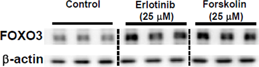
Erlotinib purchased from MedChemExpress. Usage Cited in: Oncogene. 2017 May 11;36(19):2643-2654. [Abstract]
Effects of anti-cancer agents Oxaliplatin, Erlotinib (25 μM) and cAMP inducer Forskolin (25 μM) on FOXO3 expression in Panc-1 cells are assessed for 72 h (n=3).
-
Cell Death Discov
MSLN-mediated activation of EGFR-ERK1/2 signaling drives liver metastasis in breast cancer. [Abstract]2026 Jan 9;12(1):11. PMID: 41513618 -
Cell Death Discov
Matrix metalloproteinase-10 promotes kidney fibrosis by transactivating β-catenin signaling. [Abstract]2025 May 17;11(1):241. PMID: 40382334 -
Cell Rep
Endocytic activation and exosomal secretion of matriptase stimulate the second wave of EGF signaling to promote skin and breast cancer invasion. [Abstract]2024 Mar 27;43(4):114002. PMID: 38547126 -
Cell Rep
Kinetics of RTK activation determine ERK reactivation and resistance to dual BRAF/MEK inhibition in melanoma. [Abstract]2023 May 29;42(6):112570. PMID: 37252843 -
Br J Cancer
Fibroblast growth factor signals drive the metastatic behavior in small cell lung cancer. [Abstract]2025 Dec 13. PMID: 41390896 -
Br J Cancer
Drug response profiles in patient-derived cancer cells across histological subtypes of ovarian cancer: real-time therapy tailoring for a patient with low-grade serous carcinoma. [Abstract]2023 Feb;128(4):678-690. PMID: 36476658 -
Ecotoxicol Environ Saf
Protective role of ghrelin against 6PPD-quinone-induced neurotoxicity in zebrafish larvae (Danio rerio) via the GHSR pathway. [Abstract]2024 Oct 15:285:117031. PMID: 39341137 -
Ecotoxicol Environ Saf
Hyperphosphorylation of EGFR/ERK signaling facilitates long-term arsenite-induced hepatocytes epithelial-mesenchymal transition and liver fibrosis in sprague-dawley rats. [Abstract]2023 Jan 1:249:114386. PMID: 36508792 -
Biomater Adv
Development of a HER1/CP2c dual-targeting biopharmaceutical for HER1-overexpressing head and neck cancer. [Abstract]2026 Feb 18:183:214782. PMID: 41722284 -
Anal Chim Acta
Development of novel affinity reagents for detecting protein tyrosine phosphorylation based on superbinder SH2 domain in tumor cells. [Abstract]2018 Nov 22:1032:138-146. PMID: 30143211
Erlotinib purchased from MedChemExpress. Usage Cited in: Anal Chim Acta. 2018 Nov 22:1032:138-146. [Abstract]
30 min after EGF(100 ng/mL) and EGFP-sSH2(Arg)9 (5 μM) are added into the MDA-MB-468 cell culture medium, Erlotinib is added into the cell culture medium (100 nM), and the picture was collected at 30 min after Erlotinib treatment.
-
Cell Biol Toxicol
TRPV3 enhances skin keratinocyte proliferation through EGFR-dependent signaling pathways. [Abstract]2021 Apr;37(2):313-330. PMID: 32535744 -
J Invest Dermatol
2019 Jan;139(1):224-234. PMID: 30081003 -
Biochem Pharmacol
Pitavastatin overcomes multi-drug resistance in CRC and NSCLC by targeting the NRP1-ZFX axis. [Abstract]2025 Jul 18:241:117183. PMID: 40684995 -
Neurobiol Dis
ErbB1-dependent signalling and vesicular trafficking in primary afferent nociceptors associated with hypersensitivity in neuropathic pain. [Abstract]2020 Aug;142:104961. PMID: 32531343 -
J Enzyme Inhib Med Chem
Natural product sennoside B disrupts liquid-liquid phase separation of SARS-CoV-2 nucleocapsid protein by inhibiting its RNA-binding activity. [Abstract]2025 Dec;40(1):2501743. PMID: 40371698 -
Mar Drugs
New Fusarochromanone Derivatives from the Marine Fungus Fusarium equiseti UBOCC-A-117302. [Abstract]2024 Sep 28;22(10):444. PMID: 39452852 -
Virulence
Adaptor complex-mediated trafficking of Newcastle disease virus fusion protein is regulated by the YLMY motif of its cytoplasmic tail. [Abstract]2022 Dec;13(1):1849-1867. PMID: 36258290 -
Commun Biol
A small-molecule compound D6 overcomes EGFR-T790M-mediated resistance in non-small cell lung cancer. [Abstract]2021 Dec 13;4(1):1391. PMID: 34903832 -
CNS Neurosci Ther
Pharmacological inhibition of epidermal growth factor receptor attenuates intracranial aneurysm formation by modulating the phenotype of vascular smooth muscle cells. [Abstract]2022 Jan;28(1):64-76. PMID: 34729926 -
Int J Mol Sci
Carcinoembryonic Antigen-Related Cell Adhesion Molecule 5 as a Biomarker for Predicting Response to Erlotinib and Gefitinib in Lung Adenocarcinoma: An Integrative Analysis of Transcriptomic Data of PC-9 and Drug-Resistant PC-9 Cell Lines. [Abstract]2026 Mar 28;27(7):3092. PMID: 41977278 -
PLoS Pathog
EGFR-MEK1/2 cascade negatively regulates bactericidal function of bone marrow macrophages in mice with Staphylococcus aureus osteomyelitis. [Abstract]2024 Aug 5;20(8):e1012437. PMID: 39102432 -
J Nutr Biochem
High glucose enhances the aggressiveness of lung adenocarcinoma via activating epidermal growth factor receptor/signal transducer and activator of transcription 3 pathways. [Abstract]2023 Sep:119:109399. PMID: 37271322 -
Pharmaceuticals (Basel)
Bufadienolides from Chansu Injection Synergistically Enhances the Antitumor Effect of Erlotinib by Inhibiting the KRAS Pathway in Pancreatic Cancer. [Abstract]2024 Dec 16;17(12):1696. PMID: 39770538 -
Front Pharmacol
Transcriptomics and Metabolomics Identify Drug Resistance of Dormant Cell in Colorectal Cancer. [Abstract]2022 Apr 8;13:879751. PMID: 35462906 -
Front Pharmacol
2018 Jun 21:9:660. PMID: 29977207
Erlotinib purchased from MedChemExpress. Usage Cited in: Front Pharmacol. 2018 Jun 21:9:660. [Abstract]
By Western blotting, the α-SMA and palladin proteins are determined in cells that are pretreated with NF-κB (JSH-23), Wnt/β-catenin (XAV939), EGFR (erlotinib), p38 MAPK (TAK-715), and Smad3 (SIS3) inhibitors and are followed by TWEAK stimulation.
-
Bioorg Chem
A pomalidomide-based gefitinib PROTAC degrader effectively inhibits lung cancer progression in EGFR-TKIs-acquired resistant models by targeting EGFR degradation and ETFA-mediated ATP generation. [Abstract]2025 Aug 9:164:108864. PMID: 40815992 -
Mol Cancer Res
TAS6417/CLN-081 Is a Pan-Mutation-Selective EGFR Tyrosine Kinase Inhibitor with a Broad Spectrum of Preclinical Activity against Clinically Relevant EGFR Mutations. [Abstract]2019 Nov;17(11):2233-2243. PMID: 31467113 -
Cell Rep Methods
RECOVER identifies synergistic drug combinations in vitro through sequential model optimization. [Abstract]2023 Oct 23;3(10):100599. PMID: 37797618 -
Mol Oncol
Activation of the EGFR/PI3K/AKT pathway limits the efficacy of trametinib treatment in head and neck cancer. [Abstract]2023 Dec;17(12):2618-2636. PMID: 37501404 -
Cell Rep Methods
2023 Feb 21;3(2):100411. PMID: 36936075 -
Mol Pharm
Combining Gemcitabine-Loaded Macrophage-like Nanoparticles and Erlotinib for Pancreatic Cancer Therapy. [Abstract]2021 Jul 5;18(7):2495-2506. PMID: 34078087 -
Mol Oncol
TGF-β transactivates EGFR and facilitates breast cancer migration and invasion through canonical Smad3 and ERK/Sp1 signaling pathways. [Abstract]2018 Mar;12(3):305-321. PMID: 29215776
Erlotinib purchased from MedChemExpress. Usage Cited in: Mol Oncol. 2018 Mar;12(3):305-321. [Abstract]
Western blot analysis of total and phosphorylated EGFR expression in two breast cancer cells pretreated with different concentration Erlotinib for 4h.
-
Lung Cancer
HMGB1 Expression Levels Correlate with Response to Immunotherapy in Non-Small Cell Lung Cancer. [Abstract]2024 May 9:15:55-67. PMID: 38741920 -
Cancers (Basel)
SCAMP3 Regulates EGFR and Promotes Proliferation and Migration of Triple-Negative Breast Cancer Cells through the Modulation of AKT, ERK, and STAT3 Signaling Pathways. [Abstract]2022 Jun 5;14(11):2807. PMID: 35681787 -
ACS Omega
Association Study of OATP1B3 Polymorphisms on Hepatic Uptake and Drug-Drug Interaction In Vitro. [Abstract]2025 Oct 29;10(44):52562-52575. PMID: 41244417 -
J Cell Mol Med
PRMT5 promotes epithelial-mesenchymal transition via EGFR-β-catenin axis in pancreatic cancer cells. [Abstract]2020 Jan;24(2):1969-1979. PMID: 31851779 -
-
Oncol Rep
Repurposing of a monoamine oxidase A inhibitor‑heptamethine carbocyanine dye conjugate for paclitaxel‑resistant non‑small cell lung cancer. [Abstract]2021 Mar;45(3):1306-1314. PMID: 33650669 -
Oncol Rep
Upregulated CTHRC1 promotes human epithelial ovarian cancer invasion through activating EGFR signaling. [Abstract]2016 Dec;36(6):3588-3596. PMID: 27779718 -
Mol Divers
Design, synthesis and biological evaluation of thienopyridine derivatives as c-Met kinase inhibitors. [Abstract]2025 Jun;29(3):2391-2405. PMID: 39356364 -
Cancer Res Treat
Establishment of Patient-Derived Organoids Using Ascitic or Pleural Fluid from Cancer Patients. [Abstract]2023 Oct;55(4):1077-1086. PMID: 37309112 -
Cancer Res Treat
MLL4 Regulates the Progression of Non-Small-Cell Lung Cancer by Regulating the PI3K/AKT/SOX2 Axis. [Abstract]2023 Jul;55(3):778-803. PMID: 36701842 -
Bioengineering (Basel)
Precision Oncology for High-Grade Gliomas: A Tumor Organoid Model for Adjuvant Treatment Selection. [Abstract]2025 Oct 19;12(10):1121. PMID: 41155119 -
Proteomes
Mass Spectrometry and Pharmacological Approaches to Measuring Cooption and Reciprocal Activation of Receptor Tyrosine Kinases. [Abstract]2023 Jun 2;11(2):20. PMID: 37368466 -
Stem Cells
DAPK1 Interacts with the p38 Isoform MAPK14, Preventing Its Nuclear Translocation and Stimulation of Bone Marrow Adipogenesis. [Abstract]2022 May 27;40(5):508-522. PMID: 35403694 -
Mol Med Rep
ISX promotes tumor migration and invasion in lung cancer by upregulating COL1A1 in vitro. [Abstract]2026 Feb;33(2):77. PMID: 41480691 -
Exp Cell Res
HPRT promotes proliferation and metastasis in head and neck squamous cell carcinoma through direct interaction with STAT3. [Abstract]2021 Feb 1;399(1):112424. PMID: 33340493 -
Viruses
Broad-Spectrum Host-Based Antivirals Targeting the Interferon and Lipogenesis Pathways as Potential Treatment Options for the Pandemic Coronavirus Disease 2019 (COVID-19). [Abstract]2020 Jun 10;12(6):628. PMID: 32532085 -
Exp Cell Res
Network-based analysis with primary cells reveals drug response landscape of acute myeloid leukemia. [Abstract]2020 Aug 1;393(1):112054. PMID: 32376287 -
Saudi Pharm J
Augmented antitumor effects of erlotinib and cabozantinib on A549 non-small cell lung cancer: In vitro and in vivo studies. [Abstract]2023 Oct;31(10):101756. PMID: 37705877 -
J Cancer
2024 Mar 25;15(10):2891-2899. PMID: 38706904 -
Mol Carcinog
Epidermal growth factor receptor signaling in precancerous keratinocytes promotes neighboring head and neck cancer squamous cell carcinoma cancer stem cell-like properties and phosphoinositide 3-kinase inhibitor insensitivity. [Abstract]2022 Jul;61(7):664-676. PMID: 35417043 -
Cancer Med
Proton pump inhibitors stabilize the expression of PD-L1 on cell membrane depending on the phosphorylation of GSK3β. [Abstract]2024 May;13(10):e7083. PMID: 38752436
Erlotinib purchased from MedChemExpress. Usage Cited in: Cancer Med. 2024 May;13(10):e7083. [Abstract]
FACS analysis was used to perform the MFI of cell membrane of PD‐L1 in H441 cells with control or multiply small molecular inhibitors (Seen in the Table 1, 24 h) (n = 3). AV412 (HY‐10346; 20, 50 nM), ERK1/2 inhibitor 1 (HY‐112287; 20, 50 nM), AR‐A014418 (HY‐10512; 100, 200 nM), Erlotinib (HY‐50896; 50, 100 nM).
-
Cancer Med
MYST1/KAT8 contributes to tumor progression by activating EGFR signaling in glioblastoma cells. [Abstract]2019 Dec;8(18):7793-7808. PMID: 31691527
Erlotinib purchased from MedChemExpress. Usage Cited in: Cancer Med. 2019 Dec;8(18):7793-7808. [Abstract]
Western blots of proteins expression in EGFR signaling pathways in LN229 and U87 cells with MYST1/GFP overexpression after erlotinib (200 ng/mL)/DMSO treatment.
-
Breast Cancer Res Treat
2025 Jun;211(2):467-478. PMID: 40055251 -
-
PLoS One
A novel small molecule screening assay using normal human chondrocytes toward osteoarthritis drug discovery. [Abstract]2024 Nov 1;19(11):e0308647. PMID: 39485774 -
Cancer Manag Res
Deguelin Restores Paclitaxel Sensitivity in Paclitaxel-Resistant Ovarian Cancer Cells via Inhibition of the EGFR Signaling Pathway. [Abstract]2024 May 28:16:507-525. PMID: 38827785 -
PLoS One
Validated UPLC-MS/MS method for the quantification of dasatinib in plasma: Application to pharmacokinetic interaction studies with nutraceuticals in Wistar rats. [Abstract]2018 Jun 14;13(6):e0199208. PMID: 29902246 -
PeerJ
2023 Oct 16:11:e16121. PMID: 37868048 -
Thorac Cancer
Potential monoamine oxidase A inhibitor suppressing paclitaxel-resistant non-small cell lung cancer metastasis and growth. [Abstract]2020 Oct;11(10):2858-2866. PMID: 32875729 -
Biomed Res Int
A Pharmacokinetic Interaction Study of Sorafenib and Iced Teas in Rats Using UPLC-MS/MS: An Illustration of Beverage-Drug Interaction. [Abstract]2019 Nov 28;2019:2410845. PMID: 31871933 -
-
Biochem Biophys Res Commun
Investigating NanoLuc-EGFR engineered cell lines for real-time monitoring of EGFR protein dynamics in live cells. [Abstract]2024 Sep 19:733:150711. PMID: 39312880 -
Biochem Biophys Res Commun
A Beclin 1-targeting stapled peptide synergizes with erlotinib to potently inhibit proliferation of non-small-cell lung cancer cells. [Abstract]2022 Dec 25;636(Pt 1):125-131. PMID: 36332474 -
J Med Food
Naturally Occurring Hair Growth Peptide: Water-Soluble Chicken Egg Yolk Peptides Stimulate Hair Growth Through Induction of Vascular Endothelial Growth Factor Production. [Abstract]2018 Jul;21(7):701-708. PMID: 29583066 -
Biomed Chromatogr
A novel, sensitive, and fast ultra-high-performance liquid chromatography tandem mass spectrometry method for TNG908 determination in dog plasma and pharmacokinetic study. [Abstract]2024 Nov 7:e6039. PMID: 39510096 -
Biomed Chromatogr
Simultaneous and rapid determination of gefitinib, erlotinib and afatinib plasma levels using liquid chromatography/tandem mass spectrometry in patients with non-small-cell lung cancer. [Abstract]2016 Jul;30(7):1150-4. PMID: 26525154 -
Am J Transl Res
Ellagic acid inhibits cell proliferation, migration, and invasion in melanoma via EGFR pathway. [Abstract]2020 May 15;12(5):2295-2304. PMID: 32509220 -
Biol Methods Protoc
Optimizing drug sensitivity assays in patient-derived tumor organoids: a comparison of IC50 estimation methods and experimental parameters. [Abstract]2025 Feb 13;10(1):bpaf012. PMID: 40060949 -
-
-
-
-
-
-
-
Res Sq
An EGFR Co-Amplified and De Novo Long Noncoding RNA HELDR Promotes Glioblastoma Malignancy through KAT7-Driven Gene Programs. [Abstract]2025 Jun 24:rs.3.rs-6456987. PMID: 40678238 -
-
-
bioRxiv
BRAFV600 and ErbB inhibitors directly activate GCN2 in an off-target manner to limit cancer cell proliferation. [Abstract]2024 Dec 20:2024.12.19.629301. PMID: 39763857 -
-
-
bioRxiv
LKB1 suppresses growth and promotes the internalization of EGFR through the PIKFYVE lipid kinase. [Abstract]2023 Oct 20:2023.10.19.563158. PMID: 37904985 -
-
Cell Insight
Phosphorylated STYK1 restrains the inhibitory role of EGFR in autophagy initiation and EGFR-TKIs sensitivity. [Abstract]2022 Jun 16;1(4):100045. PMID: 37192859 -
-
-
-
-
Oncotarget
Molecular-genetic profiling and high-throughput in vitro drug screening in NUT midline carcinoma-an aggressive and fatal disease. [Abstract]2017 Dec 2;8(68):112313-112329. PMID: 29348827


Solvent & Solubility
DMSO : 33.33 mg/mL (84.71 mM; ultrasonic and warming and heat to 60°C; Hygroscopic DMSO has a significant impact on the solubility of product, please use newly opened DMSO)
Please refer to the solubility information to select the appropriate solvent. Once prepared, please aliquot and store the solution to prevent product inactivation from repeated freeze-thaw cycles.
Storage method and period of stock solution: -80°C, 1 year; -20°C, 6 months. When stored at -80°C, please use it within 1 year. When stored at -20°C, please use it within 6 months.
Concentration (start) × Volume (start) = Concentration (final) × Volume (final)
Select the appropriate dissolution method based on your experimental animal and administration route.
- For the following dissolution methods, please ensure to first prepare a clear stock solution using an In Vitro approach and then sequentially add co-solvents:
- To ensure reliable experimental results, the clarified stock solution can be appropriately stored based on storage conditions. As for the working solution for In Vivo experiments, it is recommended to prepare freshly and use it on the same day.
- The percentages shown for the solvents indicate their volumetric ratio in the final prepared solution. If precipitation or phase separation occurs during preparation, heat and/or sonication can be used to aid dissolution.
Add each solvent one by one: 10% DMSO 40% PEG300 5% Tween-80 45% Saline
Solubility: ≥ 1.25 mg/mL (3.18 mM); Clear solution
This protocol yields a clear solution of ≥ 1.25 mg/mL (saturation unknown).
Taking 1 mL working solution as an example, add 100 μL DMSO stock solution (12.5 mg/mL) to 400 μL PEG300, and mix evenly; then add 50 μL Tween-80 and mix evenly; then add 450 μL Saline to adjust the volume to 1 mL.
Preparation of Saline: Dissolve 0.9 g sodium chloride in ddH₂O and dilute to 100 mL to obtain a clear Saline solution.
For the following dissolution methods, please prepare the working solution directly:
It is recommended to prepare fresh solutions and use them promptly within a short period of time.
The percentages shown for the solvents indicate their volumetric ratio in the final prepared solution. If precipitation or phase separation occurs during preparation, heat and/or sonication can be used to aid dissolution.
Please enter the basic information of animal experiments:
-
-
-
-
Recommended: Prepare an additional quantity of animals to account for potential losses during experiments.
Please enter your animal formula composition:
-
%DMSO +
Recommended: Keep the proportion of DMSO in working solution below 2% if your animal is weak.
-
%+
-
+%Tween-80 + +
-
%Saline +
The co-solvents required include: DMSO, . All of co-solvents are available by MedChemExpress (MCE). , Tween 80. All of co-solvents are available by MedChemExpress (MCE).
Working solution concentration: 0.22 mg/mL
Method for preparing stock solution: mg drug dissolved in μL DMSO. Stock solution concentration: mg/mL.
1. Take μL DMSO stock solution;
2. Add μL .
μL , mix evenly;
3. Then add μL Tween 80, mix evenly;
4. Then add μL
Please ensure that the stock solution in the first step is dissolved to a clear state, and add co-solvents in sequence. You can use ultrasonic heating (ultrasonic cleaner, recommended frequency 20-40 kHz), vortexing, etc. to assist dissolution.


Purity & Documentation
-
Data Sheet (293 KB)
-
SDS (396 KB)
- English - EN (396 KB)
- Français - FR (396 KB)
- Deutsch - DE (396 KB)
- Norwegian - NO (396 KB)
- Español - ES (396 KB)
- Swedish - SV (396 KB)
- Italian - IT (396 KB)
- Korean - KR (396 KB)
- Portuguese - PT (396 KB)
-
Handling Instructions (2659 KB)
References
[1]. Li S, et al. Ephrin A1 functions as a ligand of EGFR to promote EMT and metastasis in gastric cancer. The EMBO journal. 2025 Mar;44(5):1464-1487. [Content Brief]
[2]. Ferrarone JR, et al. Genome-wide CRISPR screens in spheroid culture reveal that the tumor suppressor LKB1 inhibits growth via the PIKFYVE lipid kinase. Proceedings of the National Academy of Sciences of the United States of America. 2024 May 21;121(21):e2403685121. [Content Brief]
[3]. Rysz MA et al. Erlotinib-A substrate and inhibitor of OATP2B1: pharmacokinetics and CYP3A-mediated metabolism in rSlco2b1-/- and SLCO2B1+/+ rats. Drug Metab Dispos. 2025 May;53(5):100069. [Content Brief]
[4]. Sun X, et al. Matrix metalloproteinase-10 promotes kidney fibrosis by transactivating β-catenin signaling. Cell death discovery. 2025 May 17;11(1):241. [Content Brief]
[5]. Ito F, et al. GRHL2-HER3 and E-cadherin mediate EGFR-bypass drug resistance in lung cancer cells. Frontiers in cell and developmental biology. 2024;12:1511190. [Content Brief]


Complete Stock Solution Preparation Table
Please refer to the solubility information to select the appropriate solvent. Once prepared, please aliquot and store the solution to prevent product inactivation from repeated freeze-thaw cycles.
Storage method and period of stock solution: -80°C, 1 year; -20°C, 6 months. When stored at -80°C, please use it within 1 year. When stored at -20°C, please use it within 6 months.
| Optional Solvent | Concentration Solvent Mass | 1 mg | 5 mg | 10 mg | 25 mg |
|---|---|---|---|---|---|
| DMSO | 1 mM | 2.5417 mL | 12.7084 mL | 25.4168 mL | 63.5421 mL |
| 5 mM | 0.5083 mL | 2.5417 mL | 5.0834 mL | 12.7084 mL | |
| 10 mM | 0.2542 mL | 1.2708 mL | 2.5417 mL | 6.3542 mL | |
| 15 mM | 0.1694 mL | 0.8472 mL | 1.6945 mL | 4.2361 mL | |
| 20 mM | 0.1271 mL | 0.6354 mL | 1.2708 mL | 3.1771 mL | |
| 25 mM | 0.1017 mL | 0.5083 mL | 1.0167 mL | 2.5417 mL | |
| 30 mM | 0.0847 mL | 0.4236 mL | 0.8472 mL | 2.1181 mL | |
| 40 mM | 0.0635 mL | 0.3177 mL | 0.6354 mL | 1.5886 mL | |
| 50 mM | 0.0508 mL | 0.2542 mL | 0.5083 mL | 1.2708 mL | |
| 60 mM | 0.0424 mL | 0.2118 mL | 0.4236 mL | 1.0590 mL | |
| 80 mM | 0.0318 mL | 0.1589 mL | 0.3177 mL | 0.7943 mL |


 Powered by Bioz