



MRL-SYKi

Based on 1 Customer Validation
MRL-SYKi is a chemical probe for gain-of-function variants of spleen tyrosine kinase (SYK). MRL-SYKi reduces the catalytic activity of SYKS550Y, SYKS550F and SYKP342T, and downregulates the phosphorylation level of SYKS550Y. MRL-SYKi serves as a template for developing NanoBRET tracers targeting SYK, enabling NanoBRET cellular target engagement assays for gain-of-function variants of SYK. MRL-SYKi is applicable to research related to inflammatory and immune diseases.
For research use only. We do not sell to patients.
- Purity : 98.97%
- CAS No.: 1312534-69-2
- Formula: C24H28N4O3S
- Molecular Weight:452.57
-
Storage:Powder -20°C, 3 years , 4°C, 2 years ; In solvent -80°C, 6 months , -20°C, 1 month


To place orders, for customer services and technical support, please contact: MedChemExpress USA
Tel: 609-228-6898 E-mail: [email protected] [email protected]


-
 Biological Activity
-
Chemical Information
- Protocols
- Purity & Documentation
- References
-
 Help & FAQs


-
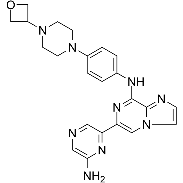
HY-109091
LanraplenibSYK Inhibitor -
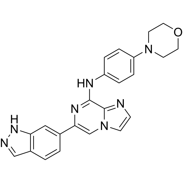
HY-15968
EntospletinibSyk Inhibitor -
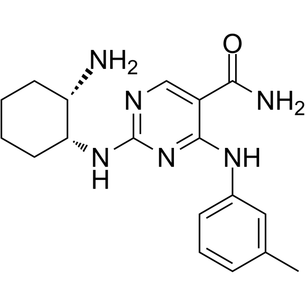
HY-12974
PRT-060318Syk Inhibitor -
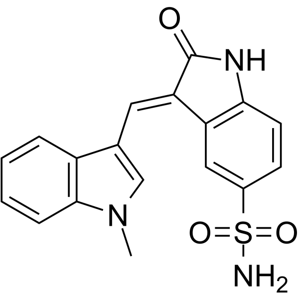
HY-112386
OXSI-2Syk Inhibitor -
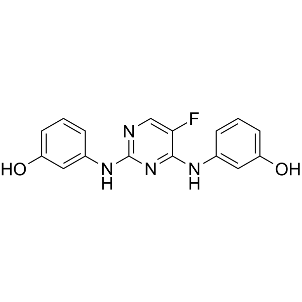
HY-16420
R112Syk Inhibitor -
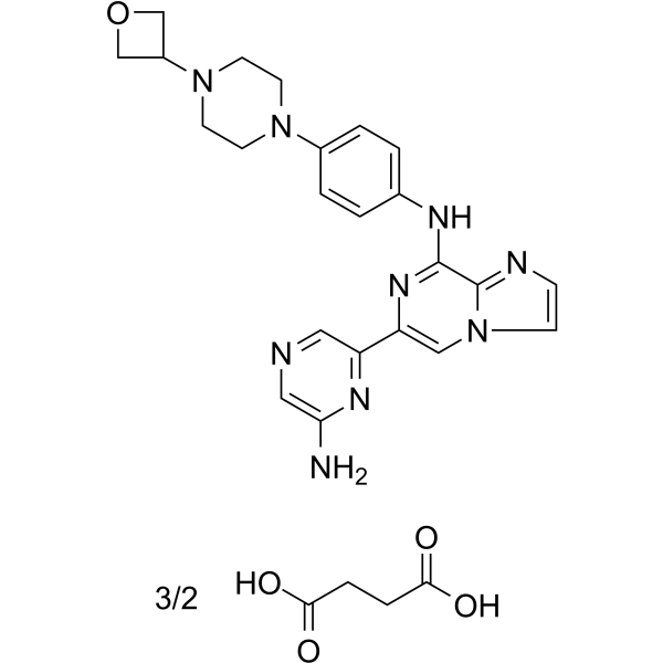
HY-109091B
Lanraplenib succinateSYK Inhibitor -
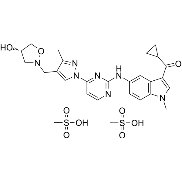
HY-109082A
Cevidoplenib dimesylateSyk Inhibitor -
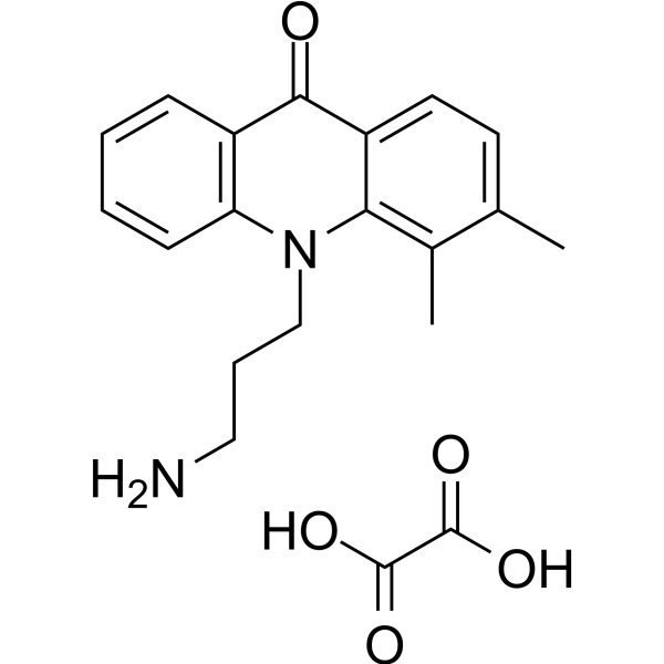
HY-138961
ER-27319SYK Iinhibitor -

HY-15968R
Entospletinib (Standard)Syk Inhibitor -

HY-109091R
Lanraplenib (Standard)Reference Standard -

HY-109082AR
Cevidoplenib dimesylate (Standard)Reference Standard


Biological Activity
Description
In Vitro
MRL-SYKi potently inhibits purified SYK protein in cell-free enzymatic assays, with an IC50 of 30 nM[1].
MRL-SYKi (20 μM) binds to all tested NL-tagged SYK variants in digitonin-permeabilized HEK293 cells, with the highest potency against SYKS550Y (IC50 = 2.1 nM) and the lowest potency against SYKM450I (IC50 = 29 nM)[1].
MRL-SYKi (1-10 μM; 2 h) directly binds to NL-tagged SYKS550Y, SYKS550F, and SYKP342T in intact living HEK293 cells, with the highest potency against SYKS550Y (IC50 = 65 nM) and the lowest potency against SYKP342T (IC50 = 410 nM)[1].
MRL-SYKi (1.75-30 μM; 30 min) binds to NL-tagged SYK (WT) in digitonin-permeabilized HEK293 cells and increases its thermal stability, with a maximum ΔTm of 3.5 °C at the concentration of 30 μM[1].
MRL-SYKi (30 min) potently inhibits the catalytic activity of purified SYKS550F (IC50 = 63 nM) and SYKP342T (IC50 = 95 nM) in cell-free kinase activity assays[1].
MRL-SYKi (100 nM-10 μM; 20 h) reduces the phosphorylation level of SYKS550Y at Y525/526 in SW480 cells stably expressing SYKS550Y[1].
MedChemExpress (MCE) has not independently confirmed the accuracy of these methods. They are for reference only.
-
Cell Line:SW480 colonic epithelial cells stably expressing SYK(S550Y)
-
Concentration:100 nM, 10 μM
-
Incubation Time:20 h
-
Result:Reduced pSYK(Y525/526) levels compared to untreated cells at 100 nM.
Reduced pSYK(Y525/526) levels compared to untreated cells at 10 μM, with a greater reduction observed than at 100 nM.


Chemical Information
-
CAS No. 1312534-69-2
-
Appearance Solid
-
Molecular Weight 452.57
-
Formula C24H28N4O3S
-
Color Off-white to light yellow
-
SMILES
O[C@@]1(C2=NC=C(C3=CC(NC4=NC=CC(C)=N4)=CC(C)=C3)S2)CC(C)([C@H](CC1)C(O)=O)C
-
Shipping
Room temperature in continental US; may vary elsewhere.
-
Storage
Powder -20°C 3 years 4°C 2 years In solvent -80°C 6 months -20°C 1 month


Purity & Documentation
-
Data Sheet (272 KB)
-
SDS (251 KB)
- English - EN (251 KB)
- Français - FR (251 KB)
- Deutsch - DE (251 KB)
- Norwegian - NO (251 KB)
- Español - ES (251 KB)
- Swedish - SV (251 KB)
- Italian - IT (251 KB)
- Korean - KR (251 KB)
- Portuguese - PT (251 KB)
-
Handling Instructions (2659 KB)
References

Calculators
Concentration (start) × Volume (start) = Concentration (final) × Volume (final)


MRL-SYKi Related Classifications
HY-178129 Related Classifications
 Powered by Bioz