



POVPC

Based on 1 Customer Validation
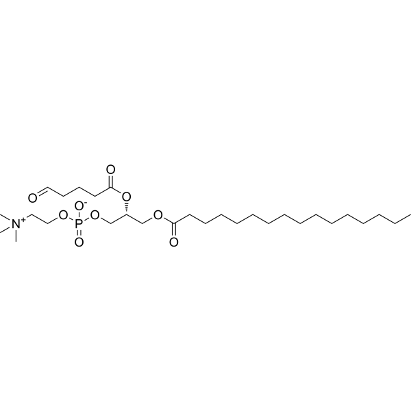
POVPC is an oxidized phospholipid can be found in in oxidatively modified low density lipoprotein (oxLDL). POVPC inhibits VSMC growth in high serum condition. POVPC induces apoptosis in low serum condition.
For research use only. We do not sell to patients.
- Purity : 99%
- CAS No.: 121324-31-0
- Formula: C29H56NO9P
- Molecular Weight:593.73
-
Storage:
Solution, -20°C, 2 years

To place orders, for customer services and technical support, please contact: MedChemExpress USA
Tel: 609-228-6898 E-mail: [email protected] [email protected]


-
 Biological Activity
-
Chemical Information
- Protocols
- Purity & Documentation
- References
-
 Help & FAQs


-
HY-126726
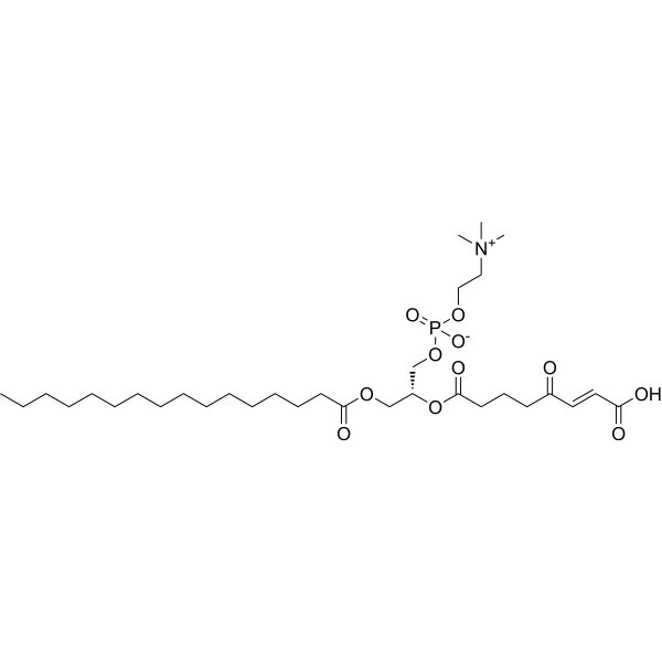
KOdiA-PCBiochemical Assay Reagent -

HY-134154
PAz-PC -
HY-113369
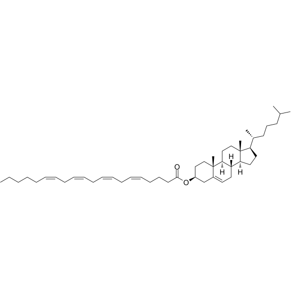
Cholesteryl arachidonateEndogenous Metabolite -
HY-130502
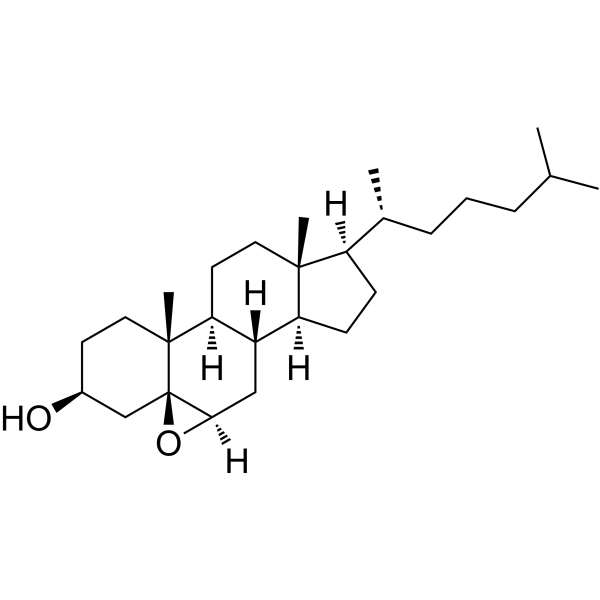
5β,6β-EpoxycholestanolOxysterol -

HY-139414
LysophosphatidylcholinesHemolytic Agent -
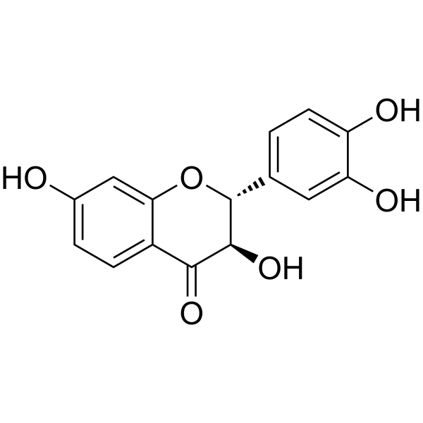
HY-N8376
FustinAβ Inhibitor -
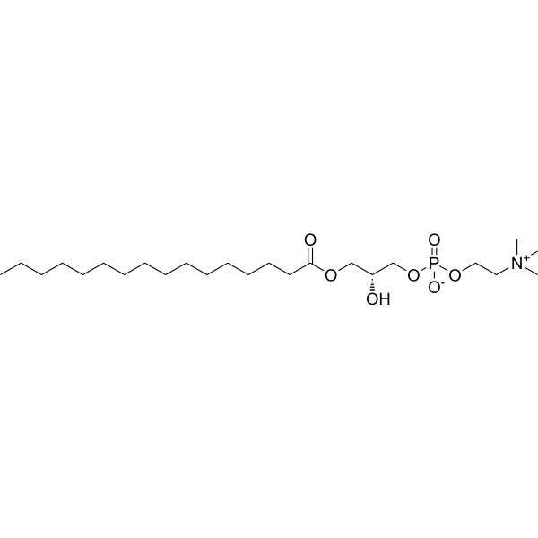
HY-125783
1-Palmitoyl-sn-glycero-3-phosphocholineLysophosphatidylcholine -
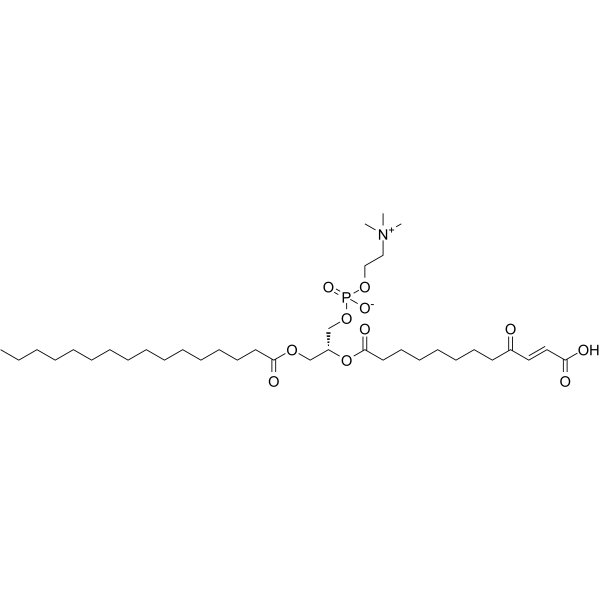
HY-125554
KDdiA-PCCD36 Ligand -
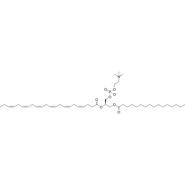
HY-126354
Palmitoyldocosahexaenoyl phosphatidylcholineEndogenous Metabolite -
HY-131389
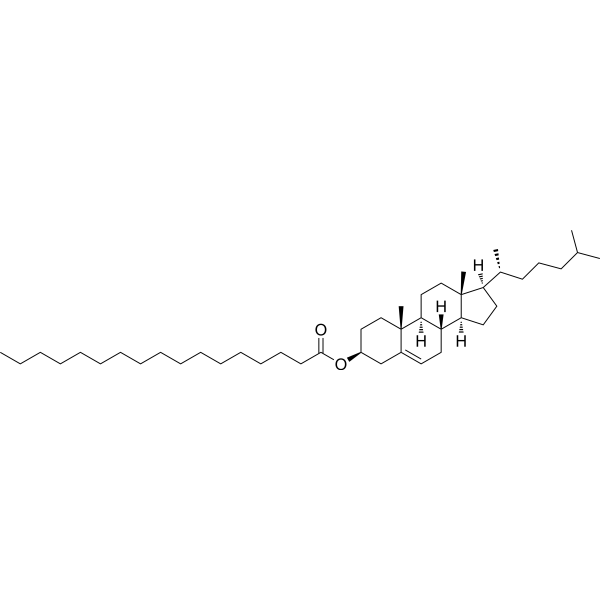
Cholesteryl heptadecanoateCholesteryl Ester Internal Standard -
HY-130675AS
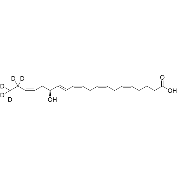
15(S)-HEPE-d5Stable Isotope -

HY-W040221
Butanoyl PAFPAF Agonist


Biological Activity
Description
In Vitro
POVPC (0-100 µM; 13 h) inhibits VSMC growth in a dose-dependent manner[1].
POVPC (50 µM; 2, 4, 6 h) induces apoptosis in a time-dependent manner[1].
MedChemExpress (MCE) has not independently confirmed the accuracy of these methods. They are for reference only.
-
Cell Line:vascular smooth muscle cells (VSMC)
-
Concentration:0-100 µM (under high serum conditions (10% FCS))
-
Incubation Time:13 h
-
Result:Inhibited VSMC growth in a dose-dependent manner.
-
Cell Line:7r5 cells
-
Concentration:50 µM (0.1% FCS)
-
Incubation Time:2, 4, 6 h
-
Result:Induced apoptosis in a time-dependent manner.


Chemical Information
-
CAS No. 121324-31-0
-
Appearance Liquid
-
Molecular Weight 593.73
-
Formula C29H56NO9P
-
Color Colorless to light yellow
-
SMILES
C[N+](C)(C)CCOP(OC[C@H](OC(CCCC=O)=O)COC(CCCCCCCCCCCCCCC)=O)([O-])=O
-
Shipping
Room temperature in continental US; may vary elsewhere.
-
Storage
Solution, -20°C, 2 years


Purity & Documentation
-
Data Sheet (269 KB)
-
SDS (393 KB)
- English - EN (393 KB)
- Français - FR (393 KB)
- Deutsch - DE (393 KB)
- Norwegian - NO (393 KB)
- Español - ES (393 KB)
- Swedish - SV (393 KB)
- Italian - IT (393 KB)
- Korean - KR (393 KB)
- Portuguese - PT (393 KB)
-
Handling Instructions (2659 KB)
References

Calculators
Concentration (start) × Volume (start) = Concentration (final) × Volume (final)


POVPC Related Classifications
HY-130576 Related Classifications
 Powered by Bioz