



APRIL/TNFSF13 Trimer Protein, Human (HEK293, His-Flag)

Based on 1 Customer Validation
APRIL protein (CD256) is a ligand in the tumor necrosis factor (TNF) family that can induce proliferation, regulate tumor cell growth, and may participate in mononuclear/macrophage-mediated immune process. APRIL protein is produced by myeloid cells and their precursors to accelerate cell maturation and peripheral rupture. APRIL protein has been widely used in the study of lymphatic malignancies. Human APRIL protein is a type II membrane protein with cytoplasmic domain, hydrophobic transmembrane domain and extracellular domain. APRIL/TNFSF13 Protein, Human (HEK293, His-Flag) is produced by HEK293 cells (K112-L250) with N-terminal His and Flag-tags.
- Species: Human
- Source: HEK293
-
Storage:Stored at -20°C for 2 years from date of receipt. After reconstitution, it is stable at 4°C for 1 week or -20°C for longer (with carrier protein). It is recommended to freeze aliquots at -20°C or -80°C for extended storage.


To place orders, for customer services and technical support, please contact: MedChemExpress USA
Tel: 609-228-6898 E-mail: [email protected] [email protected]


-
 Biological Activity
-
Technical Parameters
-
Product Properties
- Documentation
- Help & FAQs




Biological Activity
Description
APRIL protein (CD256) is a ligand in the tumor necrosis factor (TNF) family that can induce proliferation, regulate tumor cell growth, and may participate in mononuclear/macrophage-mediated immune process[1]. APRIL protein is produced by myeloid cells and their precursors to accelerate cell maturation and peripheral rupture[2]. APRIL protein has been widely used in the study of lymphatic malignancies. Human APRIL protein is a type II membrane protein with cytoplasmic domain, hydrophobic transmembrane domain and extracellular domain. APRIL/TNFSF13 Protein, Human (HEK293, His-Flag) is produced by HEK293 cells (K112-L250) with N-terminal His and Flag-tags.
Background
APRIL/TNFSF13 Protein is a cytokine and an independent secretory ligand belongs to TNF family. It binds to TNFRSF13B/TACI and to TNFRSF17/BCMA. APRIL/TNFSF13 Protein plays a role in the regulation of tumor cell growth, may involve in monocyte/macrophage-mediated immunological processes[1].
APRIL is produced by myeloid cells and their precursors in the bone marrow. APRIL is retained by surrounding tissues and via HSPG (heparan sulfate proteoglycans) is not retained. It accumulates in large amounts in the bone marrow, leading to more rapid cell maturation and peripheral burst. As for infection response, tonsil mucosa neutrophils present within the infected tissue were the main source of APRIL, whereas keratinocytes were the primary source of APRIL in tissues showing no symptoms of infection[2].
APRIL acts function by binding BCMA (B cell maturation antigen) and TACI (transmembrane activator and CAML-interactor) and competes with TALL-I (also called BLyS or BAFF) for receptor binding. Soluble BCMA and TACI specifically prevent binding of APRIL and block APRIL-stimulated proliferation of primary B cells, and soluble BCMA is a dominant-negative molecule capable of inhibiting antibody production in vivo. Thus, APRIL stimulates in vitro the proliferation of primary lymphocytes, in addition to lymphoma cell lines, and promotes in vivo the accumulation of B cells in the spleen. Therefore, APRIL-TALL-I and BCMA-TACI form a two ligands-two receptors pathway involved in stimulation of B and T cell function. Moreover, APRIL is also a stimulator of tumor cell growth although TNRF death ligand-1 (TRDL-1), which induces tumor cell apoptosis[1].
It is a type II membrane protein with a cytoplasmic domain, a hydrophobic transmembrane region, and an extracellular domain[2]. Mouse and human APRIL proteins are 82% identical in the COOH-terminal part of the extracellular domain, which contains the presumed receptor-binding domain. And the protein sequences of human and mouse are different with similarity of 80.91%. The APRIL protein is most often studied in the context of lymphoid malignancies[1].
In Vitro
APRIL (human; 1 ng/mL; 48 h; 37 ℃) prominently restores colorectal cancer (CRC) cell migration and invasion while APRIL knockdown suppresses migration and invasion[3].
APRIL (human; 25 or 100 ng/mL; 72 h; 37 ℃) results in an increase in proliferation rate of normal adult astrocytes and in four of eight cell lines tested[4].
In Vivo
APRIL-transfected NIH-3T3 cells (105 cells; s.c.; single dose) show an increased rate of tumor growth in nude mice compared with the parental cell line, and induces tumors after only 3-4 weeks[5].
Verified Bioactivity
Measured by its binding ability in a functional ELISA. When Recombinant Human APRIL Trimer Protein is immobilized at 0.1 μg/mL (100 µL/well) can bind Biotinylated Human BCMA (HY-P70827). The ED50 for this effect is <2.0 ng/mL.
MCE Validation Data
-
Purity - SDS-PAGE
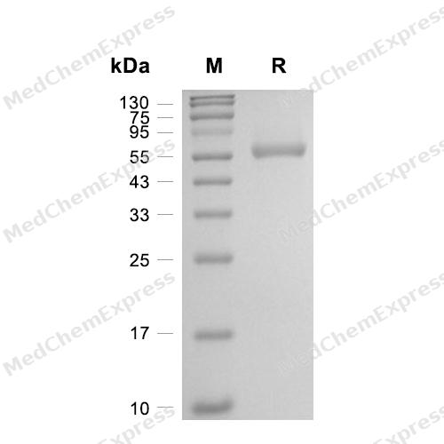
Purity - SDS-PAGE
-
Bioactivity - ELISA
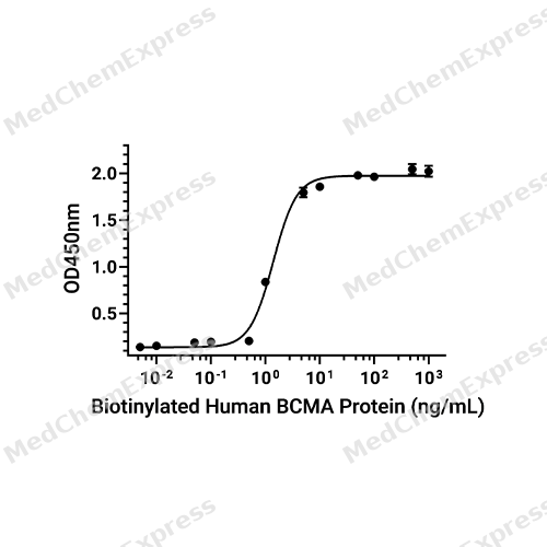
Bioactivity - ELISA


Technical Parameters
-
Species Human
-
Source HEK293
-
Tag N-6*His;N-Flag
-
Accession
O75888 (K112-L250)
-
Gene ID8741
-
Molecular Construction
-
N-term
-
6*His-Flag
-
APRIL (K112-L250)
Accession # O75888 -
C-term
-
-
Protein Length
Full Length of THD Domain
-
Synonyms
TNFSF13; ZTNF2; TNF Superfamily Member 13; Tumor Necrosis Factor Ligand Superfamily Member 13 Epsilon; APRIL; Tumor Necrosis Factor-Related Death Ligand-1; Tumor Necrosis Factor Ligand Superfamily Member 13; Tumor Necrosis Factor Superfamily Member 13; A Proliferation-Inducing Ligand; Tumor Necrosis Factor-Like Protein ZTNF2; CD256; Tumor Necrosis Factor Ligand 7B; Tumor Necrosis Factor (Ligand) Superfamily, Member 13; TNF-Related Death Ligand 1; TNF- And APOL-Related Leukocyte Expressed Ligand 2; UNQ383/PRO715; TALL-2; CD256 Antigen; TRDL-1; TNLG7B; TALL2
-
AA Sequence
KKQHSVLHLVPINATSKDDSDVTEVMWQPALRRGRGLQAQGYGVRIQDAGVYLLYSQVLFQDVTFTMGQVVSREGQGRQETLFRCIRSMPSHPDRAYNSCYSAGVFHLHQGDILSVIIPRARAKLNLSPHGTFLGFVKL
-
Predicted Molecular Mass
50.9 kDa
-
Molecular Weight
Approximately 55-60 kDa, based on SDS-PAGE under reducing conditions, due to the glycosylation.
-
Glycosylation
Yes
-
Structure/Form
Trimer
-
Purity
≥ 95%, as determined by reducing SDS-PAGE.


Product Properties
Lyophilized powder
Lyophilized from a 0.22 μm filtered solution of PBS, pH 7.4, 8% trehalose.
<1 EU/μg, determined by LAL method.
It is not recommended to reconstitute to a concentration less than 100 μg/mL in ddH2O.
Stored at -20°C for 2 years from date of receipt. After reconstitution, it is stable at 4°C for 1 week or -20°C for longer (with carrier protein). It is recommended to freeze aliquots at -20°C or -80°C for extended storage.
Room temperature in continental US; may vary elsewhere.


Documentation
-
Data Sheet (238 KB)
-
SDS (252 KB)
- English - EN (252 KB)
- Français - FR (252 KB)
- Deutsch - DE (252 KB)
- Norwegian - NO (252 KB)
- Español - ES (252 KB)
- Swedish - SV (252 KB)
- Italian - IT (252 KB)
- Korean - KR (252 KB)
- Portuguese - PT (252 KB)
-
Handling Instructions (2659 KB)

Calculators
Concentration (start) × Volume (start) = Concentration (final) × Volume (final)


 Powered by Bioz

