



Resomelagon acetate

Based on 1 Customer Validation
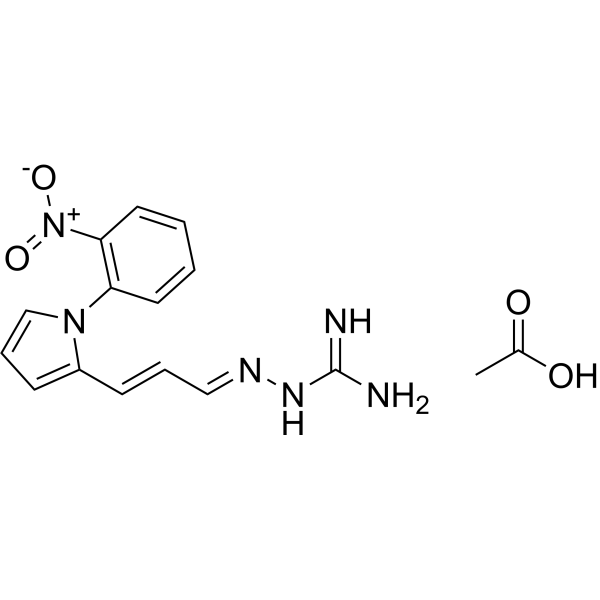
Resomelagon (AP1189) acetate is a potent, orally active melanocortin receptor (MR) agonist about MC1 and MC3. Resomelagon acetate induces ERK1/2 phosphorylation and Ca2+ mobilization. Resomelagon acetate has anti-inflammatory activity. Resomelagon acetate can be used for obesity and chronic inflammation research.
Nur für Forschungszwecke. Wir verkaufen nicht an Patienten.
- Reinheit : 99.15%
- CAS. Nr.: 1809420-72-1
- Formel: C16H18N6O4
- Molecular Weight:358.35
-
Speicherung:
4°C, sealed storage, away from moisture
* In solvent : -80°C, 6 months; -20°C, 1 month (sealed storage, away from moisture)

To place orders, for customer services and technical support, please contact: MedChemExpress USA
Tel: 609-228-6898 E-mail: [email protected] [email protected]


-
 Biologische Aktivität
-
Chemical Information
-
Lösungsmittel & Löslichkeit
- Protokoll
- Reinheit & Dokumentation
- Verweise
-
 Help & FAQs


-
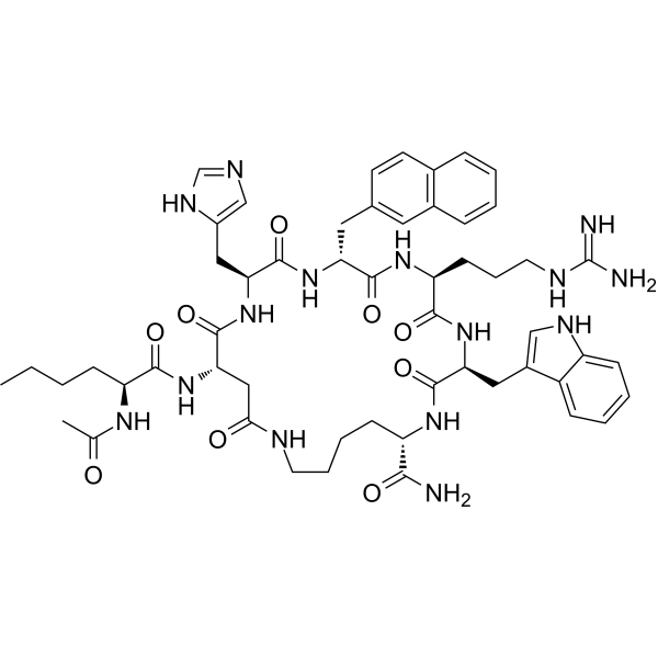
HY-P0227
SHU 9119MC3/4R Antagonist -
![[D-Trp8]-γ-MSH](https://file.medchemexpress.com/product_pic/hy-p1217.gif)
HY-P1217
[D-Trp8]-γ-MSHMC3 Agonist -
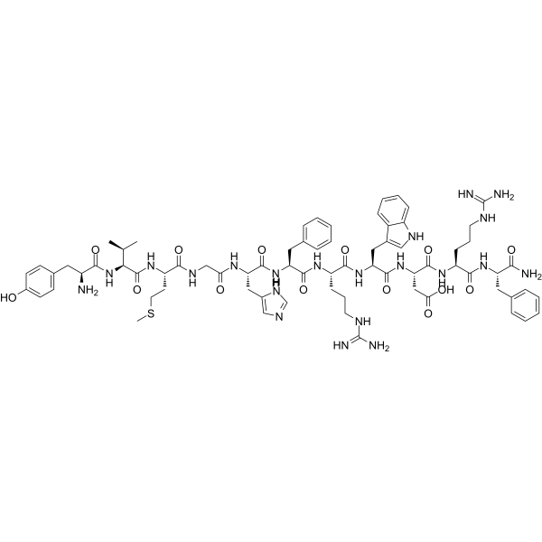
HY-P1214
γ1-MSHMC3 Receptor Agonist -
HY-P1208
PG-931Melanocortin 4 Receptor Agonist -
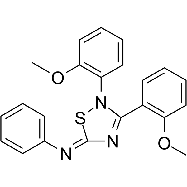
HY-107139
JNJ-10229570Melanocortin Receptor Antagonist -
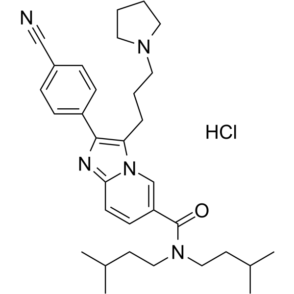
HY-11029
SNT-207707MC-4 Receptor Antagonist -
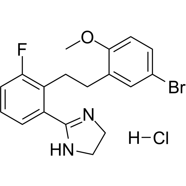
HY-110123
ML-00253764 hydrochlorideMC4R Antagonist -

HY-19870
SetmelanotideMC4R Agonist -
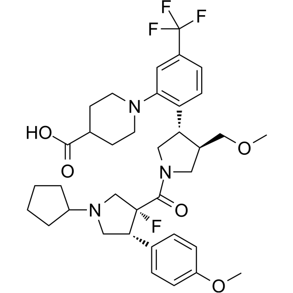
HY-109114
DersimelagonMC1R Agonist -
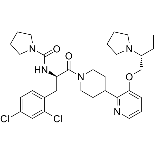
HY-11030A
SNT-207858 free baseMelanocortin Receptor Antagonist -
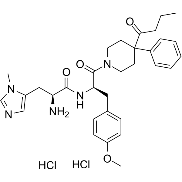
HY-115644
BMS-470539 dihydrochlorideMC-1 Receptor Agonist -
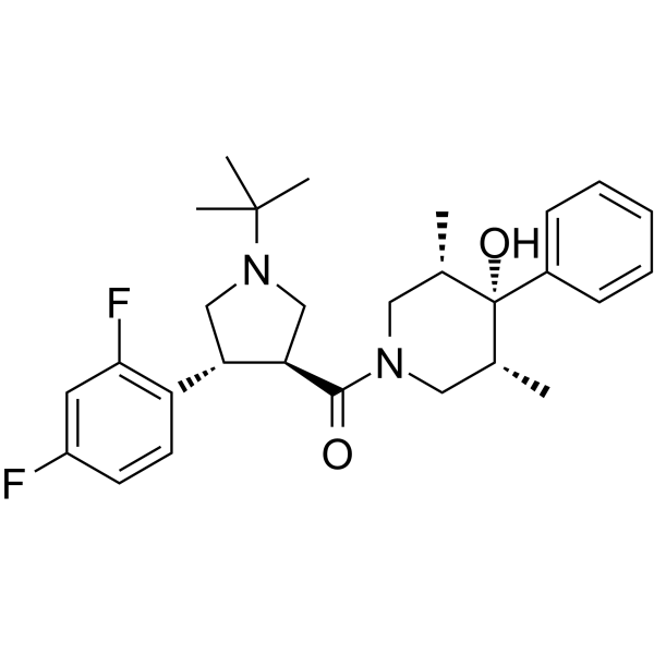
HY-10622
PF-00446687 free baseMC4R Agonist


Biologische Aktivität
Beschreibung
IC50 & Target
Targrt: MC1 and MC3[1]
In Vitro
Resomelagon acetate (0-1000 μM; 8 min; HEK293A cells) promotes melanocortin signal transduction through ERK1/2 phosphorylation and Ca2+ mobilization[1].
Resomelagon acetate (1 nM; 30 min; peritoneal macrophages) has anti-inflammatory activity and inhibits TNF-α release and efferocytosis[1].
MedChemExpress (MCE) has not independently confirmed the accuracy of these methods. They are for reference only.
In Vivo
Resomelagon acetate (25-50 mg/kg; p.o.; daily, for 8 d; male C57BL/6J wild-type (WT) and BALB/c mice) reduces arthritis in mice[1].
MedChemExpress (MCE) has not independently confirmed the accuracy of these methods. They are for reference only.


Chemical Information
-
CAS. Nr. 1809420-72-1
-
Appearance Solid
-
Molecular Weight 358.35
-
Formel C16H18N6O4
-
Color Yellow to brown
-
SMILES
N=C(N)N/N=C/C=C/C1=CC=CN1C2=C(C=CC=C2)[N+]([O-])=O.CC(O)=O
-
Synonyms
AP1189 acetate
-
Versand
Room temperature in continental US; may vary elsewhere.
-
Speicherung
4°C, sealed storage, away from moisture
* In solvent : -80°C, 6 months; -20°C, 1 month (sealed storage, away from moisture)


Lösungsmittel & Löslichkeit
In Vitro:
DMSO : 100 mg/mL (279.06 mM; Need ultrasonic; Hygroscopic DMSO has a significant impact on the solubility of product, please use newly opened DMSO)
Please refer to the solubility information to select the appropriate solvent. Once prepared, please aliquot and store the solution to prevent product inactivation from repeated freeze-thaw cycles.
Storage method and period of stock solution: -80°C, 6 months; -20°C, 1 month (sealed storage, away from moisture). When stored at -80°C, please use it within 6 months. When stored at -20°C, please use it within 1 month.
Please refer to the solubility information to select the appropriate solvent. Once prepared, please aliquot and store the solution to prevent product inactivation from repeated freeze-thaw cycles.
Storage method and period of stock solution: -80°C, 6 months; -20°C, 1 month (sealed storage, away from moisture). When stored at -80°C, please use it within 6 months. When stored at -20°C, please use it within 1 month.
Konzentration (Stammlösung) × Volumen (Stammlösung) = Konzentration (Ziellösung) × Volumen (Ziellösung)


Reinheit & Dokumentation
-
Data Sheet (273 KB)
-
SDS (252 KB)
- English - EN (252 KB)
- Français - FR (252 KB)
- Deutsch - DE (252 KB)
- Norwegian - NO (252 KB)
- Español - ES (252 KB)
- Swedish - SV (252 KB)
- Italian - IT (252 KB)
- Korean - KR (252 KB)
- Portuguese - PT (252 KB)
-
Handling Instructions (2659 KB)
Verweise
[1]. Montero-Melendez T, et, al. Biased agonism as a novel strategy to harness the proresolving properties of melanocortin receptors without eliciting melanogenic effects. J Immunol. 2015 Apr 1;194(7):3381-8. [Content Brief]
[2]. WHO Drug Information. International Nonproprietary Names for Pharmaceutical.


Complete Stock Solution Preparation Table
Please refer to the solubility information to select the appropriate solvent. Once prepared, please aliquot and store the solution to prevent product inactivation from repeated freeze-thaw cycles.
Storage method and period of stock solution: -80°C, 6 months; -20°C, 1 month (sealed storage, away from moisture). When stored at -80°C, please use it within 6 months. When stored at -20°C, please use it within 1 month.
| Optional Solvent | Concentration Solvent Mass | 1 mg | 5 mg | 10 mg | 25 mg |
|---|---|---|---|---|---|
| DMSO | 1 mM | 2.7906 mL | 13.9528 mL | 27.9057 mL | 69.7642 mL |
| 5 mM | 0.5581 mL | 2.7906 mL | 5.5811 mL | 13.9528 mL | |
| 10 mM | 0.2791 mL | 1.3953 mL | 2.7906 mL | 6.9764 mL | |
| 15 mM | 0.1860 mL | 0.9302 mL | 1.8604 mL | 4.6509 mL | |
| 20 mM | 0.1395 mL | 0.6976 mL | 1.3953 mL | 3.4882 mL | |
| 25 mM | 0.1116 mL | 0.5581 mL | 1.1162 mL | 2.7906 mL | |
| 30 mM | 0.0930 mL | 0.4651 mL | 0.9302 mL | 2.3255 mL | |
| 40 mM | 0.0698 mL | 0.3488 mL | 0.6976 mL | 1.7441 mL | |
| 50 mM | 0.0558 mL | 0.2791 mL | 0.5581 mL | 1.3953 mL | |
| 60 mM | 0.0465 mL | 0.2325 mL | 0.4651 mL | 1.1627 mL | |
| 80 mM | 0.0349 mL | 0.1744 mL | 0.3488 mL | 0.8721 mL | |
| 100 mM | 0.0279 mL | 0.1395 mL | 0.2791 mL | 0.6976 mL |


Resomelagon acetate Related Classifications
HY-147301A Related Classifications
 Powered by Bioz