



RTx-161

Based on 1 Customer Validation
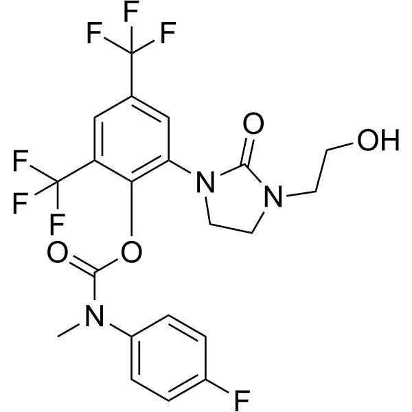
RTx-161 is a DNA polymerase θ inhibitor with an IC50 of 4.1 nM. RTx-161 induces DNA damage, PARP cleavage, apoptosis, and selectively kills homologous recombination-deficient (HRD) cells. RTx-161 acts synergistically with PARP inhibitors to suppress PARP inhibitor resistance in cancer cells. RTx-161 can be used for the research of BRCAG12C mutant cancer and HR-deficient cancers.
Nur für Forschungszwecke. Wir verkaufen nicht an Patienten.
- Reinheit : 98.0%
- CAS. Nr.: 3035072-49-9
- Formel: C21H18F7N3O4
- Molecular Weight:509.37
-
Speicherung:
4°C, protect from light
* In solvent : -80°C, 6 months; -20°C, 1 month (protect from light)

To place orders, for customer services and technical support, please contact: MedChemExpress USA
Tel: 609-228-6898 E-mail: [email protected] [email protected]


-
 Biologische Aktivität
-
Chemical Information
- Protokoll
- Reinheit & Dokumentation
- Verweise
-
 Help & FAQs



Alle DNA/RNA Synthesis Isoform-spezifische Produkte anzeigen
More

Biologische Aktivität
Beschreibung
In Vitro
RTx-161 (1 μM; 60 min) has poor metabolic stability in mouse CD1 male liver microsomes, with a half-life of <2 min[1].
RTx-161 (10-12 day) selectively inhibits colony formation in BRCA2-/- HCT116 cells with an IC50 of 1.4 μM, while having minimal effect on BRCA2+/+ HCT116 cells (IC50 >8 μM)[1].
RTx-161 binds to an allosteric pocket of PolθΔL in complex with DNA and ddGTP, forming specific hydrophobic and hydrogen bond interactions, with a structure resolved to 3.31 Å resolution[1].
RTx-161 (4-500 nM; 15 min) concentration-dependently inhibits recombinant human Polθ-pol MMEJ activity in a cell-free in vitro assay, with maximal inhibition at 500 nM[2].
RTx-161 (2 μM; 0-40 min) does not significantly inhibit recombinant human Polθ-pol ddCMP incorporation on open-conformation DNA/RNA primer-templates in a cell-free assay[2].
RTx-161 (Varying concentrations up to 16 μM; 10-12 days) selectively reduces the survival of HR-deficient cell lines (BRCA2-null DLD1, BRCA2-null HCT116, PALB2-mutant EUFA1341, Brca1cc/cc MEFs) in clonogenic assays, with minimal effect on HR-proficient controls[2].
RTx-161 (20 μM; 16 h) suppresses MMEJ repair of site-specific DSBs in U2OS cells expressing a chromosomal GFP reporter cassette[2].
RTx-161 selectively induces DNA damage (measured by γ-H2AX foci and protein levels) in BRCA2-null DLD1 cells, with minimal effect on BRCA2-WT DLD1 cells[2].
RTx-161 selectively induces apoptosis (measured by PARP cleavage) in BRCA2-null DLD1 cells, with no significant effect on BRCA2-WT DLD1 cells[2].
RTx-161 (0.5-10 μM; 10-12 days) acts synergistically with PARP inhibitors (Olaparib (HY-10162), Rucaparib (HY-10617A), Talazoparib (HY-16106)) to reduce survival of HR-deficient cell lines (BRCA2-null HCT116, BRCA2-null DLD1, PE01, VC8, CAPAN-1) and modestly potentiates Talazoparib activity in HR-proficient MDA-MB-231 cells in clonogenic assays[2].
MedChemExpress (MCE) has not independently confirmed the accuracy of these methods. They are for reference only.
-
Cell Line:BRCA2-null HCT116, BRCA2-null DLD1, PE01 (BRCA2-mutant ovarian), VC8 (BRCA2-truncating mutant hamster), CAPAN-1 (BRCA2-pathogenic mutant pancreatic), MDA-MB-231 (HR-proficient triple-negative breast)
-
Concentration:0.5; 5; 10 μM
-
Incubation Time:10-12 days
-
Result:Exhibited strong synergistic activity with Olaparib in BRCA2-null HCT116, BRCA2-null DLD1, PE01, VC8, and CAPAN-1 cells.
Modestly potentiated Talazoparib activity in HR-proficient MDA-MB-231 cells.


Chemical Information
-
CAS. Nr. 3035072-49-9
-
Appearance Solid
-
Molecular Weight 509.37
-
Formel C21H18F7N3O4
-
Color Colorless to off-white
-
SMILES
FC(F)(F)C1=CC(N2CCN(CCO)C2=O)=C(OC(N(C3=CC=C(F)C=C3)C)=O)C(C(F)(F)F)=C1
-
Versand
Room temperature in continental US; may vary elsewhere.
-
Speicherung
4°C, protect from light
* In solvent : -80°C, 6 months; -20°C, 1 month (protect from light)


Reinheit & Dokumentation
-
Data Sheet (278 KB)
-
SDS (251 KB)
- English - EN (251 KB)
- Français - FR (251 KB)
- Deutsch - DE (251 KB)
- Norwegian - NO (251 KB)
- Español - ES (251 KB)
- Swedish - SV (251 KB)
- Italian - IT (251 KB)
- Korean - KR (251 KB)
- Portuguese - PT (251 KB)
-
Handling Instructions (2659 KB)
Verweise
[1]. Chandramouly G, et al. RTx-303, an Orally Bioavailable Polθ Polymerase Inhibitor That Potentiates PARP Inhibitors in BRCA Mutant Tumors. J Med Chem. 2025;68(21):22196-22215. [Content Brief]
[2]. Fried W, et al. Discovery of a small-molecule inhibitor that traps Polθ on DNA and synergizes with PARP inhibitors. Nat Commun. 2024;15(1):2862. Published 2024 Apr 5. [Content Brief]

Calculators
Konzentration (Stammlösung) × Volumen (Stammlösung) = Konzentration (Ziellösung) × Volumen (Ziellösung)


RTx-161 Related Classifications
HY-161430 Related Classifications
 Powered by Bioz
Keywords