
34 Results for "

Fluorescence quantification

" in MedChemExpress (MCE) Product Catalog:
Products (34)

34 Results for "Fluorescence quantification" in MCE Product Catalog:

Cat. No.: HY-D2817
Target:  

Fluorescent Dye

Research Areas:  

Others

FITC-PLL is a fluorescently labeled polycationic polypeptide probe composed of polylysine (PLL) and FITC (HY-66019), with a typical Ex/Em of 495/520 nm. The fluorescence intensity and lifetime of FITC-PLL are sensitive to pH and FITC density. FITC-PLL can be used for quantifying hapten receptor binding and endocytosis kinetics in macrophages, as a patternable substrate for neuron guidance, for negatively charged phospholipid membrane binding and transmembrane transport, and for live-cell imaging of starch nanoparticle conjugates and lysosome labeling .
loading...
    loading...
Cat. No.: HY-149028
Target:  

Fluorescent Dye

Research Areas:  

Others

Mem-C1C18 is a polarity-sensitive fluorescent probe with excellent plasma membrane anchoring, high brightness and a sensitive response to environmental polarity by altering the fluorescence lifetime. Mem-C1C18 can be used to quantify changes in the polarity of the plasma membrane during iron death .
loading...
    loading...
Cat. No.: HY-N16300
CAS No.: 3091879-75-0
Target:  

Fluorescent Dye

Research Areas:  

Others

Mito-laurdan (bromide) is a Fluorescent dye for mitochondrial membrane fluidity and packing measurement. It depends on membrane potential for targeting: it contains a cationic triphenylphosphonium moiety that accumulates at the inner mitochondrial membrane due to the membrane's negative potential, connected via a 3-carbon linker. As a solvatochromic dye derived from Laurdan (HY-D0080), it retains the characteristic mechanism where fluorescence emission shifts based on membrane packing: in more fluid, loosely packed membranes, greater water penetration leads to increased dipolar relaxation and a bathochromic red shift in emission, while more ordered membranes show a blue-shifted emission; these shifts are quantified using generalized polarization, calculated from emission intensities in ordered and disordered spectral windows. Its excitation wavelength is 352 nm, with emission detected in ordered (409-463 nm) and disordered (473-516 nm) spectral windows. It localizes specifically to mitochondria in HeLa cells, showing high signal overlap with MitoTracker Deep Red, and can detect changes in inner mitochondrial membrane fluidity .
loading...
    loading...
Cat. No.: HY-D1769
CAS No.: 1115867-62-3
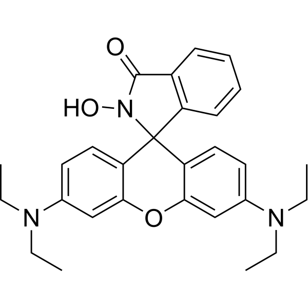
N-hydroxy Rhodamine B amide is a ClO - indicator, which is hydrolyzed to prduce fluorescence in the presence of ClO -. The fluorescence intensity of N-hydroxy Rhodamine B amide is proportional to the product, thus can be used to quantify ClO -.
loading...
    loading...
Cat. No.: HY-135070B
Synonyms: Ac-Arg-Leu-Arg-AMC TFA
Target:  

Proteasome

Research Areas:  

Others

Ac-RLR-AMC (Ac-Arg-Leu-Arg-AMC) TFA is a fluorogenic substrate for the 26S proteasome (Ex/Em: 380/440-460 nm). AMC is released upon cleavage, and its fluorescence can be used to quantify the trypsin-like activity of 26S proteasomes .
loading...
    loading...
Cat. No.: HY-P2176A
Synonyms: Z-Leu-Arg-Gly-Gly-AMC TFA
Target:  

Fluorescent Dye

Research Areas:  

Others

Z-LRGG-AMC TFA is the trifluoroacetate salt of Z-LRGG-AMC (HY-P2176). Z-LRGG-AMC TFA is a fluorogenic substrate for isopeptidase T. Upon cleavage of Z-LRGG-AMC TFA by isopeptidase T, 7-amino-4-methylcoumarin (AMC) is released, and its fluorescence can be used to quantify isopeptidase T activity (Ex/Em : 360/460 nm) .
loading...
    loading...
Cat. No.: HY-P4401A
Target:  

Fluorescent Dye Caspase

Research Areas:  

Others

Z-VEID-AFC TFA is the trifluoroacetate salt of Z-VEID-AFC (HY-P4401). Z-VEID-AFC TFA is a fluorescent substrate for caspase-6. When Z-VEID-AFC TFA is cleaved by caspase-6, 7-amino-4-trifluoromethylcoumarin (AFC) is released, and its fluorescence can be used to quantify caspase-6 activity (Ex/Em: 400/505 nm) .
loading...
    loading...
Cat. No.: HY-D2758
CAS No.: 1911598-65-6
TAMRA DBCO, 5-isomer is a derivative of TAMRA fluorescent isomer with a DBCO functional group. TAMRA DBCO, 5-isomer undergoes strain-promoted azide-alkyne cycloaddition (SPAAC) under copper-free catalytic conditions. TAMRA DBCO, 5-isomer serves as a FRET acceptor for FAM fluorophores. TAMRA DBCO, 5-isomer can label proteins, peptides, nucleic acids and all azide-modified biomolecules, and is suitable for fluorescence imaging and flow cytometry to quantify intracellular SPAAC reactions .
loading...
    loading...
Cat. No.: HY-D3159
CAS No.: 2304942-78-5
Research Areas:  

Others

BTC1070 is an anti-quenching pentamethine cyanine NIR-II fluorescent probe. BTC1070 exhibits high brightness and excellent photostability in aqueous solution (Ex/Em = 1015/1065 nm). BTC1070 can clearly visualize lymphatic structures after intradermal injection, and produces a ratiometric fluorescence response in the acidic range to non-invasively quantify gastric pH. BTC1070 is applicable to studies related to in vivo deep tissue imaging and gastric pH sensing .
loading...
    loading...
Cat. No.: HY-DY1089
CAS No.: 797785-10-5
Di-4-ANEPPDHQ (solution) is a polarity-sensitive membrane probe with a doubly positive charge that can be used to visualize membrane microdomains in living Arabidopsis cells. Di-4-ANEPPDHQ produces a fluorescence spectrum shift by changing the polarity of the lipid environment after embedding into the cell membrane, thereby quantifying the degree of lipid accumulation. The excitation wavelength of Di-4-ANEPPDHQ is 488 nm, and the emission wavelengths are 560 nm and 650 nm in the liquid ordered and disordered phases, respectively. Di-4-ANEPPDHQ is often used for fluorescence imaging studies of membrane microstructure, lipid order, and membrane tension response in living cells[1][2][3].
Solvent and concentration: DMSO: 5 mM
loading...
    loading...
Cat. No.: HY-DY2037
Target:  

Fluorescent Dye

Research Areas:  

Others

Lignin Staining Solution (Safranin Method) is a histochemical staining solution for lignin with Safranin O as the active component. Lignin Staining Solution (Safranin Method) enables lignified cell walls to appear red to deep red (pinkish in regions with low lignification) via the high affinity of the dye for lignin. Lignin Staining Solution (Safranin Method) can be used to visualize lignin distribution in ex vivo thick sections of annual shoots from woody plants and vibratome sections of stems from herbaceous plants (such as Arabidopsis and flax), as well as for in situ quantification of cell wall substructural regions (cell corners, intercellular layers, secondary walls) based on Safranin O ratiometric fluorescence .
loading...
    loading...
Cat. No.: HY-184777
CAS No.: 94367-34-7
Target:  

Aminopeptidase

Research Areas:  

Others

Met-AMC is a fluorescent substrate used to detect the activity of methionine aminopeptidase (MetAP). Hydrolysis of the substrate releases the fluorophore AMC, with detection performed at an excitation wavelength of 360 nm and an emission wavelength of 460 nm. Met-AMC can distinguish the effects of different metal cofactors on the substrate preference of MetAP1: Zn (II)-MetAP1 barely hydrolyzes Met-AMC, while Co (II)-, Mn (II)-, and Ni (II)-MetAP1 can stably cleave this substrate. Met-AMC can be used to establish an intracellular MetAP activity cell-based assay system, which quantifies the intracellular MetAP inhibition level by monitoring the fluorescence signal from substrate hydrolysis in living cells .
loading...
    loading...
Cat. No.: HY-D3103
CAS No.: 1027058-07-6
Target:  

Fluorescent Dye

Research Areas:  

Others

CMTDP is a Fluorescent probe for detecting local polarity changes around the Cys34 domain of bovine serum albumin during pH-induced N→B conformational transition. It is a thiol-specific and polarity-sensitive probe; it covalently labels the free cysteine residue at position 34 (Cys34) of bovine serum albumin, and its fluorescence maximum emission wavelength shifts in response to solvent polarity, but not to pH and temperature. When labeled to BSA, as the local polarity around the Cys34 domain increases with rising pH, the probe shows a bathchromic shift in fluorescence emission wavelength and a decrease in fluorescence intensity, which allows quantification of the local polarity change via the relationship between emission wavelength shift and dielectric constant. CMTDP itself has absorption peaks at ~392 and 508 nm, and CMTDP-labeled BSA has excitation maxima at 376 and 470 nm at pH 6.0, with the shorter-wavelength excitation peak red-shifting as pH increases while the longer one remains stable; when excited at 470 nm, the fluorescence emission maximum shifts from 570 nm at pH 6.0 to 577 nm at pH 9.1, while free CMTDP at pH 7.4 has an emission maximum at 621 nm. The excitation/emission wavelengths for CMTDP-labeled BSA are Ex/Em = 376/570 nm at pH 6.0, Ex/Em = ~376+/573 nm at pH 7.4, Ex/Em = ~376+/575 nm at pH 8.0, Ex/Em = ~376+/577 nm at pH 9.1, and for free CMTDP at pH 7.4, Ex/Em (when excited at 470 nm) = 470/621 nm[1].
loading...
    loading...
Cat. No.: HY-D3073
CAS No.: 2477607-64-8
Target:  

Fluorescent Dye

Research Areas:  

Others

DXB-NIR is a push-pull dioxaboron fluorescent probe with solvatochromic properties, featuring high fluorescence brightness, photostability, and a large two-photon absorption cross-section. DXB-NIR exhibits Ex/Em of 550/620–780 nm (with two-photon excitation at 930 nm). DXB-NIR quantifies local polarity via the intensity ratio I (>640)/I (<640) between the far-red channel (<640 nm) and the near-infrared channel (>640 nm), and it can simultaneously label the plasma membrane, endoplasmic reticulum, and lipid droplets in live cells. DXB-NIR monitors the increase in local polarity of various cell compartments under conditions of cholesterol depletion, starvation, and oxidative stress. DXB-NIR can be used for polarity imaging of biological membranes and lipid droplets, as well as studies related to cellular stress .
loading...
    loading...

