



AKT1 Antibody (YA834)
(Synonyms: KIAA0600, HDAC5, Histone deacetylase 5, HD5, Antigen NY-CO-9)Based on 1 publication(s) in Google Scholar
AKT1 Antibody (YA834) is a Mouse-derived and non-conjugated IgG2b monoclonal antibody, targeting to AKT1.
-
Host:
Mouse
-
Isotype:
IgG
-
Application:
WB, IHC-P, ICC/IF
-
Reactivity :
Human
-
Formulation:
Supplied in 1*PBS (pH7.4), 0.2% BSA and 40% Glycerol. Preservative: 0.05% Sodium Azide.
-
Conjugation:
Non-conjugated


To place orders, for customer services and technical support, please contact: MedChemExpress USA
Tel: 609-228-6898 E-mail: [email protected] [email protected]



Publications Citing Use of MedChemExpress (MCE) AKT1 Antibody (YA834)
More

Applications
| Application |
WB

WB: Western Blot
|
IHC-P
IHC-P: Immunohistochemistry-Paraffin
|
ICC/IF
ICC/IF: Immunocytochemistry/
Immunofluorescence |
|---|---|---|---|
| Dilution Ratio | 1:2000 | 1:100-1:200 | 1:100-200 |


Product Details
AKT1 Antibody (YA834) is a Mouse-derived and non-conjugated IgG2b monoclonal antibody, targeting to AKT1.
-
Host Mouse

-
Clonality Monoclonal

-
Species ReactivityHuman
-
Observed Molecular WeightObserved band size: 56 kDa
Note: Due to possible protein modifications or aggregation, the molecular weight should be confirmed by actual measurement, and the predicted value is for reference only.

-
Calculated Molecular Weight Predicted band size: 56 kDa

Synthetic peptide corresponding to Human AKT1.AA range:1-80.
Endogenous
Protein A affinity purified.
Non-conjugated
Unmodified
IgG


Product Properties
-
Appearance
Solution
-
Formulation
Supplied in 1*PBS (pH7.4), 0.2% BSA and 40% Glycerol. Preservative: 0.05% Sodium Azide.
-
Concentration
Batch-dependent, Please check the COA for the concentration of each lot. Check Lot Concentration
-
Storage & Stability
Stored at -20°C for 1 year. Avoid repeated freeze / thaw cycles.
-
Shipping
Shipping with blue ice.


Publications (1)
-
Journal Impact Factor
-
Most Recent
-
ACS Chem Biol
Targeted O-GlcNAcylation of CK2α Triggers Its Ubiquitin-Proteasome Degradation and Alters Downstream Phosphorylation. [Abstract]2025 Jul 18;20(7):1646-1659. PMID: 40521680


Verification Images
-
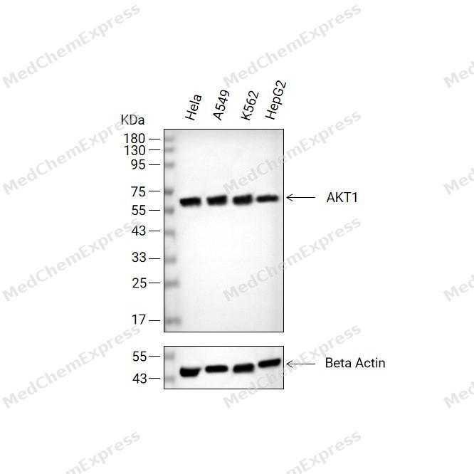 Western blot analysis of extracts from Hela (lane2(20μg), A549(lane3(20μg), K562(lane4(20μg) and HepG2(lane5(20μg) using AKT1 Antibody (HY-P80007). Proteins were transferred to a PVDF membrane and blocked with 5% non-fat milk in TBST at 4°C overnight. The primary antibody (1/2000) and Loading control antibody (Beta Actin, HY-P80993, 1/10,000) was used in 5% non-fat milk in TBST for 2 hour at room temperature. Goat Anti-Mouse IgG-HRP Secondary Antibody (HY-P8004, 1/10,000) was used for 1 hour at room temperature.
 -
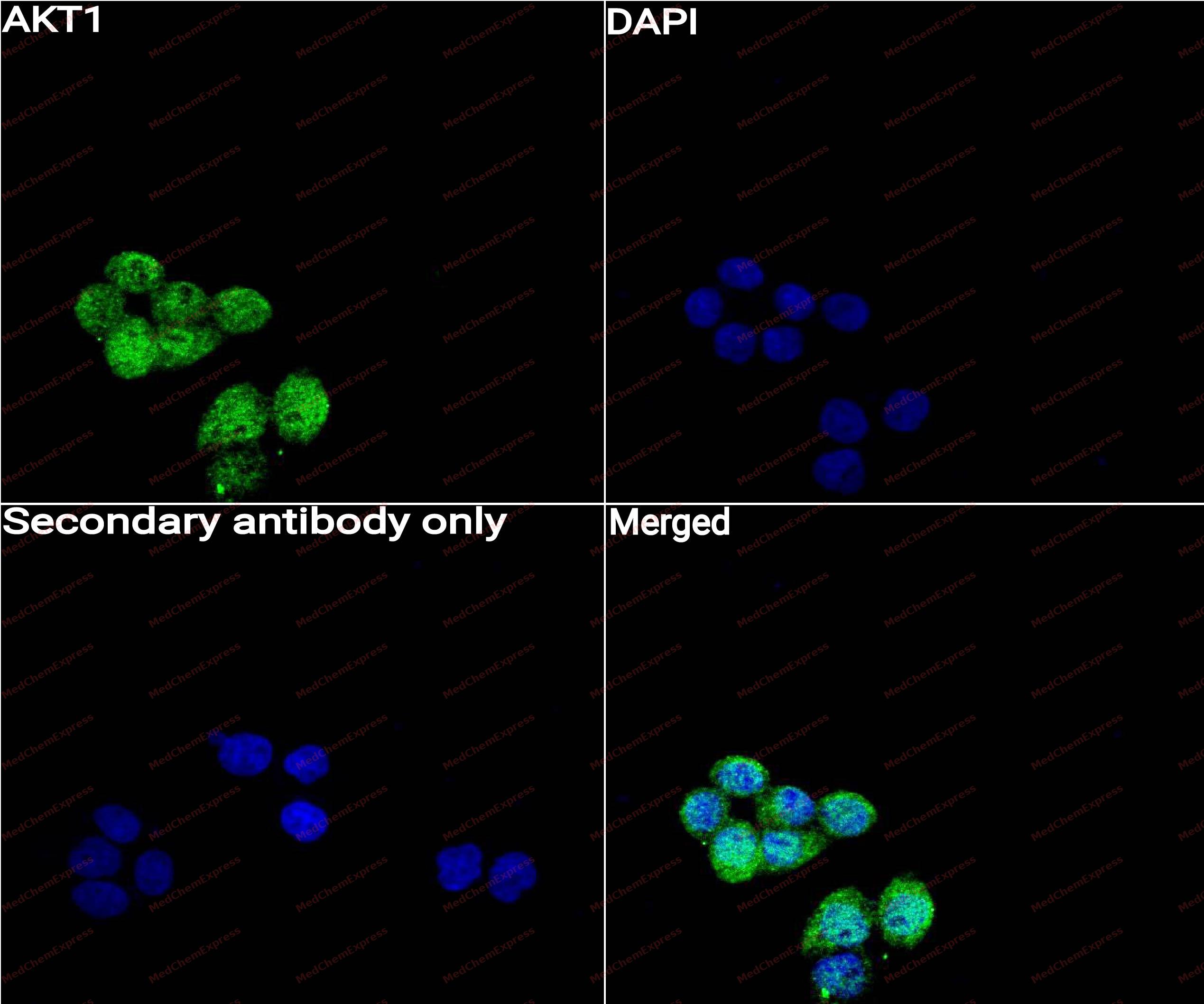 Immunocytochemistry analysis of Hela cells labeling AKT1 with Cleaved AKT1 Antibody (HY-P80007) at 1/50 dilution. Cells were fixed in 4% paraformaldehyde for 15 minutes at room temperature, permeabilized with 0.1% Triton X-100 for 10 minutes at room temperature, then blocked with QuickBlock™ Blocking Buffer for Immunol Staining for 10 min at room temperature. Cells were then incubated with AKT1 Antibody (HY-P80007) at 1/50 dilution in QuickBlock™ Blocking Buffer for Immunol Staining at 4 ℃. Alexa Fluor® 488-conjugated AffiniPure Goat Anti-Mouse IgG H&L(HY-P8005, Green) was used as the secondary antibody at 1/1,000 dilution. PBS instead of the primary antibody was used as the secondary antibody only control. The Nuclear counterstain was DAPI (Blue).
 -
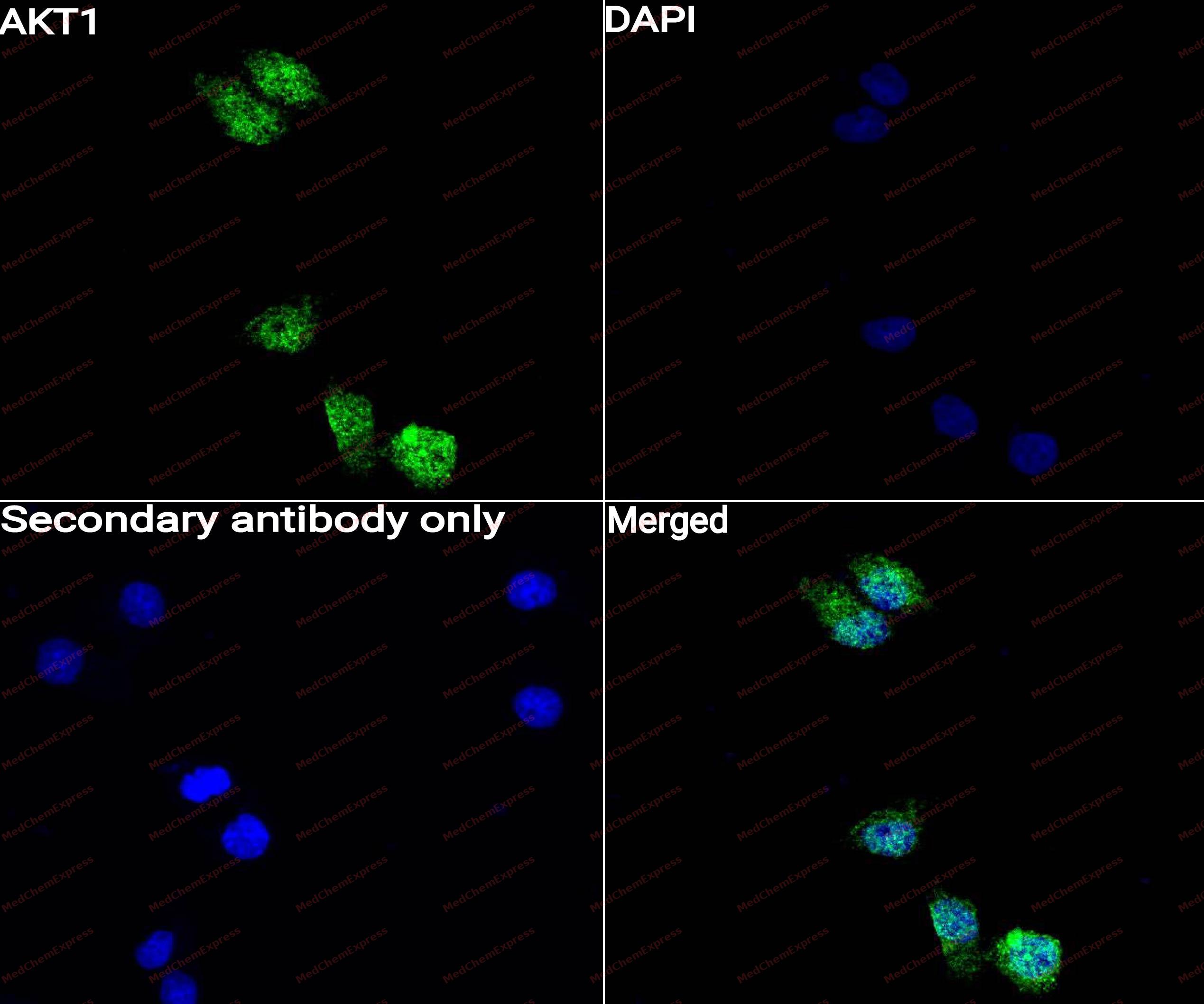 Immunocytochemistry analysis of Hela cells labeling AKT1 with AKT1 Antibody (HY-P80007) at 1/100 dilution. Cells were fixed in 4% paraformaldehyde for 15 minutes at room temperature, permeabilized with 0.1% Triton X-100 for 10 minutes at room temperature, then blocked with QuickBlock™ Blocking Buffer for Immunol Staining for 10 min at room temperature. Cells were then incubated with AKT1 Antibody (HY-P80007)at 1/100 dilution in QuickBlock™ Blocking Buffer for Immunol Staining at 4 ℃. Alexa Fluor® 488-conjugated AffiniPure Goat Anti-Mouse IgG H&L(HY-P8005, Green) was used as the secondary antibody at 1/1,000 dilution. PBS instead of the primary antibody was used as the secondary antibody only control. The Nuclear counterstain was DAPI (Blue).


Background
-
Function
AKT1 (protein kinase Bα, PKBα) is a serine/threonine kinase that functions as a central effector of the PI3K/AKT/mTOR signaling pathway, regulating cellular growth, survival, metabolism, proliferation, and differentiation across multiple tissues and cell types[1][2][3]. Activation of AKT1 occurs downstream of PI3K through phosphoinositide-dependent kinase-1 (PDK1) -mediated phosphorylation, enabling downstream regulation of targets involved in protein synthesis, apoptosis control, and metabolic adaptation[4][5]. Mechanistically, AKT1 contributes to angiogenesis, physiological tissue growth, immune-cell function, and stress-response signaling, making it a critical node in both normal physiology and disease-associated signaling networks[2][4][5]. In disease models, dysregulated AKT signaling is strongly associated with cancer, metabolic disorders, cardiovascular pathology, and inflammatory conditions, and constitutive activation of AKT-dependent pathways promotes oncogenic progression and therapeutic resistance[2][6][7]. Compared with related isoforms, AKT1 exhibits distinct biological functions despite substantial structural homology with AKT2 and AKT3; genetic studies demonstrate that AKT1 deficiency primarily causes growth retardation, whereas AKT2 deficiency induces insulin resistance and diabetes, and AKT3 loss predominantly affects brain development[4][5]. Isoform-specific investigations further show that AKT1 and AKT2 can exert non-redundant or even opposing effects on migration, invasion, inflammation, and fibrogenesis depending on cellular context[2][6][8]. For experimental applications, increasing efforts focus on developing AKT1-selective inhibitors and isoform-selective cellular models, which provide valuable tools for dissecting AKT isoform-specific signaling and for evaluating targeted therapeutic strategies with improved selectivity profiles[6][7][9].
-
Subcellular Localization
Cytoplasm; Nucleus; Cell membrane; Mitochondrion intermembrane space
-
Expression
Tissue_specificity:This protein is expressed in prostate cancer, and its expression level gradually increases from normal to malignant (protein level) . It is expressed in all human cell types analyzed to date. The phosphorylated form of Tyr-176 is significantly increased in the progressive stages of breast cancer (i.e., from normal to hyperplastic (ADH) , ductal carcinoma in situ (DCIS) , invasive ductal carcinoma (IDC) , and lymph node metastasis (LNMM) stages) . -
Isoforms & Post-Translational Modification
AKT1 has 2 isomers: P31749-1: 55686 Da (predicted); P31749-2: 48347 Da (predicted).
O-GlcNAcylation at Thr-305 and Thr-312 inhibits activating phosphorylation at Thr-308 via disrupting the interaction between AKT1 and PDPK1. O-GlcNAcylation at Ser-473 also probably interferes with phosphorylation at this site;Phosphorylation on Thr-308, Ser-473 and Tyr-474 is required for full activity. Phosphorylation of the activation loop at Thr-308 by PDPK1/PDK1 is a prerequisite for full activation. Phosphorylation by mTORC2 in response to growth factors plays a key role in AKT1 activation: mTORC2 phosphorylates different sites depending on the context, such as Thr-450, Ser-473, Ser-477 or Thr-479, thereby facilitating subsequent phosphorylation of the activation loop by PDPK1/PDK1. -
Subunit
Interacts (via the C-terminus) with CCDC88A (via its C-terminus). Interacts with AGAP2 (isoform 2/PIKE-A); the interaction occurs in the presence of guanine nucleotides. Interacts with AKTIP. Interacts (via PH domain) with MTCP1, TCL1A and TCL1B. Interacts with CDKN1B; the interaction phosphorylates CDKN1B promoting 14-3-3 binding and cell-cycle progression. Interacts with MAP3K5 and TRAF6. Interacts with BAD, PPP2R5B, STK3 and STK4. Interacts (via PH domain) with SIRT1. Interacts with SRPK2 in a phosphorylation-dependent manner. Interacts with RAF1. Interacts with TRIM13; the interaction ubiquitinates AKT1 leading to its proteasomal degradation. Interacts with TNK2 and CLK2. Interacts (via the C-terminus) with THEM4 (via its C-terminus). Interacts with and phosphorylated by PDPK1.
-
SwissProt ID
-
Synonyms
KIAA0600, HDAC5, Histone deacetylase 5, HD5, Antigen NY-CO-9
-
Research Field
Signal Transduction
Documentation
-
Data Sheet (263 KB)
-
SDS (251 KB)
- English - EN (251 KB)
- Français - FR (251 KB)
- Deutsch - DE (251 KB)
- Norwegian - NO (251 KB)
- Español - ES (251 KB)
- Swedish - SV (251 KB)
- Italian - IT (251 KB)
- Korean - KR (251 KB)
- Portuguese - PT (251 KB)
-
User Guide for Antibodies (1077 KB)
References
[1]. Guerau-de-Arellano M, et al. Akt isoforms in the immune system. Front Immunol. 2022 Aug 23;13:990874. [Content Brief]
[2]. Reyes-Gordillo K, et al. Akt1 and Akt2 Isoforms Play Distinct Roles in Regulating the Development of Inflammation and Fibrosis Associated with Alcoholic Liver Disease. Cells. 2019 Oct 29;8(11):1337. [Content Brief]
[3]. O'Neill BT, et al. Akt1 in the cardiovascular system: friend or foe? J Clin Invest. 2005 Aug;115(8):2059-64. [Content Brief]
[4]. Tellai AD, et al. The complex post-transcriptional regulation of genes coding for methionine adenosyl transferase: New insights for liver cancer. Biochimie. 2025 Nov;238(Pt A):19-28. [Content Brief]
[5]. Quambusch L, et al. Cellular model system to dissect the isoform-selectivity of Akt inhibitors. Nat Commun. 2021 Sep 6;12(1):5297. [Content Brief]

 Powered by Bioz
