



Phospho-AKT1/2/3 (Thr308) Antibody
(Synonyms: AKT1;AKT2;AKT3; PKB; RAC; RAC-alpha serine/threonine-protein kinase; Protein kinase B; PKB; Protein kinase B alpha; PKB alpha; Proto-oncogene c-Akt; RAC-PK-alpha)Based on 2 publication(s) in Google Scholar
Phospho-AKT1/2/3 (Thr308) Antibody is a Rabbit-derived and non-conjugated IgG polyclonal antibody, targeting to Phospho-AKT1/2/3 (Thr308).
-
Host:
Rabbit
-
Isotype:
IgG
-
Application:
WB, IHC-P, ICC/IF, ELISA
-
Reactivity :
Human, Mouse, Rat
-
Formulation:
Supplied in 1*PBS (pH 7.3), 50% glycerol and 0.5% BSA. Preservative: 0.02% sodium azide.
-
Conjugation:
Non-conjugated


To place orders, for customer services and technical support, please contact: MedChemExpress USA
Tel: 609-228-6898 E-mail: [email protected] [email protected]



-

HY-P80535
AKT1+AKT2+AKT3 Antibody (YA635) -
HY-P80846
Phospho-PI3 Kinase p85/p55 (Tyr467/Tyr199) Antibody -
HY-P80276
Phospho-AKT1(Ser473) Antibody (YA227) -
HY-P8001
HRP-conjugated Goat Anti-Rabbit IgG H&L -
HY-P80788
Phospho-AKT1 (Ser473) Antibody (YA228) -
HY-P80867
PI3 Kinase p85 alpha Antibody (YA689) -
HY-12815
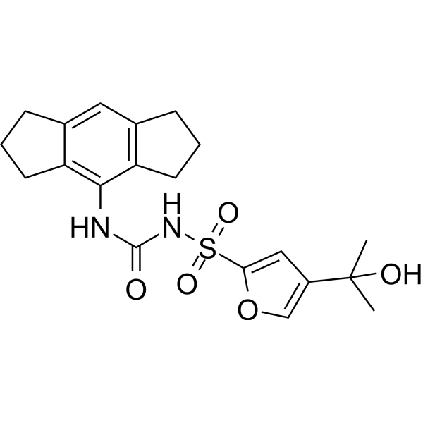
MCC950NLRP3 Inhibitor -
HY-10108
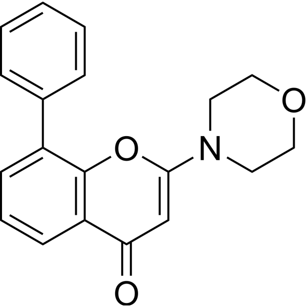
LY294002PI3K Inhibitor -
HY-P86645
PI3 Kinase p85 alpha Antibody (YA6337) -
HY-P8004
HRP-conjugated Goat Anti-Mouse IgG H&L -
HY-P80137
GAPDH Antibody (YA418) -

HY-K0601
JC-1 Mitochondrial Membrane Potential Assay Kit
Publications Citing Use of MedChemExpress (MCE) Phospho-AKT1/2/3 (Thr308) Antibody
More
 2 Publications Citing Use of MCE Phospho-AKT1/2/3 (Thr308) Antibody (1)
-
WB


Applications
| Application |
WB

WB: Western Blot
|
IHC-P
IHC-P: Immunohistochemistry-Paraffin
|
ICC/IF
ICC/IF: Immunocytochemistry/
Immunofluorescence |
ELISA
ELISA: Enzyme Linked Immunosorbent Assay
|
|---|---|---|---|---|
| Dilution Ratio | 1:500-1:1000 | 1:50-1:100 | 1:50-1:200 | 1:10000 |


Product Details
Phospho-AKT1/2/3 (Thr308) Antibody is a Rabbit-derived and non-conjugated IgG polyclonal antibody, targeting to Phospho-AKT1/2/3 (Thr308).
-
Host Rabbit

-
Clonality Polyclonal

-
Species ReactivityHuman, Mouse, Rat
-
Observed Molecular WeightObserved band size: 56 kDa
Note: Due to possible protein modifications or aggregation, the molecular weight should be confirmed by actual measurement, and the predicted value is for reference only.

-
Calculated Molecular Weight Predicted band size: 56 kDa

Entrez Gene: 207 Human ; 11651 Mouse ; 24185 Rat
SwissProt: P31749 Human ; P31750 Mouse ; P47196 Rat
OMIM: 114480 Human
Synthetic phosphopeptide corresponding to residues surrounding Thr308 of Human Akt.The exact sequence is proprietary to MCE.
Endogenous
affinity purified
Non-conjugated
Phosphorylated
IgG


Product Properties
-
Appearance
Solution
-
Formulation
Supplied in 1*PBS (pH 7.3), 50% glycerol and 0.5% BSA. Preservative: 0.02% sodium azide.
-
Concentration
Batch-dependent, Please check the COA for the concentration of each lot. Check Lot Concentration
-
Shipping
Shipping with blue ice.


Publications (2)
-
Journal Impact Factor
-
Most Recent
-
Int J Biol Macromol
Integrin β8: A novel prognostic biomarker for ovarian cancer identified through multi-omics-based prognostic models and experimental validation. [Abstract]2026 Jan;335(Pt 1):149046. PMID: 41297810
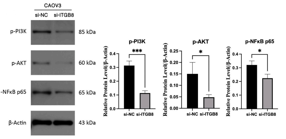
Phospho-AKT1/2/3 (Thr308) Antibody purchased from MedChemExpress. Usage Cited in: Int J Biol Macromol. 2026 Jan;335(Pt 1):149046. [Abstract]
Results of Western Blot analysis. They respectively display the relative expression levels of p-PI3K, p-AKT, and p-NFκB p65 proteins in CAOV3 and OVCAR3 cells (Cell lines were obtained as described in the Methods) when ITGB8 is knocked down compared with the negative control.


Verification Images
-
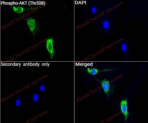 Immunocytochemistry analysis of C2C12 cells labeling Phospho-AKT (Thr308) with Phospho-AKT (Thr308) Antibody (HY-P80789) at 1/200 dilution. Cells were fixed in 4% paraformaldehyde for 15 minutes at room temperature, permeabilized with 0.1% Triton X-100 for 10 minutes at room temperature, then blocked with QuickBlock™ Blocking Buffer for Immunol Staining for 10 min at room temperature. Cells were then incubated withPhospho-AKT (Thr308) (HY-P80789) at 1/200 dilution in QuickBlock™ Blocking Buffer for Immunol Staining at 4 ℃. Alexa Fluor® 488-conjugated AffiniPure Goat Anti-Rabbit IgG H&L(HY-P8002, Green) was used as the secondary antibody at 1/1,000 dilution. PBS instead of the primary antibody was used as the secondary antibody only control. The Nuclear counterstain was DAPI (Blue).
 -
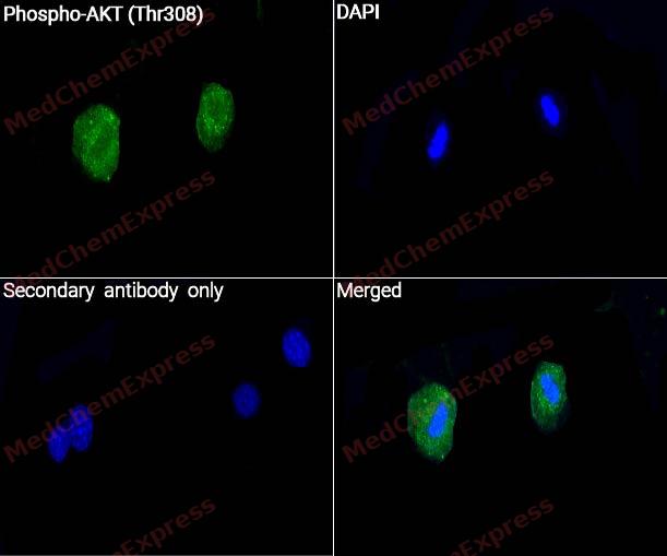 Immunocytochemistry analysis of C2C12 cells labeling Phospho-AKT (Thr308) with Phospho-AKT (Thr308) Antibody (HY-P80789) at 1/50 dilution. Cells were fixed in 4% paraformaldehyde for 15 minutes at room temperature, permeabilized with 0.1% Triton X-100 for 10 minutes at room temperature, then blocked with QuickBlock™ Blocking Buffer for Immunol Staining for 10 min at room temperature. Cells were then incubated withPhospho-AKT (Thr308) (HY-P80789) at 1/50 dilution in QuickBlock™ Blocking Buffer for Immunol Staining at 4 ℃. Alexa Fluor® 488-conjugated AffiniPure Goat Anti-Rabbit IgG H&L(HY-P8002, Green) was used as the secondary antibody at 1/1,000 dilution. PBS instead of the primary antibody was used as the secondary antibody only control. The Nuclear counterstain was DAPI (Blue).
 -
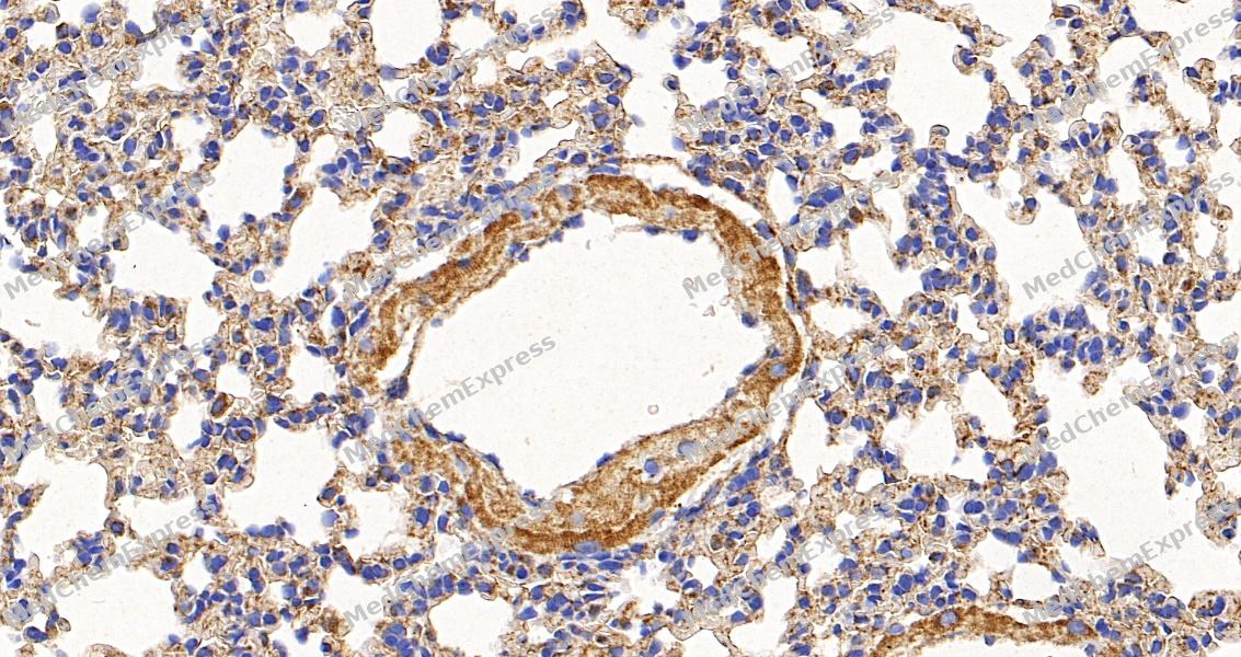 Immunohistochemical analysis of paraffin-embedded mouse lung tissue using Phospho-AKT (Thr308) Antibody. The section was pre-treated using heat mediated antigen retrieval with sodium citrate buffer (pH 6.0) for 8 minutes. The tissues were blocked in QuickBlock for 20 minutes at room temperature, washed with ddH2O and PBS, and then probed with the primary antibody at 1/100 dilution in 4℃ overnight. The detection was performed using an HRP conjugated compact polymer system. DAB was used as the chromogen. Tissues were counterstained with hematoxylin and mounted with DPX.
 -
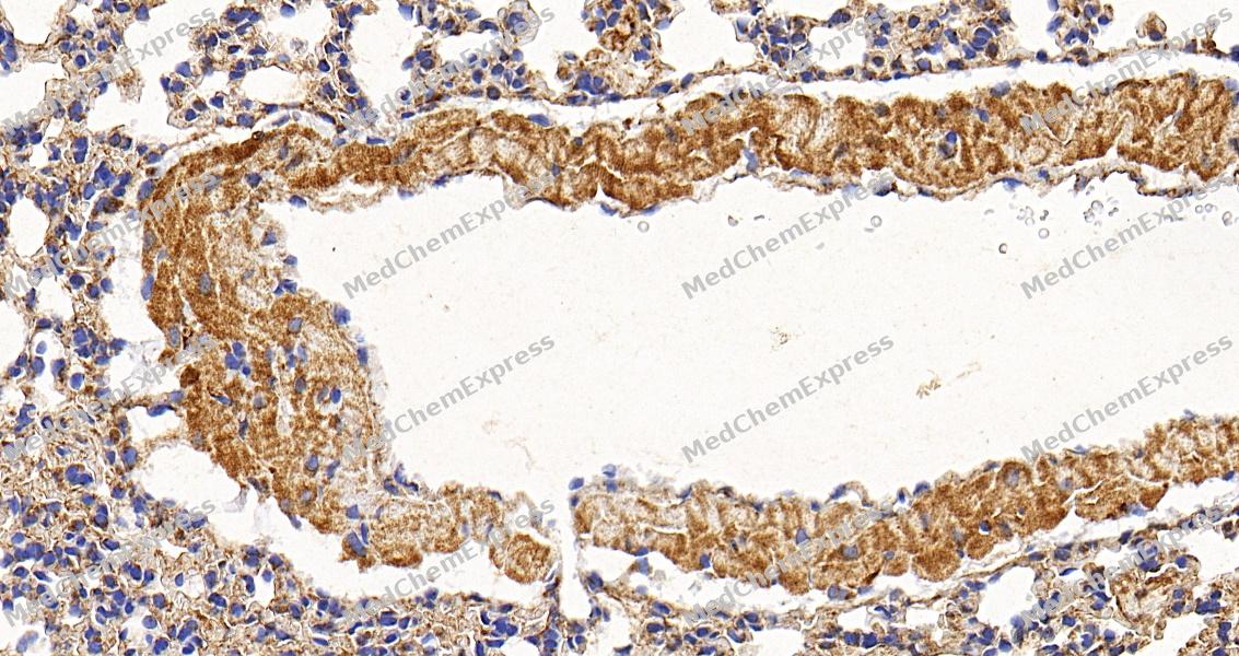 Immunohistochemical analysis of paraffin-embedded mouse lung tissue using Phospho-AKT (Thr308) Antibody. The section was pre-treated using heat mediated antigen retrieval with sodium citrate buffer (pH 6.0) for 8 minutes. The tissues were blocked in QuickBlock for 20 minutes at room temperature, washed with ddH2O and PBS, and then probed with the primary antibody at 1/100 dilution in 4℃ overnight. The detection was performed using an HRP conjugated compact polymer system. DAB was used as the chromogen. Tissues were counterstained with hematoxylin and mounted with DPX.


Background
-
Function
Akt/PKB (Protein kinase B), a serine/threonine protein kinase with antiapoptotic activity, is one of the major downstream targets of PtdIns (3, 4, 5) P3 signaling pathway. It contains a pleckstrin homology domain (PH domain) that specifically binds PtdIns (3, 4, 5) P3 on the plasma membrane. Akt phosphorylation and activation are directly determined by the level of PtdIns (3, 4, 5) P3 on the plasma membrane, which is regulated by PI3K. Akt consists of three isoforms: PKBα/Akt1, PKBβ/Akt2 and PKBγ/Akt3. Akt isoforms have an N-terminal PH (pleckstrin homology) domain and a kinase domain, which are separated by a 39-amino-acid hinge region. Catalytically active Akt regulates the function of numerous substrates involved in cell survival, growth, proliferation, metabolism and protein synthesis. Akt is a crucial mediator of cell survival and its deactivation is implicated in various stress-induced pathological cell death and degenerative diseases.
-
Subcellular Localization
Cytoplasm; Nucleus; Cell membrane; Mitochondrion intermembrane space
-
Expression
Tissue_specificity:This protein is expressed in prostate cancer, and its expression level gradually increases from normal to malignant (protein level) . It is expressed in all human cell types analyzed to date. The phosphorylated form of Tyr-176 is significantly increased in the progressive stages of breast cancer (i.e., from normal to hyperplastic (ADH) , ductal carcinoma in situ (DCIS) , invasive ductal carcinoma (IDC) , and lymph node metastasis (LNMM) stages) . -
Isoforms & Post-Translational Modification
P31749 has 2 isomers: P31749-1: 55686 Da (predicted); P31749-2: 48347 Da (predicted).
O-GlcNAcylation at Thr-305 and Thr-312 inhibits activating phosphorylation at Thr-308 via disrupting the interaction between AKT1 and PDPK1. O-GlcNAcylation at Ser-473 also probably interferes with phosphorylation at this site;Phosphorylation on Thr-308, Ser-473 and Tyr-474 is required for full activity (PubMed:12149249, PubMed:15047712, PubMed:15262962, PubMed:16266983, PubMed:18456494, PubMed:20481595, PubMed:20978158, PubMed:8978681, PubMed:9512493, PubMed:9736715). Phosphorylation of the activation loop at Thr-308 by PDPK1/PDK1 is a prerequisite for full activation (PubMed:9512493). Phosphorylation by mTORC2 in response to growth factors plays a key role in AKT1 activation: mTORC2 phosphorylates different sites depending on the context, such as Thr-450, Ser-473, Ser-477 or Thr-479, thereby facilitating subsequent phosphorylation of the activation loop by PDPK1/PDK1 (PubMed:15718470, PubMed:24670654). Phosphorylation at Ser-473 by mTORC2 promotes ubiquitination and degradation by the proteasome (By similarity). Also phosphorylated at Ser-477 and Thr-479 by CDK2, facilitating subsequent phosphorylation of the activation loop by PDPK1/PDK1 (PubMed:24670654). Activated TNK2 phosphorylates it on Tyr-176 resulting in its binding to the anionic plasma membrane phospholipid PA (PubMed:20333297). This phosphorylated form localizes to the cell membrane, where it is targeted by PDPK1 and PDPK2 for further phosphorylations on Thr-308 and Ser-473 leading to its activation (PubMed:20333297). Phosphorylated at Thr-308 and Ser-473 by IKBKE and TBK1 (PubMed:21464307). Ser-473 phosphorylation is enhanced by interaction with AGAP2 isoform 2 (PIKE-A) (PubMed:14761976). Ser-473 phosphorylation is enhanced in focal cortical dysplasias with Taylor-type balloon cells (PubMed:17013611). Ser-473 phosphorylation is enhanced by signaling through activated FLT3 (PubMed:16266983). Ser-473 is dephosphorylated by PHLPP (PubMed:28147277). Dephosphorylated at Thr-308 and Ser-473 by PP2A phosphatase (PubMed:21329884). The phosphorylated form of PPP2R5B is required for bridging AKT1 with PP2A phosphatase (PubMed:21329884). Ser-473 is dephosphorylated by CPPED1, leading to termination of signaling (PubMed:23799035). AIM2 acts as an inhibitor of AKT1 by inhibiting phosphorylation Ser-473: AIM2 acts both by inhibiting the activity of PRKDC/DNA-PK kinase and promoting dephosphorylation by PP2A phosphatase (By similarity);Ubiquitinated; undergoes both 'Lys-48'- and 'Lys-63'-linked polyubiquitination. TRAF6-induced 'Lys-63'-linked AKT1 ubiquitination is critical for phosphorylation and activation (PubMed:19713527). When ubiquitinated, it translocates to the plasma membrane, where it becomes phosphorylated (PubMed:20059950). When fully phosphorylated and translocated into the nucleus, undergoes 'Lys-48'-polyubiquitination catalyzed by TTC3, leading to its degradation by the proteasome (PubMed:20059950). Also ubiquitinated by TRIM13 leading to its proteasomal degradation (PubMed:21333377). Phosphorylated, undergoes 'Lys-48'-linked polyubiquitination preferentially at Lys-284 catalyzed by MUL1, leading to its proteasomal degradation (PubMed:22410793). Ubiquitinated via 'Lys-48'-linked polyubiquitination by ZNRF1, leading to its degradation by the proteasome (By similarity);Acetylated on Lys-14 and Lys-20 by the histone acetyltransferases EP300 and KAT2B. Acetylation results in reduced phosphorylation and inhibition of activity. Deacetylated at Lys-14 and Lys-20 by SIRT1. SIRT1-mediated deacetylation relieves the inhibition;Cleavage by caspase-3/CASP3 (By similarity). Cleaved at the caspase-3 consensus site Asp-462 during apoptosis, resulting in down-regulation of the AKT signaling pathway and decreased cell survival (PubMed:23152800) -
Subunit
Interacts with BTBD10 (By similarity). Interacts with KCTD20 (By similarity). Interacts (via the C-terminus) with CCDC88A (via its C-terminus). Interacts with GRB10; the interaction leads to GRB10 phosphorylation thus promoting YWHAE-binding (By similarity). Interacts with AGAP2 (isoform 2/PIKE-A); the interaction occurs in the presence of guanine nucleotides. Interacts with AKTIP. Interacts (via PH domain) with MTCP1, TCL1A and TCL1B. Interacts with CDKN1B; the interaction phosphorylates CDKN1B promoting 14-3-3 binding and cell-cycle progression. Interacts with MAP3K5 and TRAF6. Interacts with BAD, PPP2R5B, STK3 and STK4. Interacts (via PH domain) with SIRT1. Interacts with SRPK2 in a phosphorylation-dependent manner. Interacts with RAF1. Interacts with TRIM13; the interaction ubiquitinates AKT1 leading to its proteasomal degradation. Interacts with TNK2 and CLK2. Interacts (via the C-terminus) with THEM4 (via its C-terminus). Interacts with and phosphorylated by PDPK1. Interacts with PA2G4 (By similarity). Interacts with KIF14; the interaction is detected in the plasma membrane upon INS stimulation and promotes AKT1 phosphorylation (PubMed:24784001). Interacts with FAM83B; activates the PI3K/AKT signaling cascade (PubMed:23676467). Interacts with WDFY2 (via WD repeats 1-3) (PubMed:16792529). Forms a complex with WDFY2 and FOXO1 (By similarity). Interacts with FAM168A (PubMed:23251525). Interacts with SYAP1 (via phosphorylated form and BSD domain); this interaction is enhanced in a mTORC2-mediated manner in response to epidermal growth factor (EGF) stimulation and activates AKT1 (PubMed:23300339). Interacts with PKHM3 (By similarity). Interacts with FKBP5/FKBP51; promoting interaction between Akt/AKT1 and PHLPP1, thereby enhancing dephosphorylation and subsequent activation of Akt/AKT1 (PubMed:28147277). Interacts with TMEM175; leading to formation of the lysoK(GF) complex (PubMed:32228865). Acts as a negative regulator of the cGAS-STING pathway by mediating phosphorylation of CGAS during mitosis, leading to its inhibition (PubMed:26440888)
-
Synonyms
AKT1; AKT2; AKT3; PKB; RAC; RAC-alpha serine/threonine-protein kinase; Protein kinase B; PKB; Protein kinase B alpha; PKB alpha; Proto-oncogene c-Akt; RAC-PK-alpha
-
Research Field
Signal Transduction
Documentation
-
Data Sheet (264 KB)
-
SDS (252 KB)
- English - EN (252 KB)
- Français - FR (252 KB)
- Deutsch - DE (252 KB)
- Norwegian - NO (252 KB)
- Español - ES (252 KB)
- Swedish - SV (252 KB)
- Italian - IT (252 KB)
- Korean - KR (252 KB)
- Portuguese - PT (252 KB)
-
User Guide for Antibodies (1077 KB)
References
[1]. Khalili M, et al. Underserved Does Not Mean Undeserved: Unfurling the HCV Care in the Safety Net. Dig Dis Sci. 2018 Dec;63(12):3250-3252. [Content Brief]
[2]. Manola A, et al. Mastocytosis causing refractory hypotension after coronary angiography. Int J Cardiol. 2012 Apr 19;156(2):e43-4. [Content Brief]
[3]. Nitulescu GM, et al. The Akt pathway in oncology therapy and beyond (Review). Int J Oncol. 2018 Dec;53(6):2319-2331. [Content Brief]
[4]. Guerau-de-Arellano M, et al. Akt isoforms in the immune system. Front Immunol. 2022 Aug 23;13:990874. [Content Brief]
[5]. Hassan D, et al. AKT kinases as therapeutic targets. J Exp Clin Cancer Res. 2024 Nov 29;43(1):313. [Content Brief]
[6]. Wang Y, et al. Corrigendum to \"Mechanical stress protects against chondrocyte pyroptosis through TGF-β1-mediated activation of Smad2/3 and inhibition of the NF-κB signaling pathway in an osteoarthritis model\" [Biomed. Pharmacother., 2023 Mar:159:114216. [Content Brief]

 Powered by Bioz
