



ATM Antibody (YA597)
(Synonyms: Serine-protein kinase ATM, Ataxia telangiectasia mutated, A-T mutated, ATM)
Based on 1 Customer Validation
ATM Antibody (YA597) is a Rabbit-derived and non-conjugated IgG monoclonal antibody, targeting to ATM.
-
Host:
Rabbit
-
Isotype:
IgG
-
Application:
WB, ICC/IF, IHC-P
-
Reactivity :
Human
-
Formulation:
Supplied in 1*TBS (pH7.4), 0.05% BSA and 40% Glycerol. Preservative: 0.05% Sodium Azide.
-
Conjugation:
Non-conjugated


To place orders, for customer services and technical support, please contact: MedChemExpress USA
Tel: 609-228-6898 E-mail: [email protected] [email protected]





Applications
| Application |
WB

WB: Western Blot
|
ICC/IF
ICC/IF: Immunocytochemistry/
Immunofluorescence |
IHC-P
IHC-P: Immunohistochemistry-Paraffin
|
|---|---|---|---|
| Dilution Ratio | 1:500-1:5000 | 1:50-1:200 | 1:50-1:1000 |


Product Details
ATM Antibody (YA597) is a Rabbit-derived and non-conjugated IgG monoclonal antibody, targeting to ATM.
-
Host Rabbit

-
Clonality Recombinant,Monoclonal

-
Species ReactivityHuman
-
Observed Molecular WeightObserved band size: 370 kDa
Note: Due to possible protein modifications or aggregation, the molecular weight should be confirmed by actual measurement, and the predicted value is for reference only.

-
Calculated Molecular Weight Predicted band size: 351 kDa

Synthetic peptide corresponding to Human ATM.AA range:1951-2000.
Endogenous
Protein A affinity purified.
Non-conjugated
Unmodified
IgG


Product Properties
-
Appearance
Solution
-
Formulation
Supplied in 1*TBS (pH7.4), 0.05% BSA and 40% Glycerol. Preservative: 0.05% Sodium Azide.
-
Concentration
Batch-dependent, Please check the COA for the concentration of each lot. Check Lot Concentration
-
Storage & Stability
Stored at -20°C for 1 year. Avoid repeated freeze / thaw cycles.
-
Shipping
Shipping with blue ice.


Verification Images
-
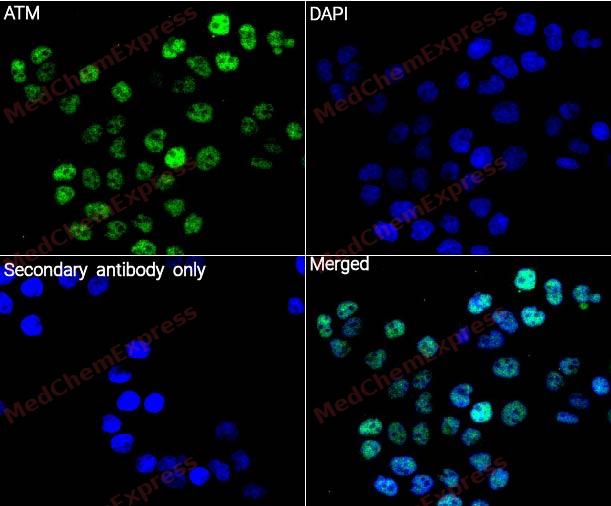 Immunocytochemistry analysis of Hela cells labeling ATM with ATM Antibody (HY-P80023)at 1/50 dilution. Cells were fixed in 4% paraformaldehyde for 15 minutes at room temperature, permeabilized with 0.1% Triton X-100 for 10 minutes at room temperature, then blocked with QuickBlock™ Blocking Buffer for Immunol Staining for 10 min at room temperature. Cells were then incubated with ATM Antibody (HY-P80023) at 1/50 dilution in QuickBlock™ Blocking Buffer for Immunol Staining at 4 ℃. Alexa Fluor® 488-conjugated AffiniPure Goat Anti-Rabbit IgG H&L(HY-P8002, Green) was used as the secondary antibody at 1/1,000 dilution. PBS instead of the primary antibody was used as the secondary antibody only control. The Nuclear counterstain was DAPI (Blue).
 -
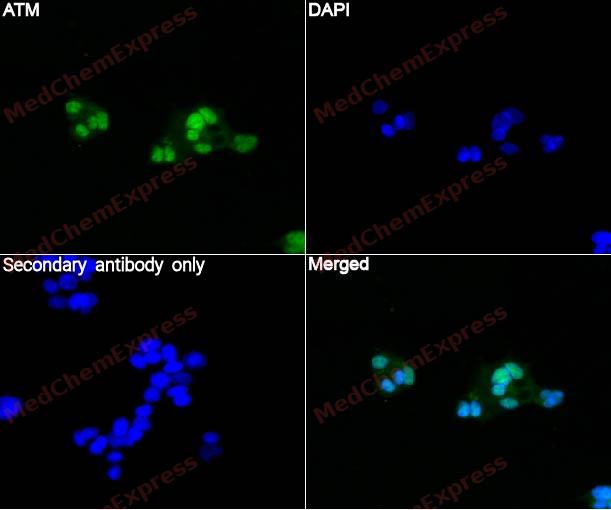 Immunocytochemistry analysis of HepG2 cells labeling ATM with ATM Antibody (HY-P80023)at 1/50 dilution. Cells were fixed in 4% paraformaldehyde for 15 minutes at room temperature, permeabilized with 0.1% Triton X-100 for 10 minutes at room temperature, then blocked with QuickBlock™ Blocking Buffer for Immunol Staining for 10 min at room temperature. Cells were then incubated with ATM Antibody (HY-P80023) at 1/50 dilution in QuickBlock™ Blocking Buffer for Immunol Staining at 4 ℃. Alexa Fluor® 488-conjugated AffiniPure Goat Anti-Rabbit IgG H&L(HY-P8002, Green) was used as the secondary antibody at 1/1,000 dilution. PBS instead of the primary antibody was used as the secondary antibody only control. The Nuclear counterstain was DAPI (Blue).
 -
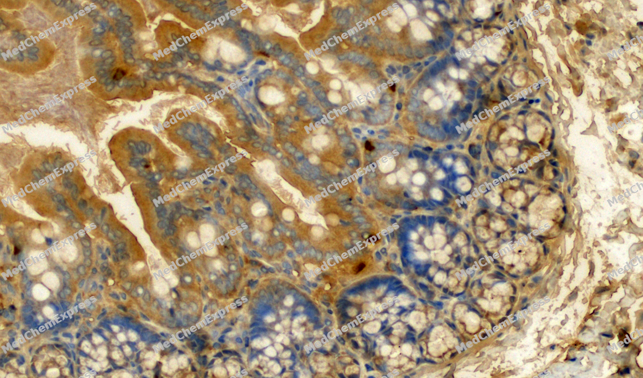 Immunohistochemical analysis of paraffin-embedded Rat colon tissue using ATM Antibody. The section was pre-treated using heat mediated antigen retrieval with sodium citrate buffer (pH 6.0) for 8 minutes. The tissues were blocked in QuickBlock for 20 minutes at room temperature, washed with ddH2O and PBS, and then probed with the primary antibody (HY-P80023, 1/500) in 4℃ overnight. The detection was performed using an HRP conjugated compact polymer system. DAB was used as the chromogen. Tissues were counterstained with hematoxylin and mounted with DPX.
 -
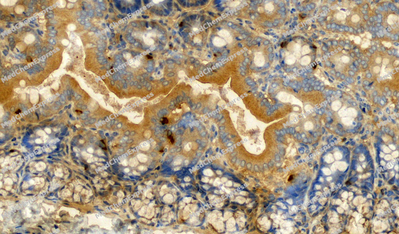 Immunohistochemical analysis of paraffin-embedded Rat colon tissue using ATM Antibody. The section was pre-treated using heat mediated antigen retrieval with sodium citrate buffer (pH 6.0) for 8 minutes. The tissues were blocked in QuickBlock for 20 minutes at room temperature, washed with ddH2O and PBS, and then probed with the primary antibody (HY-P80023, 1/500) in 4℃ overnight. The detection was performed using an HRP conjugated compact polymer system. DAB was used as the chromogen. Tissues were counterstained with hematoxylin and mounted with DPX.


Background
-
Function
ATM is a serine/threonine protein kinase and a crucial nexus for the cellular response to DNA double-stranded breaks[1]. Mechanistically, ATM signals to cell-cycle and DNA-repair components by phosphorylating downstream targets including p53, CHK2, NBS1, and BRCA1[2]. Transcription- and topoisomerase I-induced DNA double-strand breaks also activate ATM in post-mitotic neurons and lymphocytes, linking ATM signaling to transcription-associated genome stress[3]. In disease context, ATM mutations cause ataxia-telangiectasia, which includes neurodegeneration, immune dysfunction, radiosensitivity, and cancer predisposition[1]. Compared with related PIKK isoforms, ATM, ATR, and DNA-PK jointly control the DNA damage response, but ATR and DNA-PK can compensate for ATM absence in ataxia-telangiectasia[4]. DNA-PK differs functionally because DNA-PKcs and Ku are essential for double-strand break repair through non-homologous end joining[5]. For experimental applications, loss or inhibition of ATM activity increases sensitivity to ionizing radiation and chemotherapeutic agents that elicit DNA double-strand breaks[2]. KU55933 sensitized cells to the topoisomerase IIα inhibitor NK314, supporting ATM inhibition as a tool for studying DNA repair-dependent drug response[6].
-
Subcellular Localization
Nucleus; Cytoplasmic vesicle; Cytoplasm, cytoskeleton, microtubule organizing center, centrosome; Peroxisome matrix
-
Expression
Tissue_specificity:It is found in the pancreas, kidneys, skeletal muscle, liver, lungs, placenta, brain, heart, spleen, thymus, testes, ovaries, small intestine, colon, and white blood cells.
Induction:By ionizing radiation -
Subunit
Homodimer (PubMed:12556884, PubMed:15790808, PubMed:28508083). Dimers or tetramers in inactive state (PubMed:12556884, PubMed:15790808, PubMed:28508083).
-
SwissProt ID
-
Synonyms
Serine-protein kinase ATM, Ataxia telangiectasia mutated, A-T mutated, ATM
-
Research Field
Epigenetics and Nuclear Signaling


Documentation
-
Data Sheet (263 KB)
-
SDS (251 KB)
- English - EN (251 KB)
- Français - FR (251 KB)
- Deutsch - DE (251 KB)
- Norwegian - NO (251 KB)
- Español - ES (251 KB)
- Swedish - SV (251 KB)
- Italian - IT (251 KB)
- Korean - KR (251 KB)
- Portuguese - PT (251 KB)
-
User Guide for Antibodies (1077 KB)
References
[1]. McKinnon PJ. ATM and ataxia telangiectasia. EMBO Rep. 2004 Aug;5(8):772-6. doi: 10.1038/sj.embor.7400210. PMID: 15289825; PMCID: PMC1299121. et al. ATM and ataxia telangiectasia. EMBO Rep. 2004 Aug;5(8):772-6. [Content Brief]
[3]. Sordet O, et al. Ataxia telangiectasia mutated activation by transcription- and topoisomerase I-induced DNA double-strand breaks. EMBO Rep. 2009 Aug;10(8):887-93. [Content Brief]
[4]. Schlam-Babayov S, et al. It takes three to the DNA damage response tango. Mol Cell Oncol. 2021 Feb 8;8(2):1881395. [Content Brief]
[5]. Matsumoto Y, et al. DNA-Dependent Protein Kinase Catalytic Subunit: The Sensor for DNA Double-Strand Breaks Structurally and Functionally Related to Ataxia Telangiectasia Mutated. Genes (Basel). 2021 Jul 27;12(8):1143. [Content Brief]
[6]. Guo L, et al. DNA-dependent protein kinase and ataxia telangiectasia mutated (ATM) promote cell survival in response to NK314, a topoisomerase IIα inhibitor. Mol Pharmacol. 2011 Aug;80(2):321-7. [Content Brief]

 Powered by Bioz
